Cerebrum

Cerebrum

Anatomy · 65 cards · 17 labeled figures

Frontal Lobe :: Anatomy

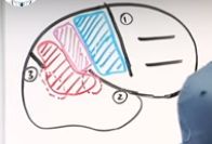
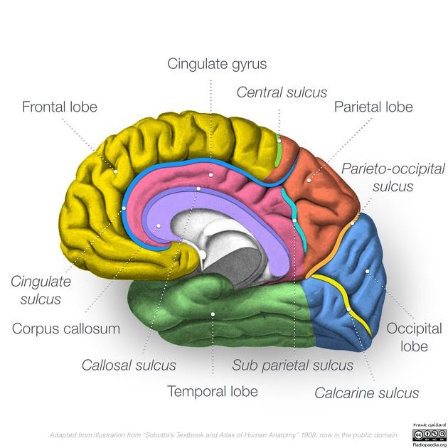
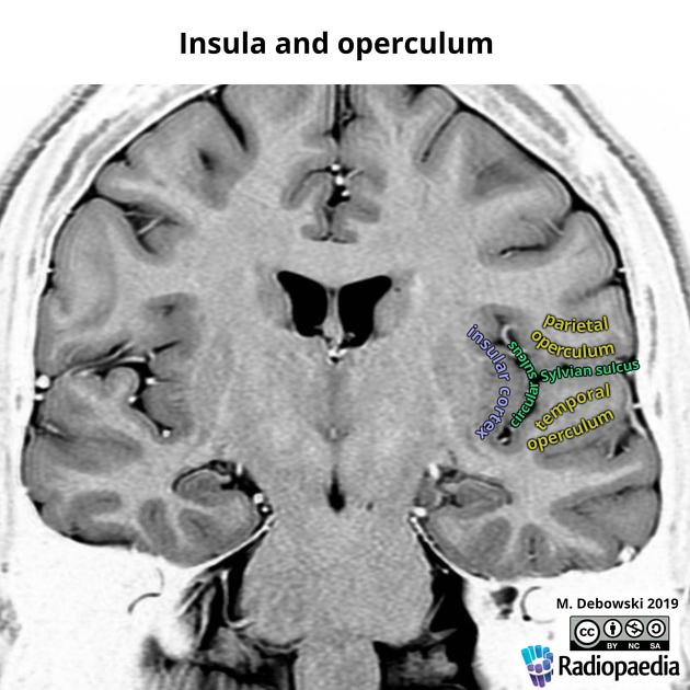
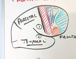
The frontal lobe is bounded posteriorly by the central sulcus[…] (separating it from parietal) and inferiorly by the Sylvian fissure[…] (separating it from temporal).

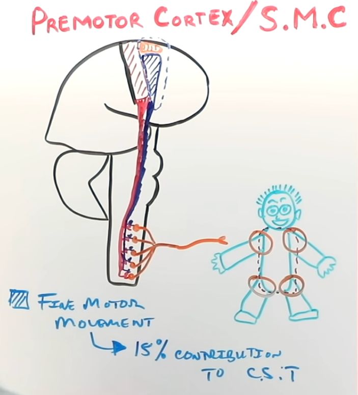
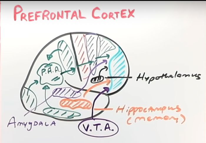
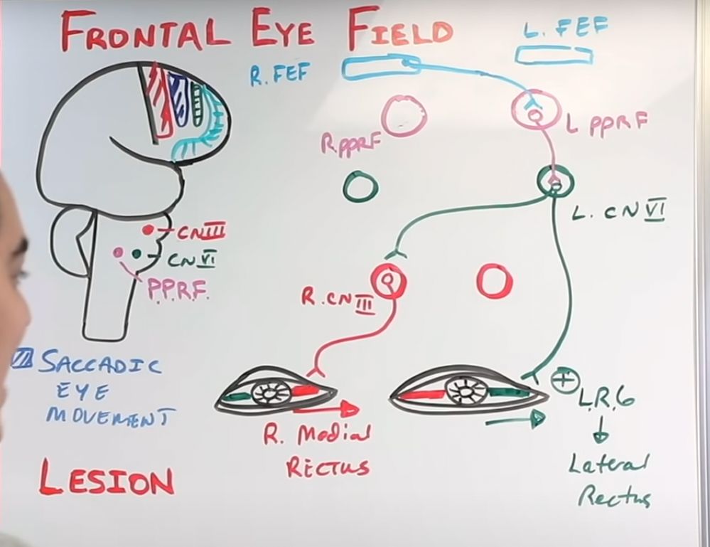
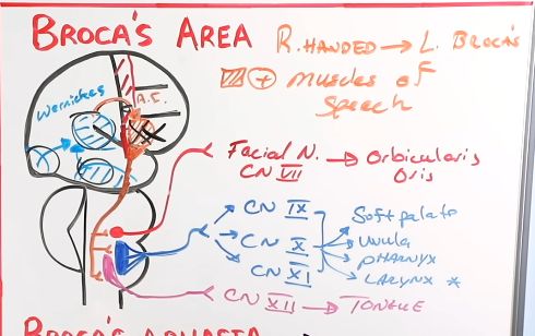
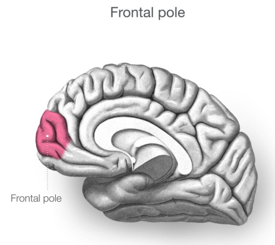
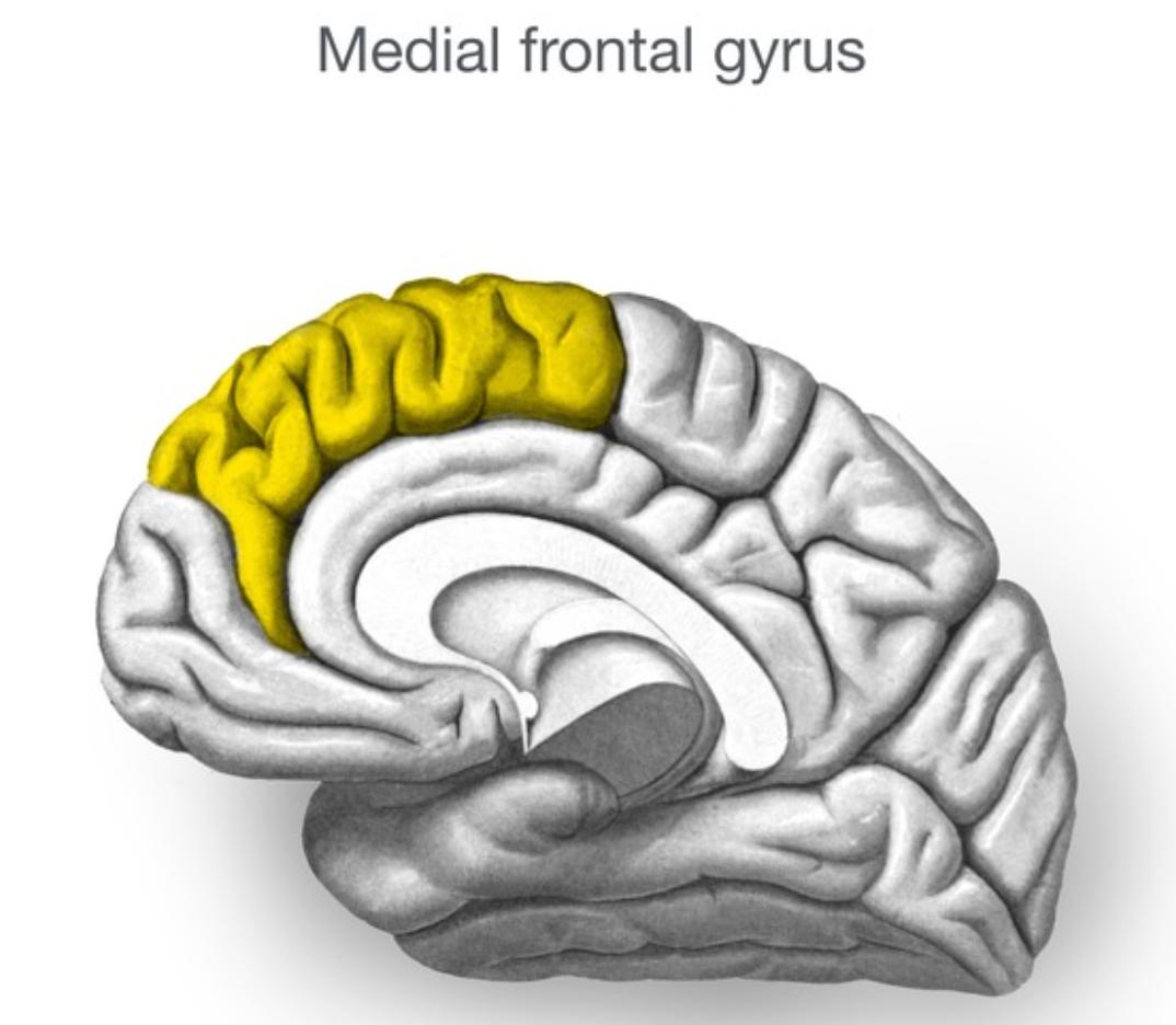
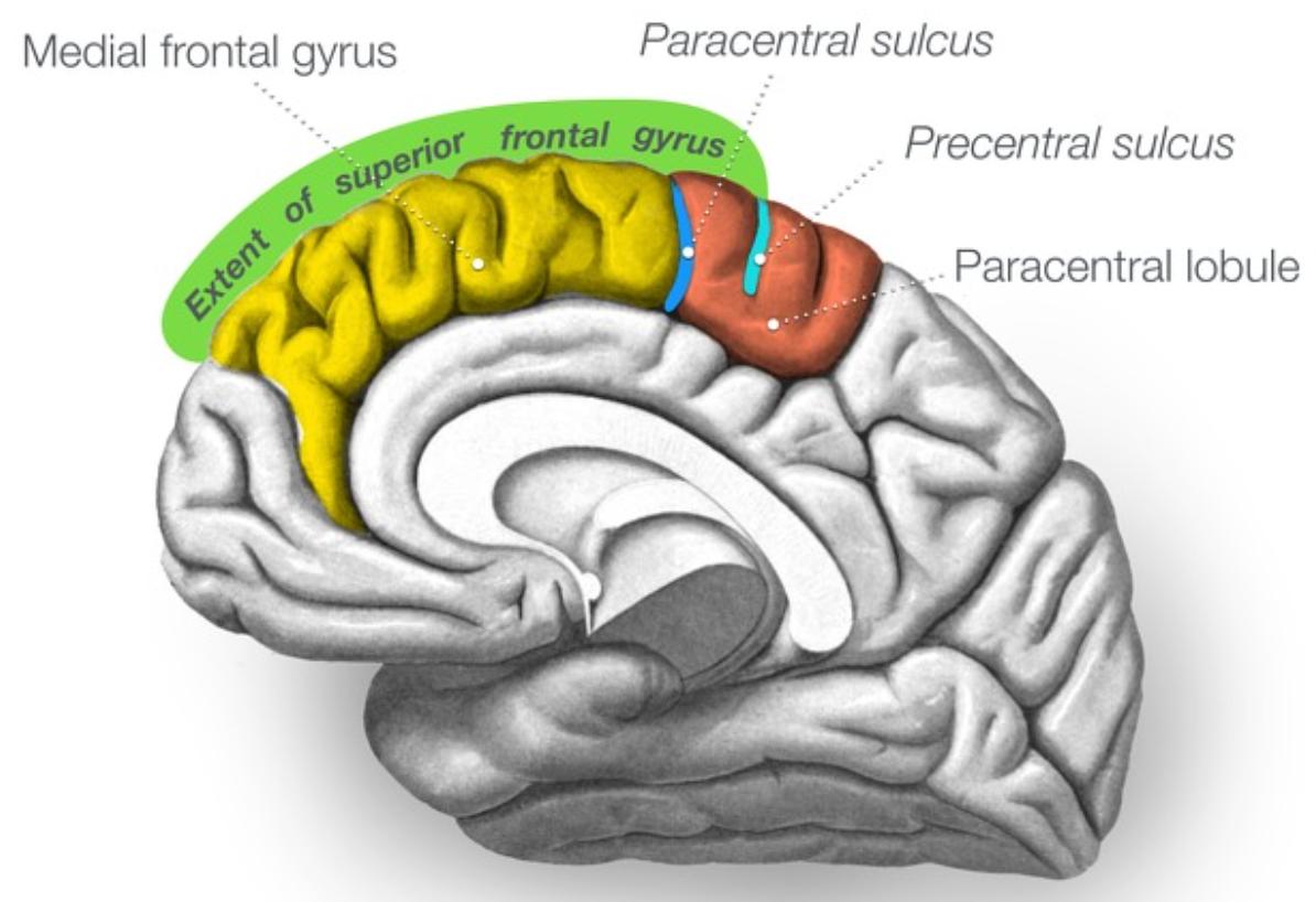
Key cortical areas include the precentral gyrus / primary motor cortex[…] (voluntary movement), the motor association cortex[…] comprising premotor cortex + supplementary motor area (planning, sequencing, fine motor of trunk/proximal extremities), the frontal eye fields[…] (voluntary saccadic eye movement), the prefrontal cortex[…] (personality, behavior, decision-making), and Broca's area[…] in the dominant inferior frontal gyrus (expressive language).
Frontal Lobe :: Descending Tract Pathways

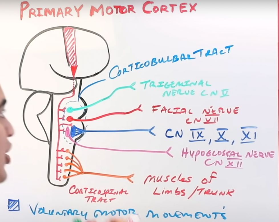
Primary motor cortex corresponds to Brodmann area 4[…] and contributes the bulk of fibers to the corticospinal[…] tract (limbs/trunk voluntary movement) and the corticobulbar[…] tract (CN 5/7/9/10 voluntary movement).

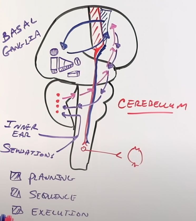
The motor association cortex (premotor + SMA) corresponds to Brodmann area 6[…] and contributes ~15[…]% of the CST's fibers — driving fine motor movements of axial and proximal extremity muscles and coordinating with the basal ganglia[…] and cerebellum[…].

Broca's area in the dominant hemisphere connects to the contralateral primary motor cortex face area via the arcuate fasciculus[…], executing speech motor output.
Primary Motor Cortex :: Homunculus and Stroke Correlation

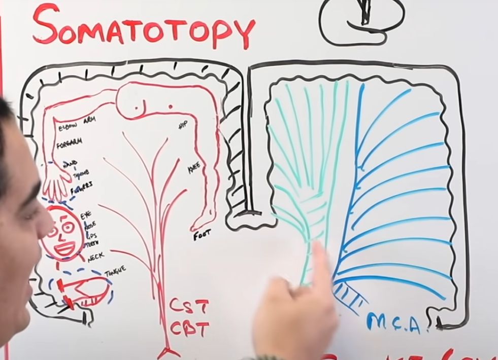
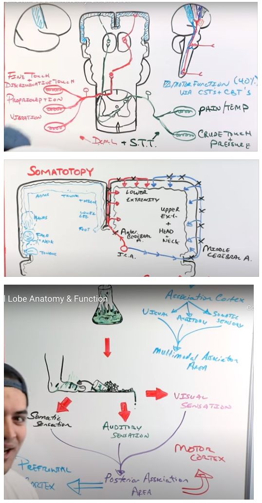
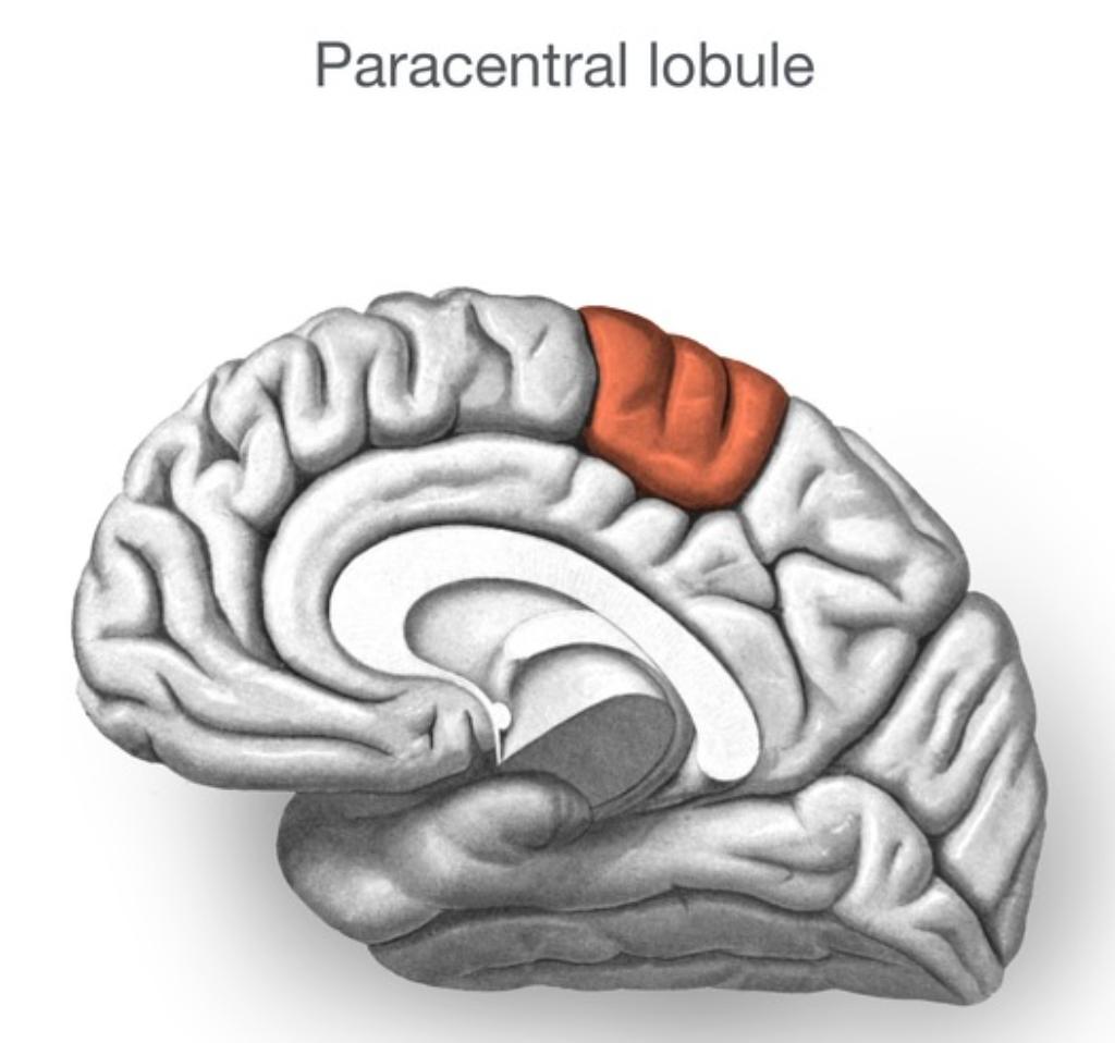
The motor homunculus is organized medial[…] (leg) to lateral[…] (arm then face).

Therefore ACA[…] strokes cause contralateral lower-extremity paralysis, while MCA[…] strokes cause contralateral upper-extremity and face paralysis.

Body parts with larger cortical representation (e.g., face, hands) reflect finer[…] muscle movements.
Parietal Lobe :: Boundaries and Key Areas

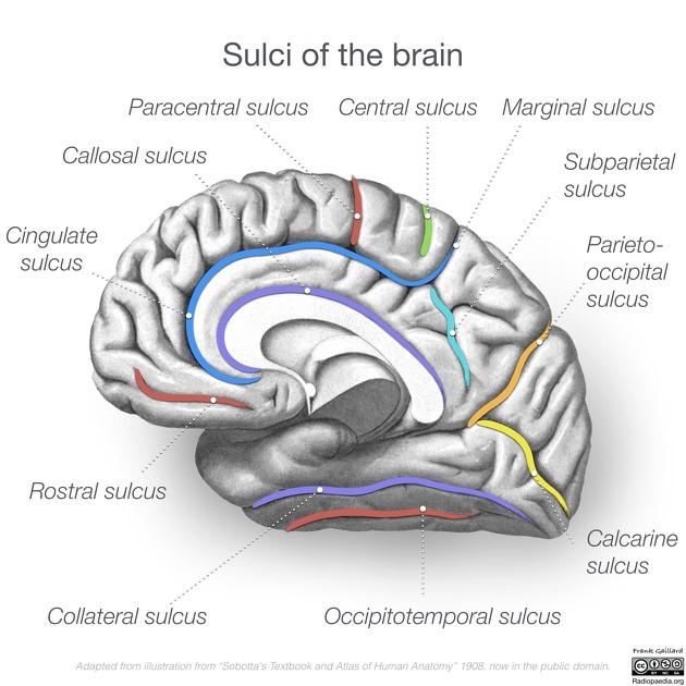
Bounded anteriorly by the central sulcus[…], inferiorly by the Sylvian fissure[…], and posteriorly by the parieto-occipital sulcus[…].

The postcentral gyrus / primary somatosensory cortex[…] (Brodmann areas 1, 2, 3) mediates conscious awareness of somatic sensation, receiving DCML[…] input (fine touch, vibration, proprioception) and spinothalamic[…] input (pain, temperature, crude touch).

It also contributes ~40[…]% of descending CBT/CST fibers.

The somatosensory association cortex[…] integrates sensation into perceptions of size, shape, texture; lesions cause astereognosis[…] and agraphesthesia[…].

The angular gyrus[…] of the dominant parietal lobe supports reading and writing; lesions produce Gerstmann syndrome — agraphia, acalculia, finger agnosia, left-right disorientation[…].

Temporal Lobe :: Boundaries and Anatomy

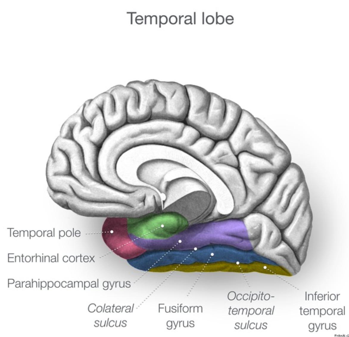
Separated from frontal and parietal lobes by the Sylvian fissure[…] and from the occipital lobe by an imaginary line from the pre-occipital notch[…] to the parieto-occipital sulcus.

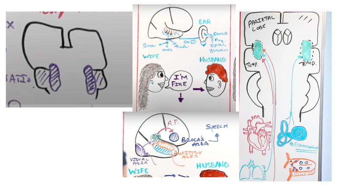
Key areas: primary auditory cortex[…] (Heschl's gyrus) — conscious awareness of sound frequency, pitch, and location; lesions cause contralateral loss of these features.

The auditory association cortex[…] gives meaning to sound by analyzing it against past memories.

Wernicke's area[…] (dominant superior temporal gyrus) mediates comprehension of written/spoken language; lesions produce receptive (fluent) aphasia[…].

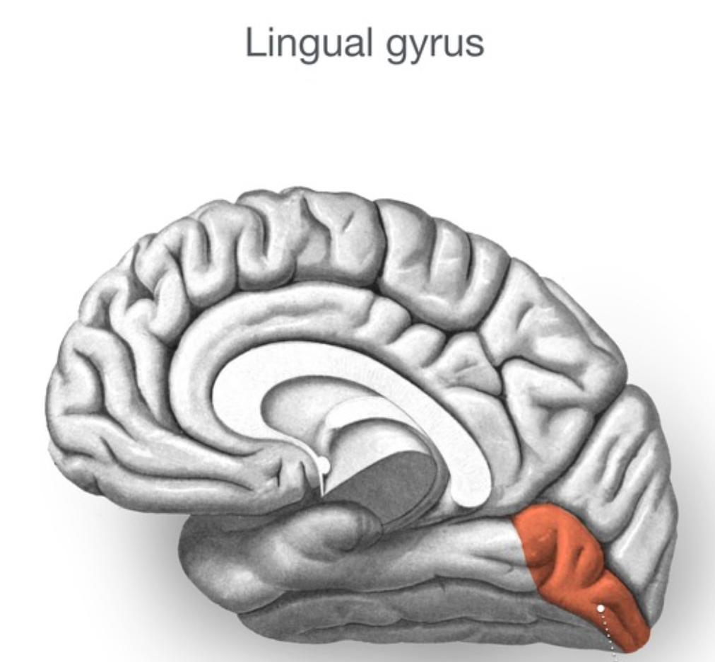
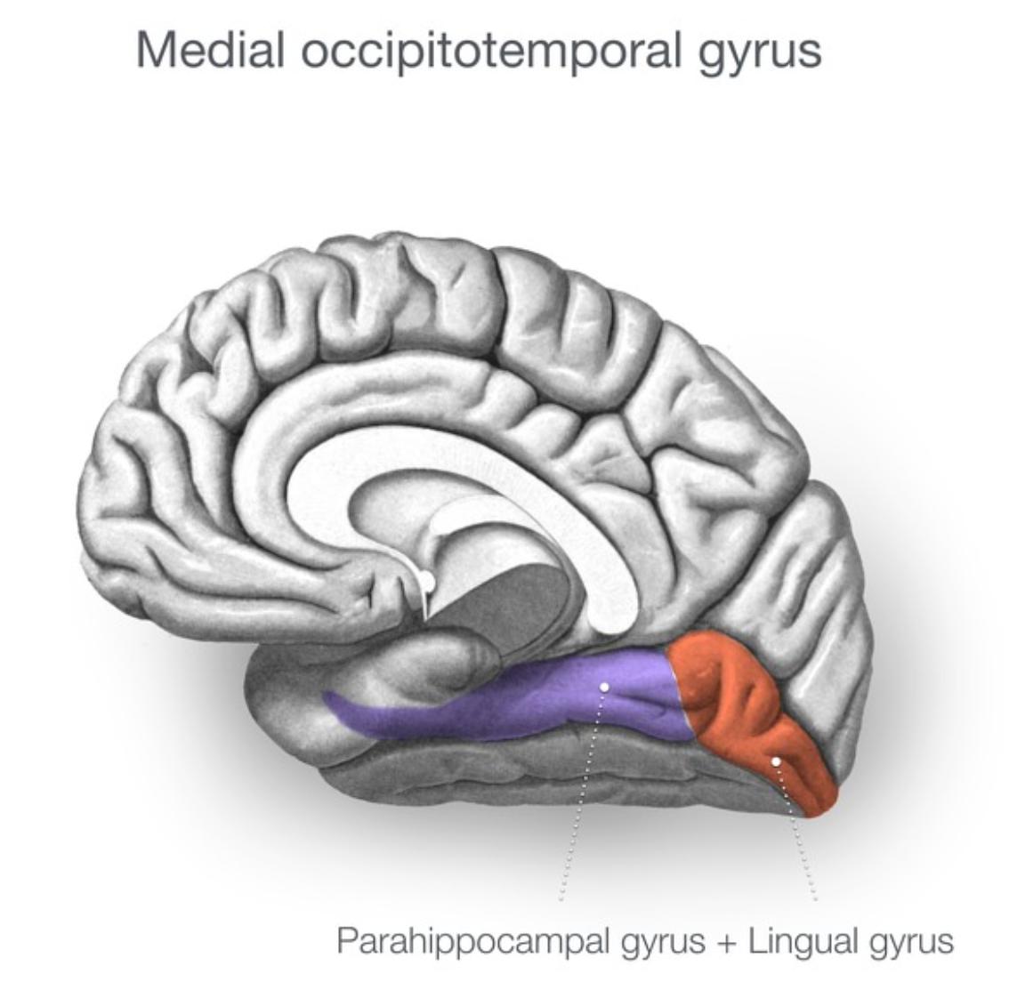
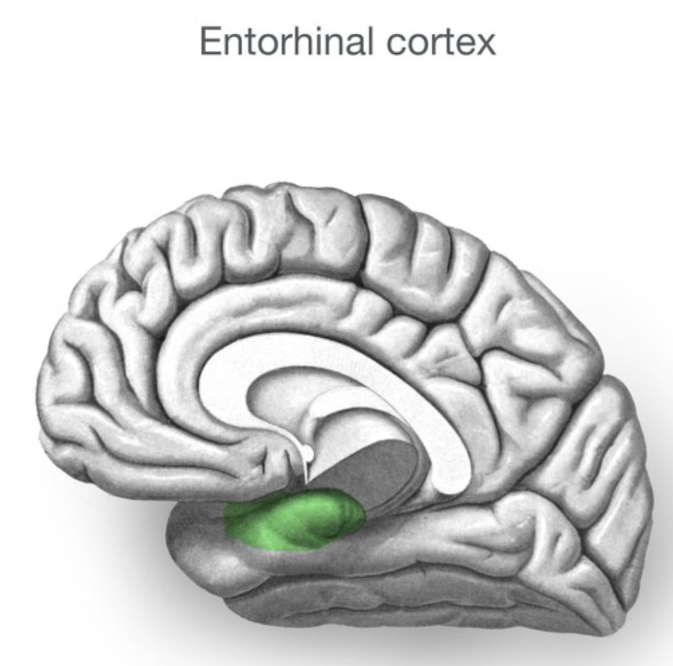
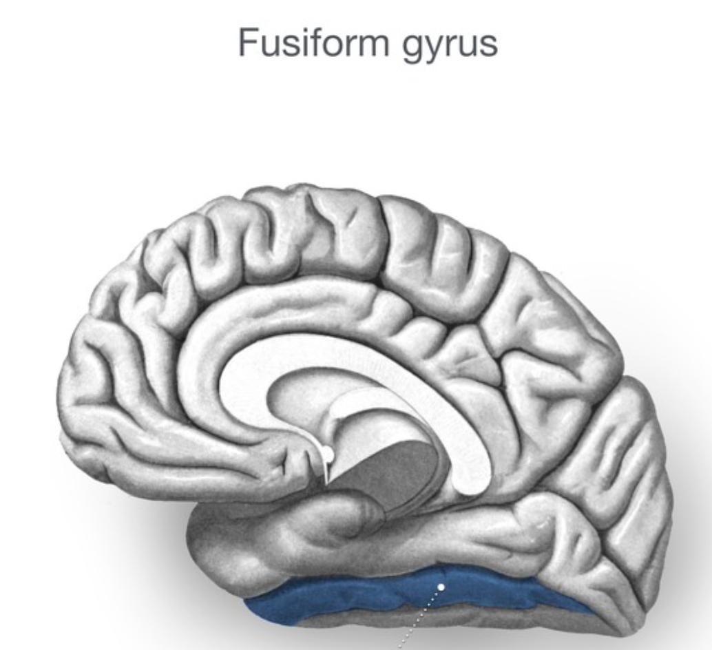
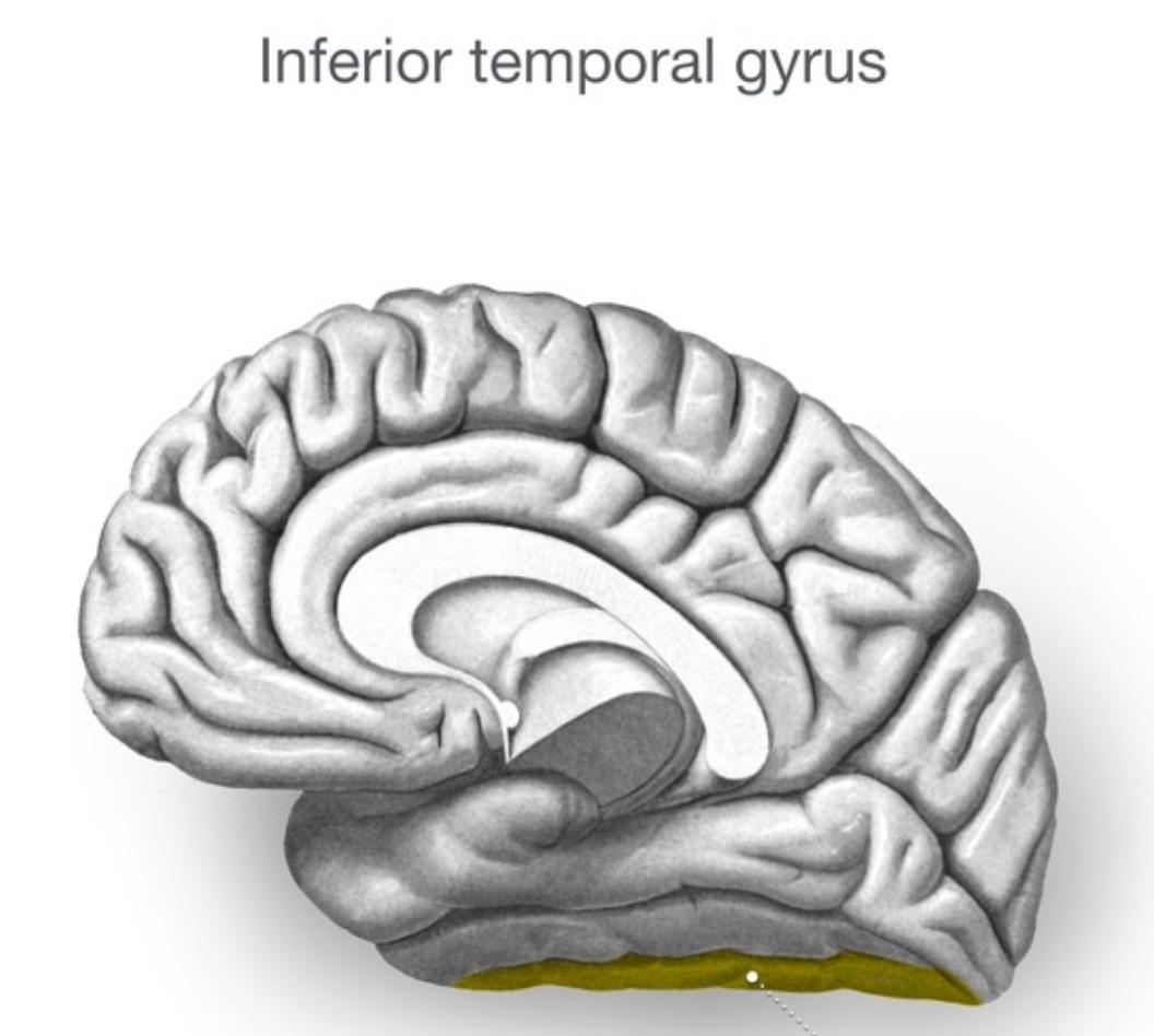
The medial temporal lobe contains the hippocampus[…] and amygdala[…], supporting memory consolidation and emotional processing.

Occipital Lobe :: Boundaries and Anatomy

Separated from the parietal lobe by the parieto-occipital sulcus[…], and from the temporal lobe by an imaginary line from the pre-occipital notch[…] to the parieto-occipital sulcus.

The primary visual cortex[…] (V1, Brodmann 17) lies along the calcarine sulcus and provides conscious awareness of visual stimuli.

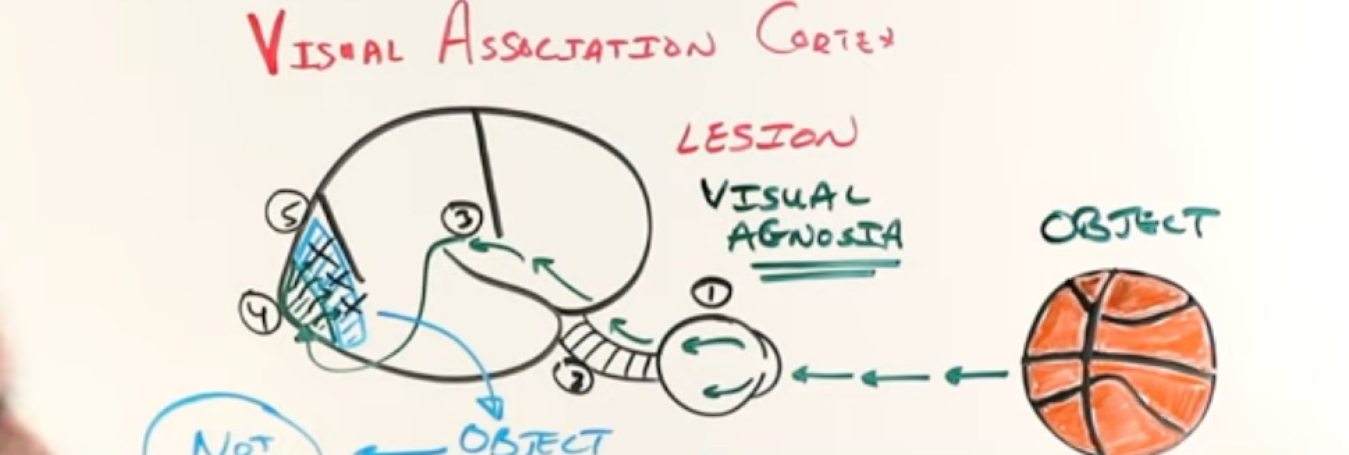
The visual association cortex[…] (V2-V5) analyzes shape, size, color, and motion — recognizing visual inputs by comparing to past memories.

Lesions of the visual association cortex produce visual agnosia[…], while bilateral occipitoparietal lesions produce Balint syndrome[…] (simultanagnosia, oculomotor apraxia, optic ataxia).

Basal Ganglia :: Anatomy

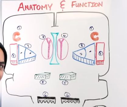
Components include the caudate nucleus[…], putamen[…] (caudate + putamen = striatum[…]), globus pallidus[…] (GPe and GPi; putamen + GP = lentiform nucleus[…]), the VA and VL nuclei of the thalamus[…], the subthalamic nucleus (STN)[…], and the substantia nigra[…] in the midbrain — divided into pars compacta (DA-packed) and pars reticulata.
Basal Ganglia :: Direct, Indirect, and Nigrostriatal Pathways

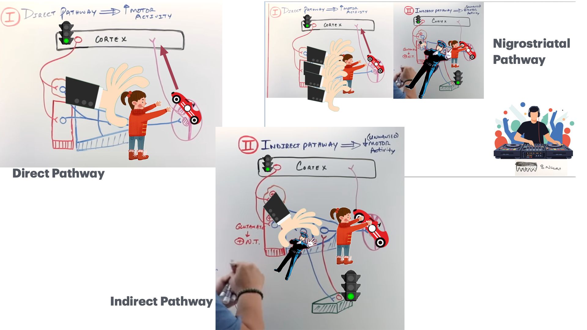
The direct[…] pathway INCREASES motor activity: Cortex (+) → Striatum (-) → GPi (-) → Thalamus (+) → end effect is (+) cortex. Damage example: Parkinson's disease[…].

The indirect[…] pathway DECREASES unwanted motor activity: Cortex (+) → Striatum (-) → GPe (-) → STN (+) → GPi (-) → Thalamus (+) → end effect is (-) cortex. Damage causes hyperkinetic disorders like Huntington's, Wilson's, Sydenham chorea, hemiballismus[…].

The nigrostriatal[…] pathway from substantia nigra pars compacta to striatum MODULATES movement via dopamine; degeneration here is the central lesion in Parkinson's disease[…].
Basal Ganglia :: Dopamine Receptors (D1 vs D2)

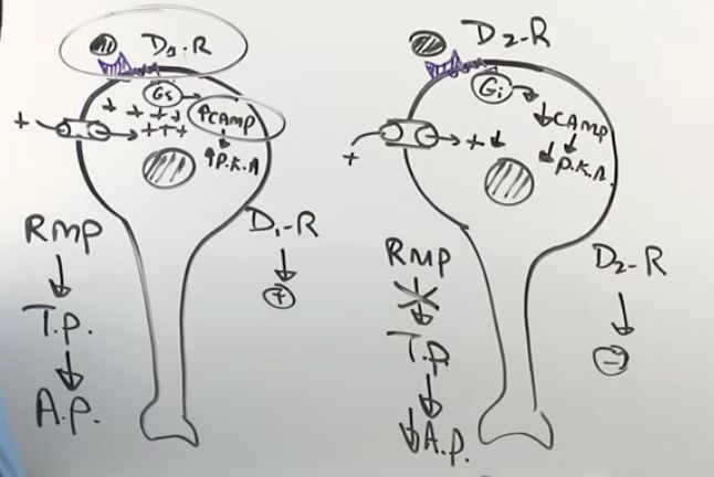
The D1[…] receptor is excitatory: DA binds a GPCR[…] → (+)cAMP → (+)PKA → positive charge inside the cell — drives the direct pathway.

The D2[…] receptor is inhibitory: DA binds a GPCR → (-)cAMP → (-)PKA → negative charge inside the cell, preventing action potentials — drives the indirect pathway (via inhibition of striatal indirect-pathway neurons).

Dopamine therefore simultaneously activates direct and inhibits indirect[…] pathways, both promoting movement — explaining why DA loss in PD produces hypokinesia.
Basal Ganglia :: Functional Loop

The primary somatosensory[…], primary motor[…], and motor association cortices (with input from Broca's area) form the voluntary motor plan.

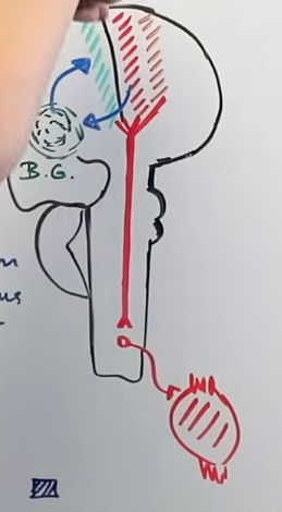
This plan is sent to the basal ganglia, which start[…] desired movement (direct pathway), stop[…] unwanted movement (indirect pathway), and modulate[…] movement (nigrostriatal pathway).

The processed plan returns via the thalamus to cortex and descends through CST/CBT[…] to body and cranial nerve muscles.