MRI

MRI

Radiology · 804 cards · 172 labeled figures

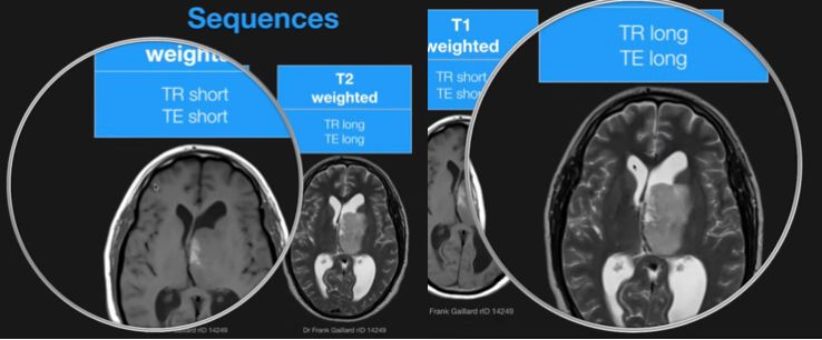
Differentiating T1 from T2
  • T1: normal: denser gray matter cortex darker than white matter subcortex[…]
  • T2: weird: less dense white matter subcortex darker than denser gray matter cortex[…]
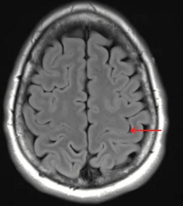
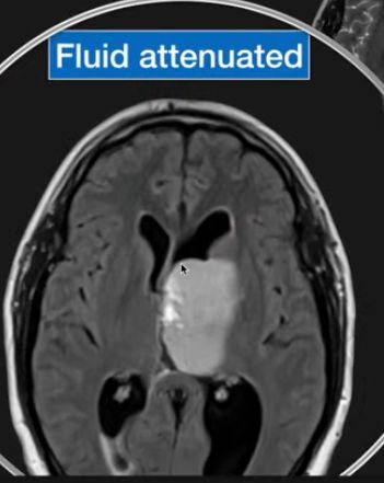
FLAIR Sequence Below. Is it T1 or T2 - how do you know? 
T2: weird, white matter subcortex darker than denser gray matter cortex[…]
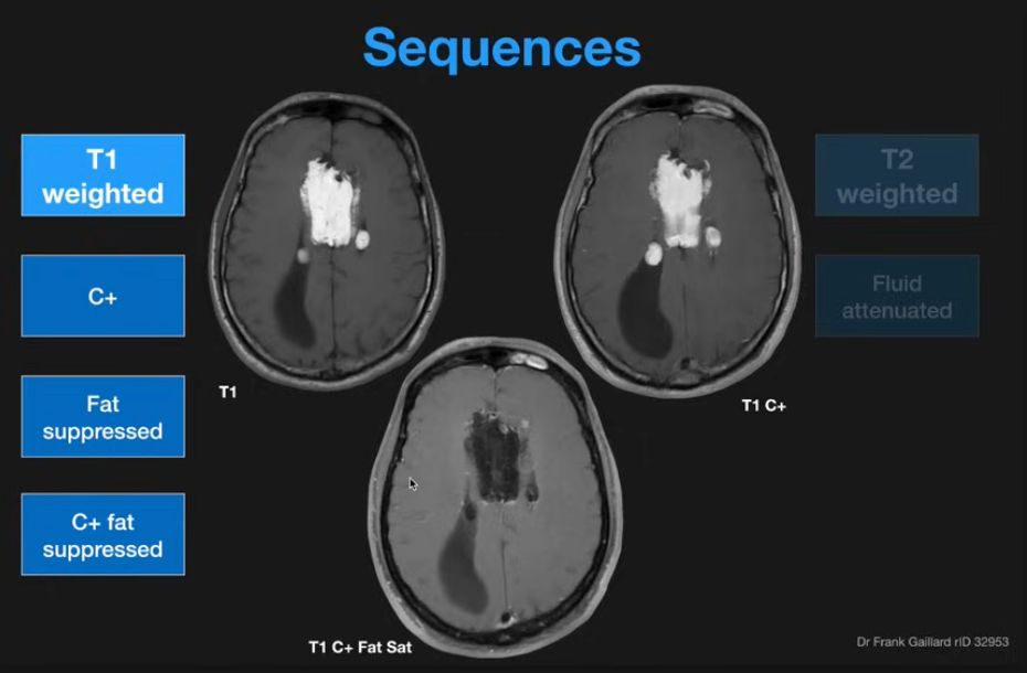
T1 Sequences
  • Regular T1
  • Contrast+[…]
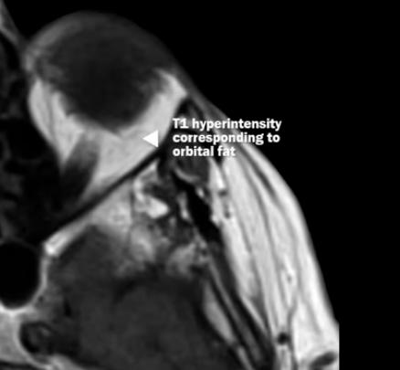
  • C+ Fat Sat[…]: fat[…] suppressed to rid artifact and better show C+[…] areas 
    • this is done for ex with pts w/IC mass[…] lesions in skull base where extracranial extension is possible (such as skull base meningiomas[…] or CPA angle tumors[…])
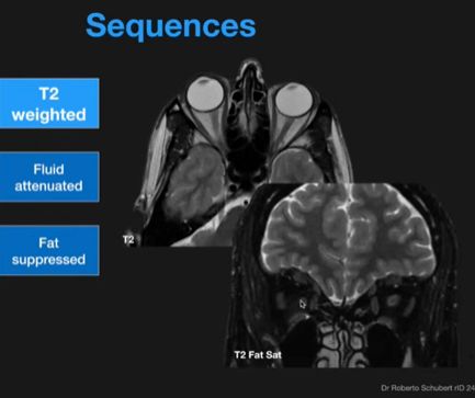
T2 Weighted Sequences
  • T2 Fat Sat[…]
    • Orbits[…]: often to look at the EOM[…] 
    • rule out of CSF leak[…]
  • FLAIR[…]
  • T2* SWI[…] or GRE[…]
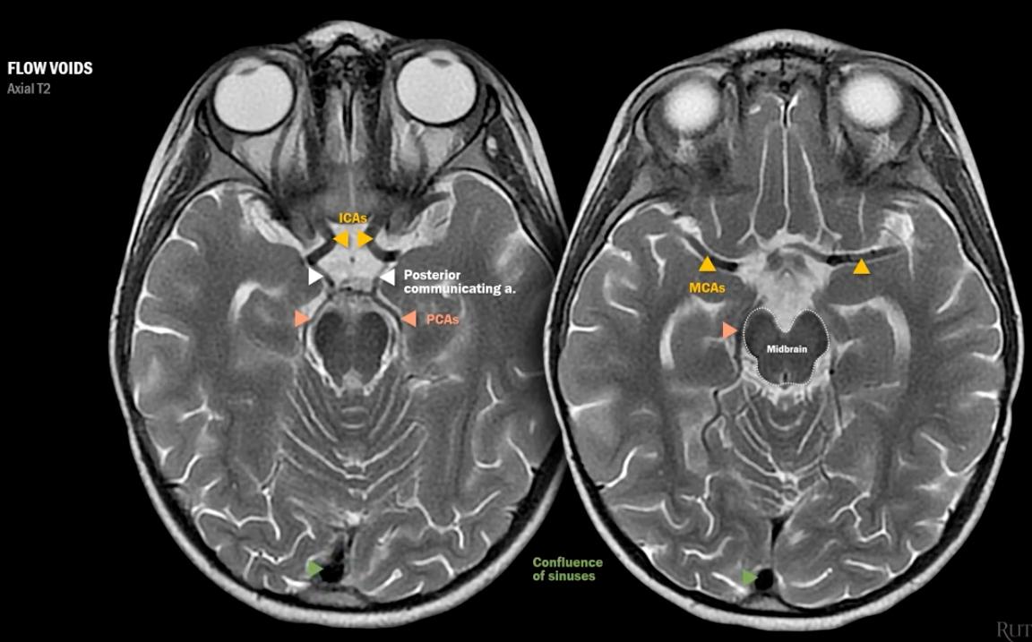
    • these look for areas of paramagnetic[…] void
  • DWI[…]
  • ADC[…]
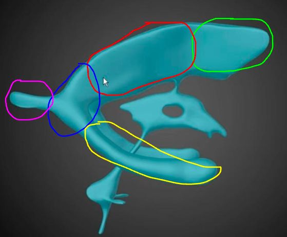
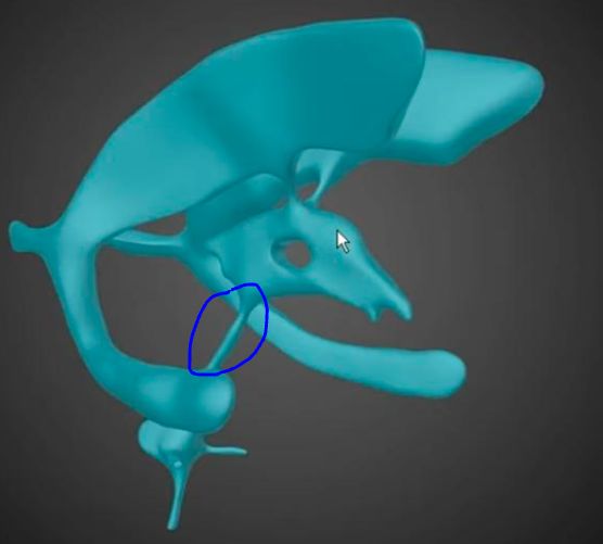
Ventricular System Anatomy Review
  • green: anterior horn, lateral ventricle[…]
  • red: body, lateral ventricle[…]
  • blue: atrium/trigone, lateral ventricle[…]
  • purple: occipital horn, lateral ventricle[…]
  • yellow: temporal horn, lateral ventricle[…]

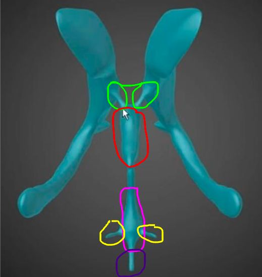
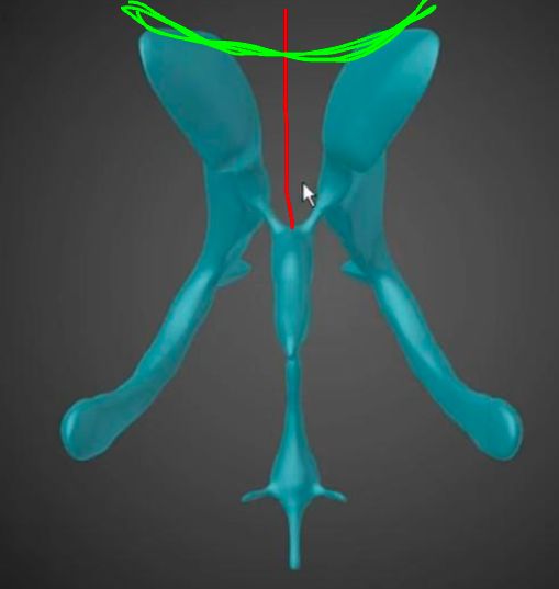
Ventricular System Anatomy Review
  • green: interventricular foramen[…] aka foramen of Monroe[…]
  • red: 3rd ventricle[…]
  • blue: cerebral aqueduct of Sylvius[…]
  • pink: 4th ventricle[…]
  • yellow: lateral apertures[…] aka foramen of Luschka[…]
  • dark purple: median aperture[…] aka foramen of Magendie[…]

Ventricular System Anatomy Review
  • red: septum pellucidum[…]
  • green: corpus callosum[…]

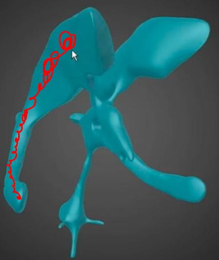
Ventricular System Anatomy Review
  • red: caudate[…] nucleus runs here

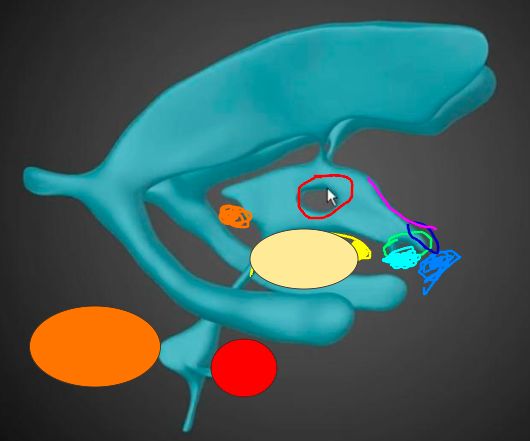
Ventricular System Anatomy Review
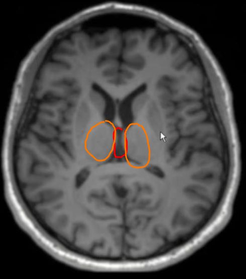
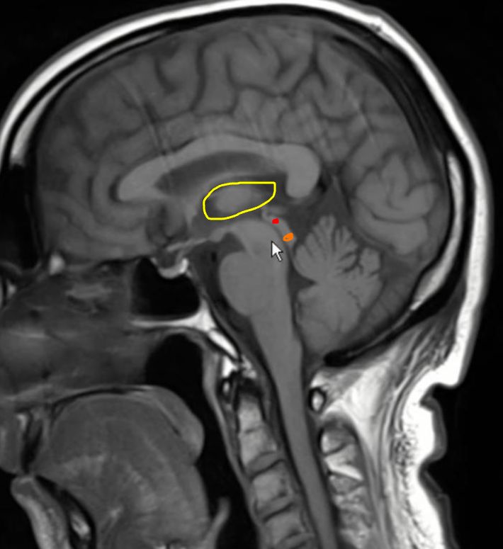
  • red hole present in some people in 3rd ventricle: where the thalami[…] touch called an interthalamic adhesion[…]
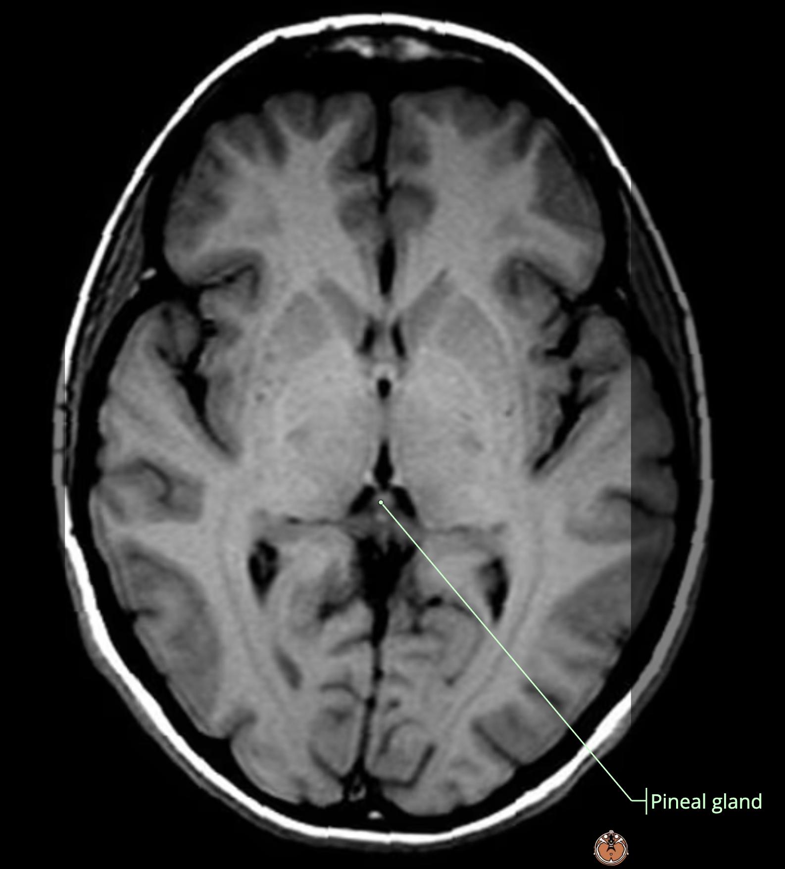
  • orange: pineal gland[…]
  • circle: midbrain[…] with yellow mamillary bodies[…]
  • green: infundibular recess[…]
  • light blue: pituitary gland[…]
  • medium blue: optic chiasm[…]
  • dark blue: optic recess[…]
  • pink: lamina terminalis[…]
  • red circle: pons[…]
  • orange circle: cerebellum[…]

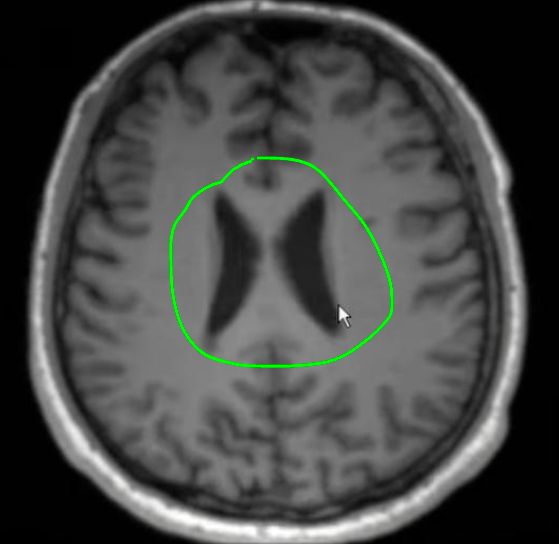
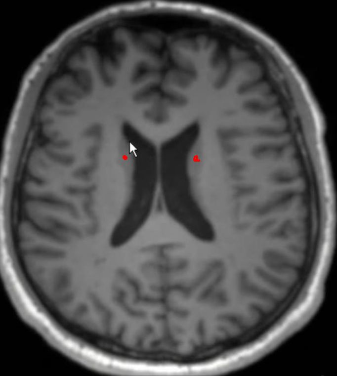
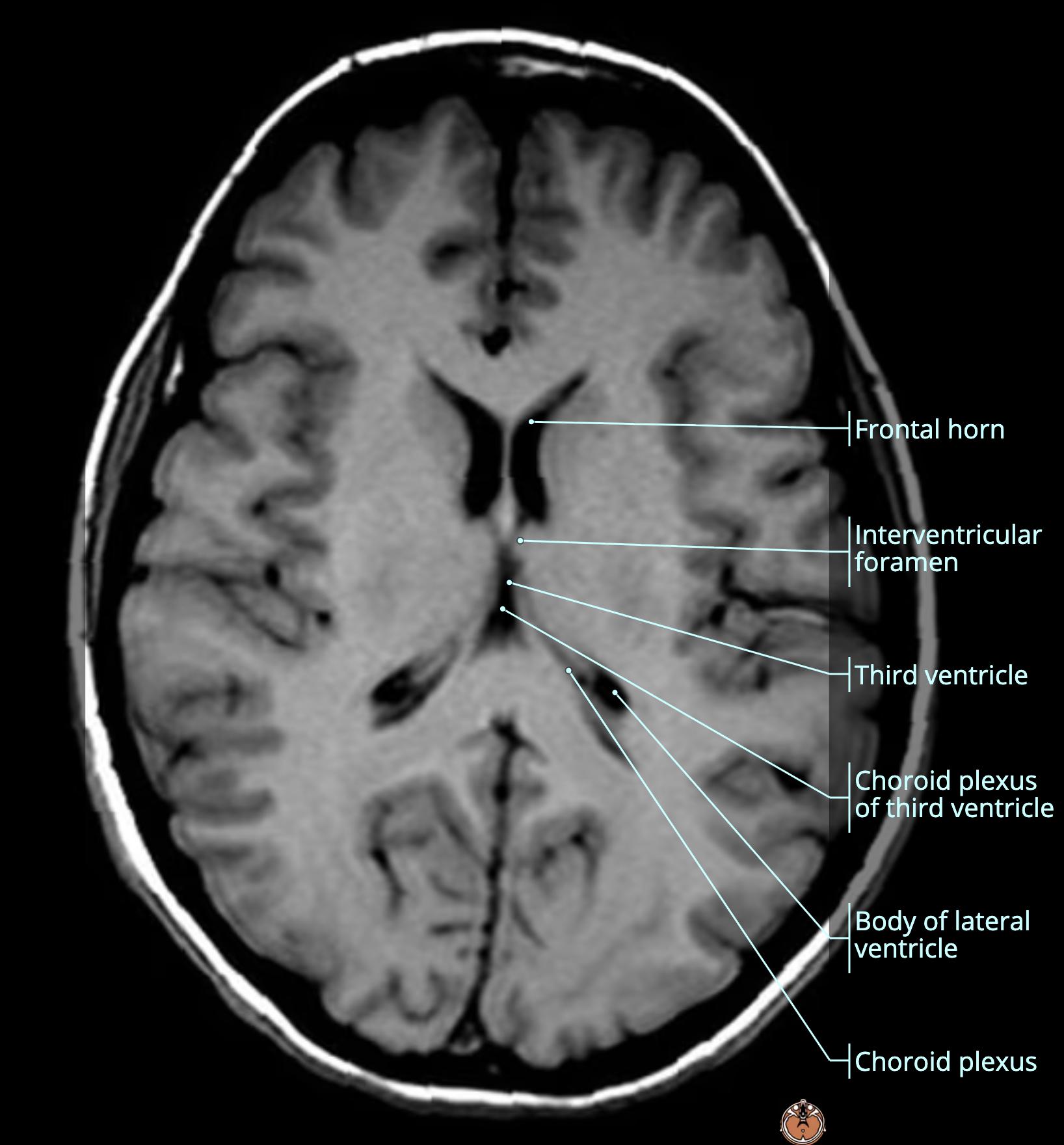
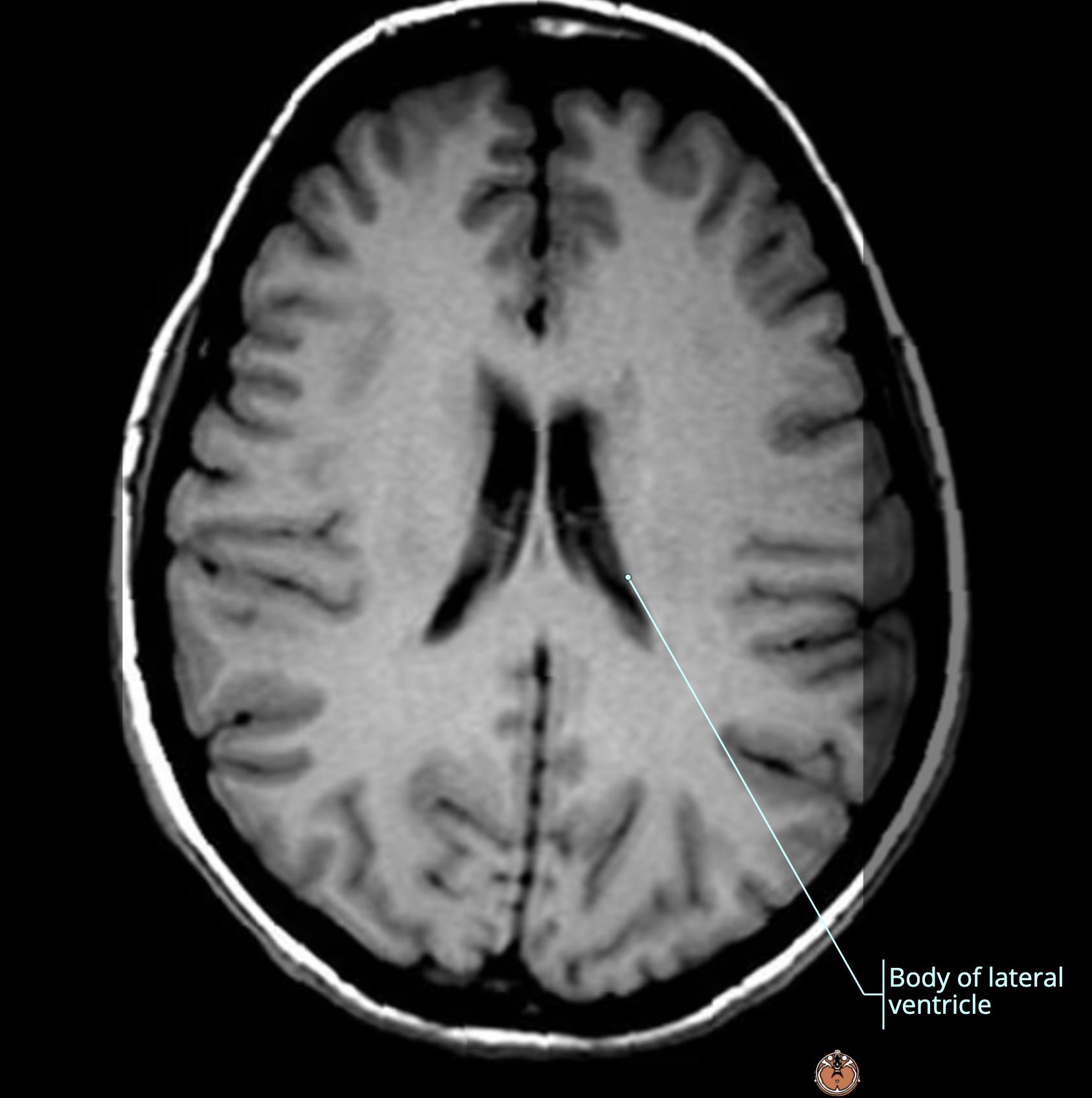
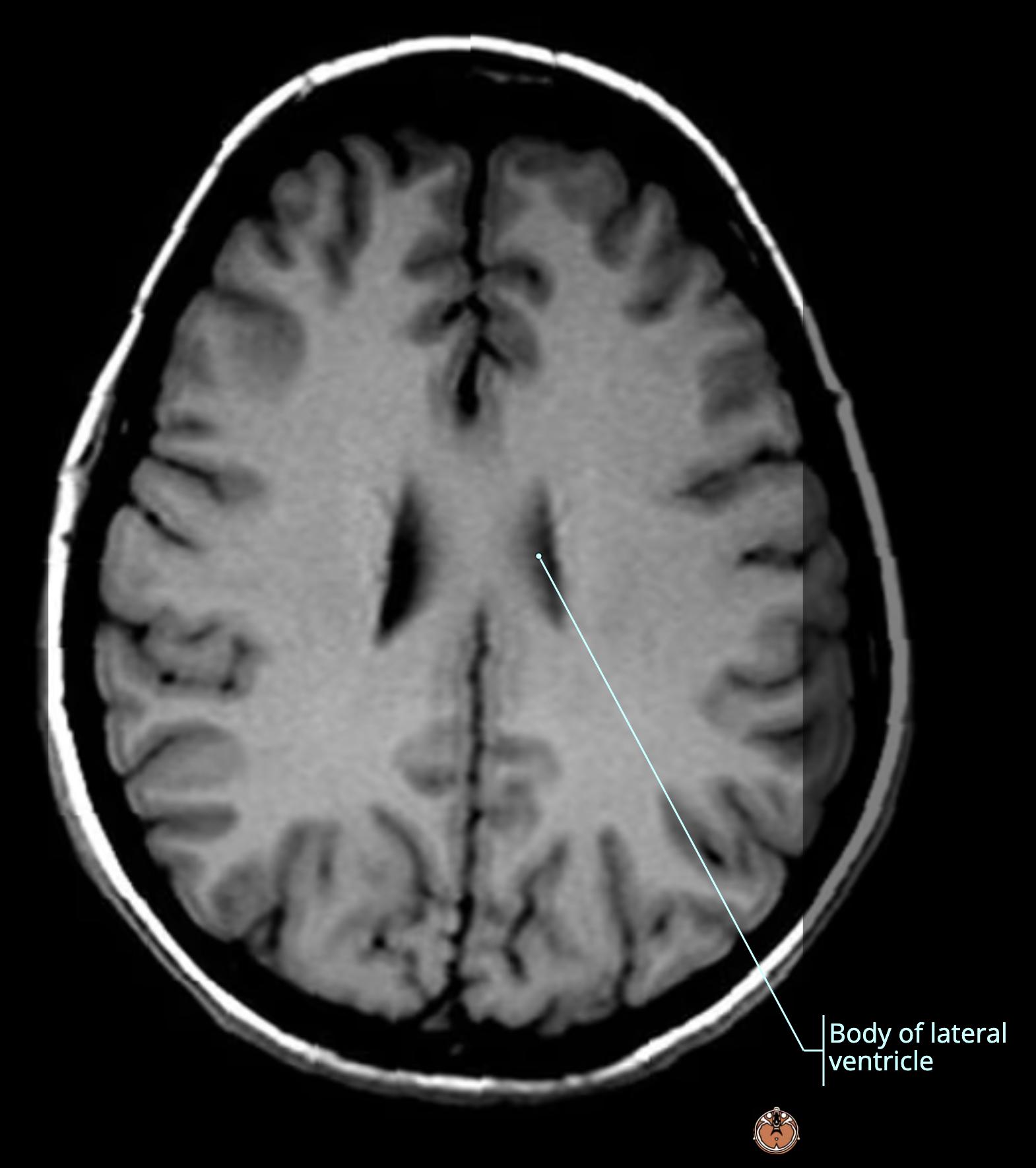
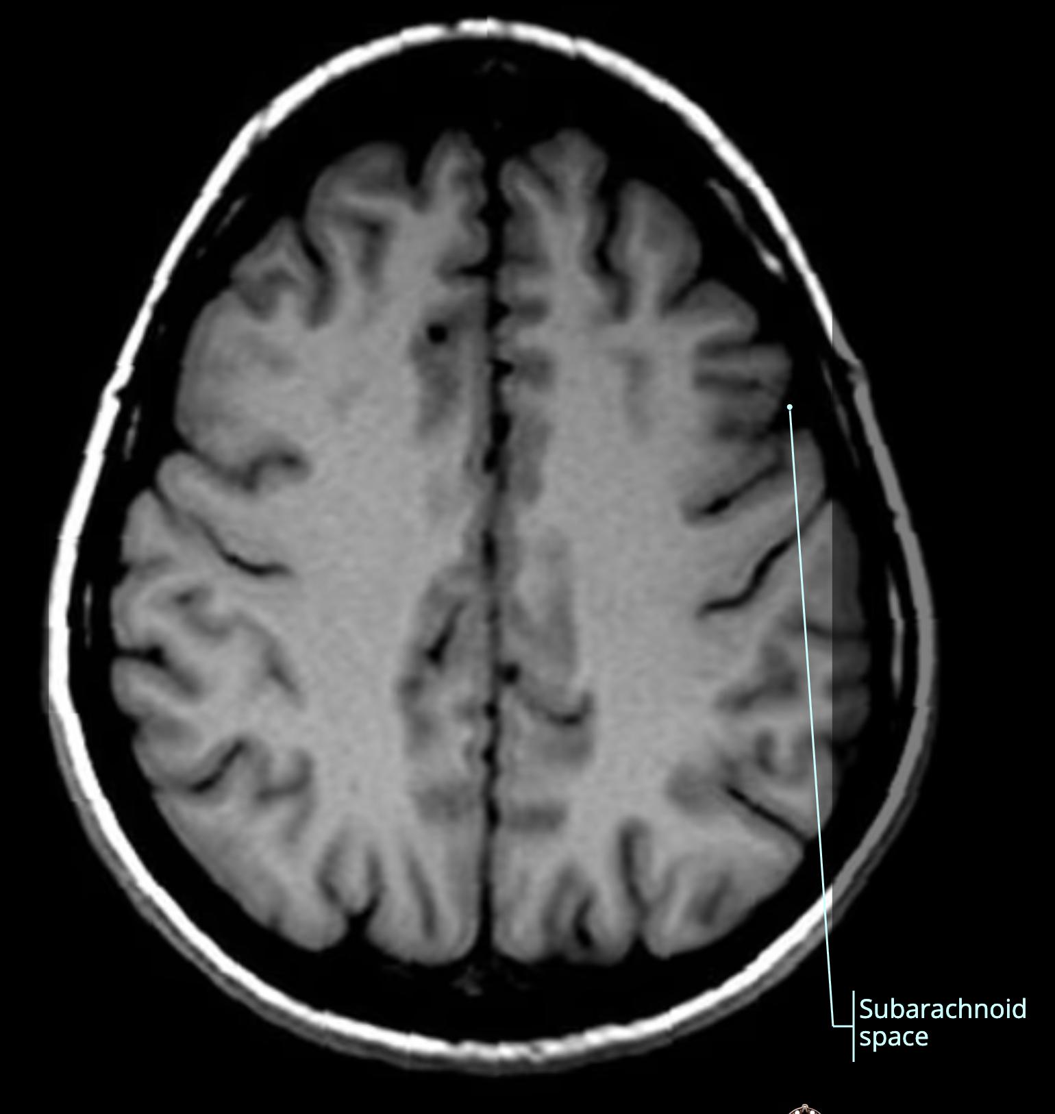
Ventricular System Anatomy Review (Top to Bottom) (1)
  • T1[T1 or T2?], Axial[cut] scan
  • These are the lateral ventricles[…]

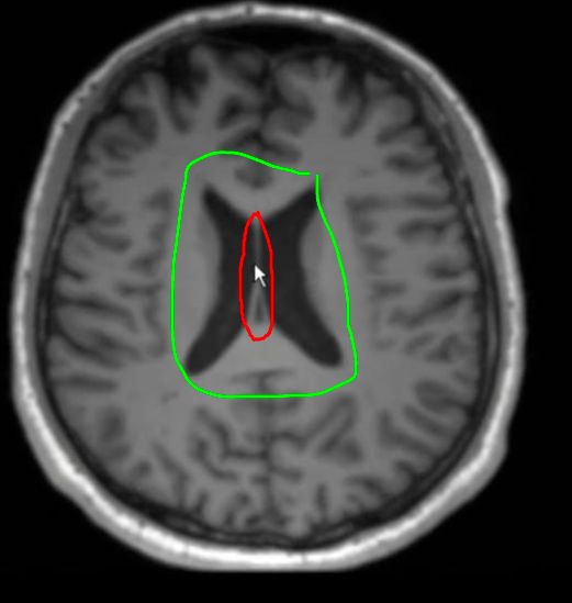
Ventricular System Anatomy Review (Top to Bottom) (2)
  • These are the lateral ventricles[…] separated by thin membrane called septum pellucidum[…]

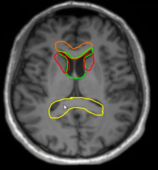
Ventricular System Anatomy Review (Top to Bottom) (3-4)
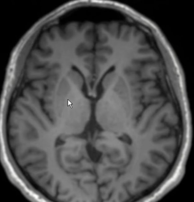
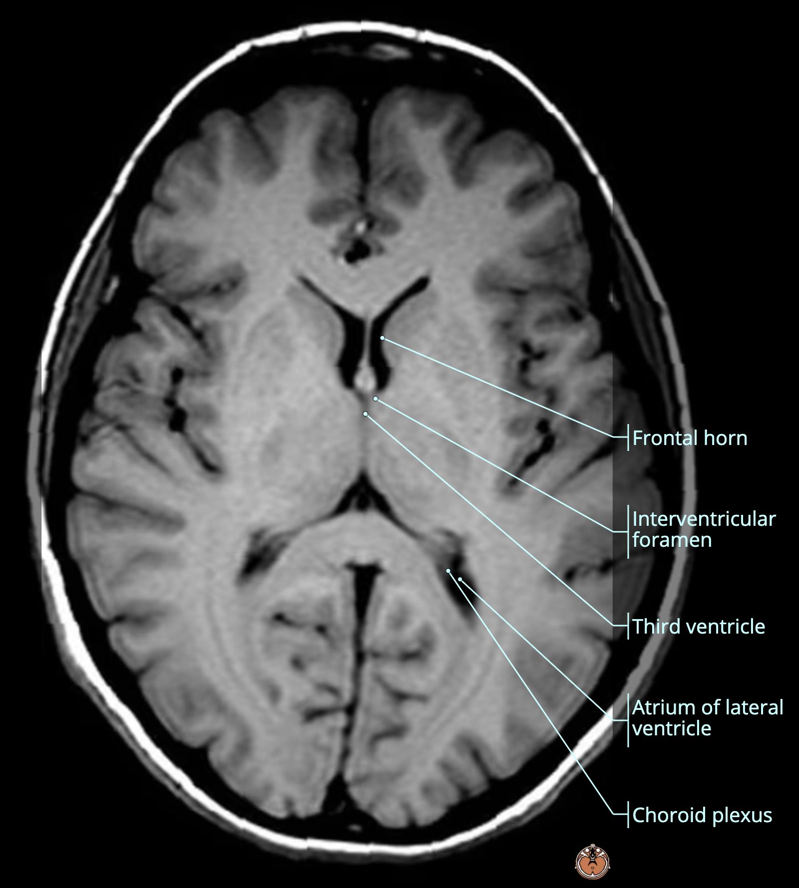
  • These are the anterior horns, lateral ventricles[…] (green) with caudate nucleus[…] (red) to the side and genu, corpus callosum[…] (orange) and splenium of corpus callosum[…] (yellow)
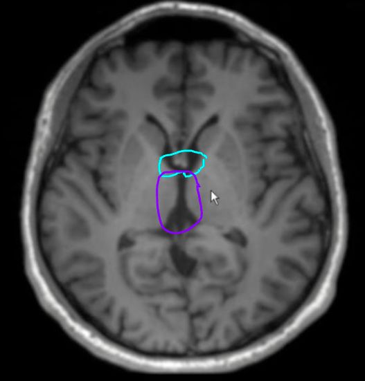
  • they connect via the interventricular foramen / foramen of monroe[…] (light blue) into the 3rd ventricle[…] (purple)

Ventricular System Anatomy Review (Top to Bottom) (5-9)
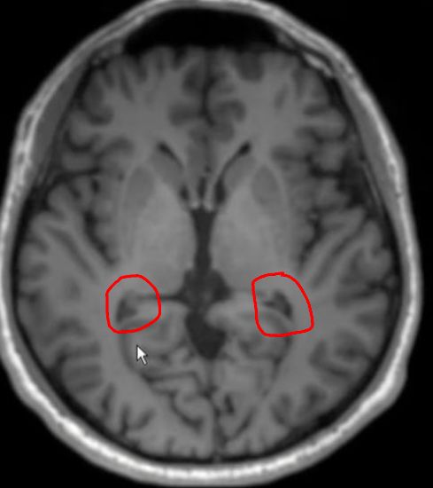
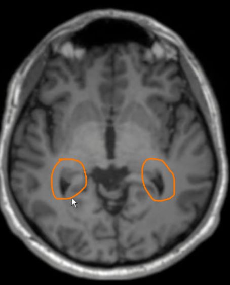
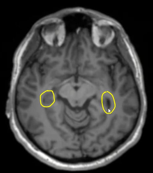
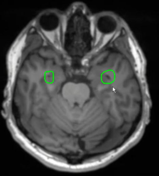
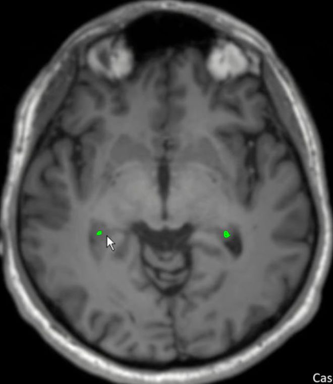
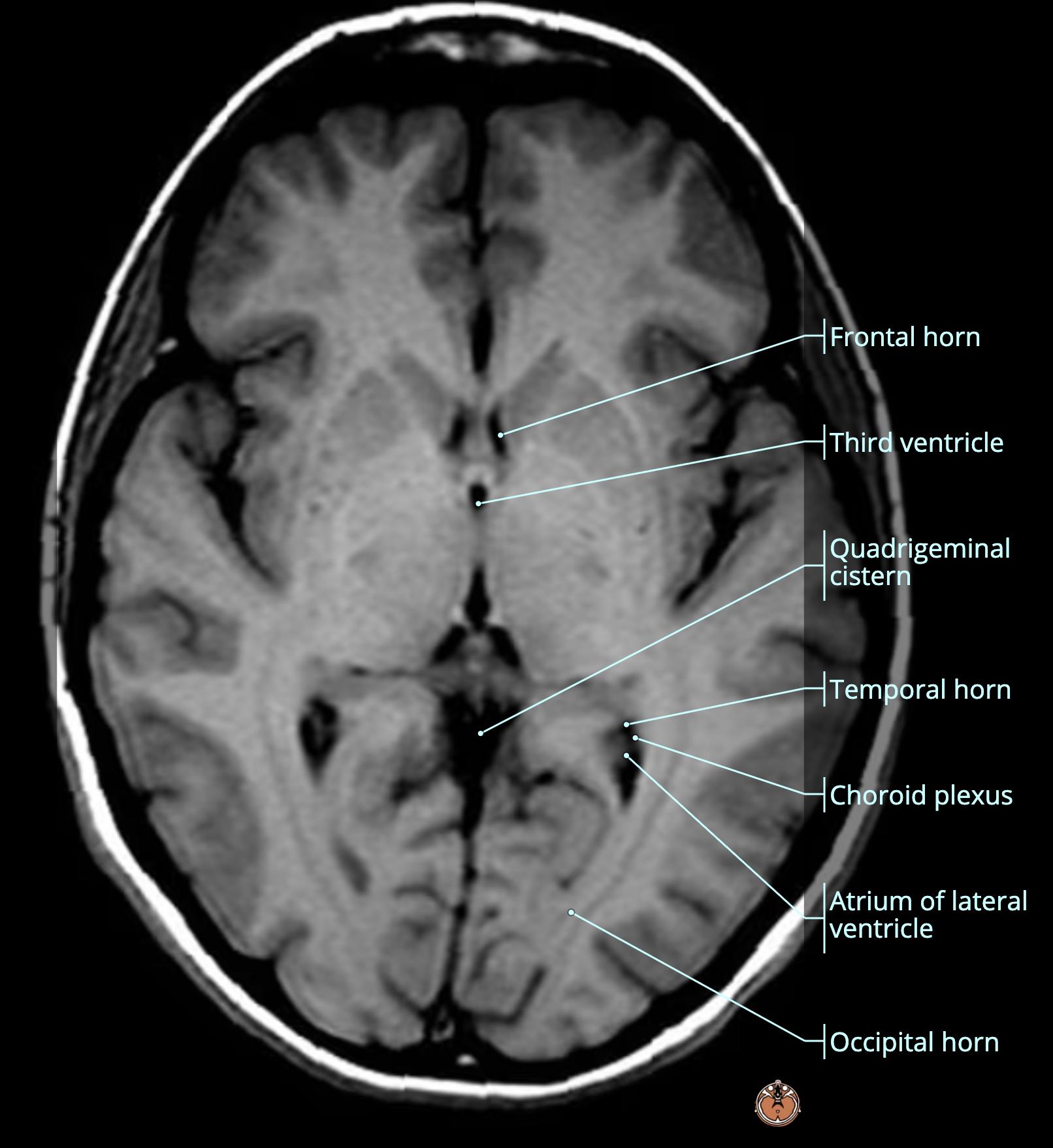
  • these are various cross sections of the occipital horn, lateral ventricles[…] descending until you reach the temporal[…] lobe where they turn into the temporal horns, lateral ventricles[…]


Ventricular System Anatomy Review (Top to Bottom) (10-13)
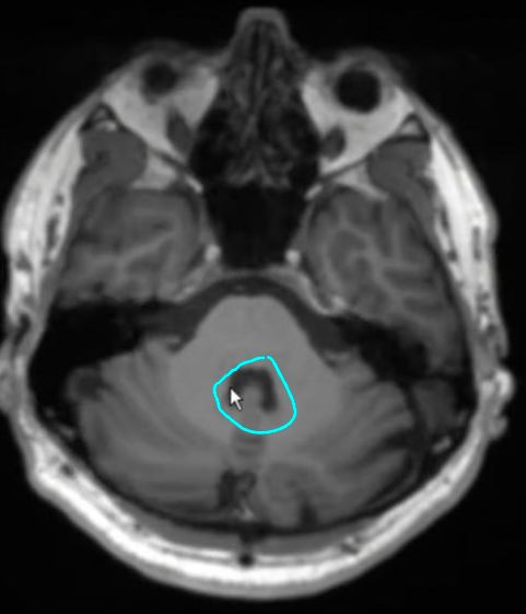
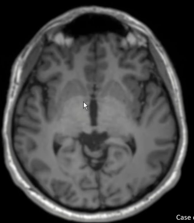
  • this is the third ventricle (red) flanked on either side by the thalami (orange) that descends (yellow) and narrows into the cerebral aqueduct[…] (green) at the level of midbrain[…] 
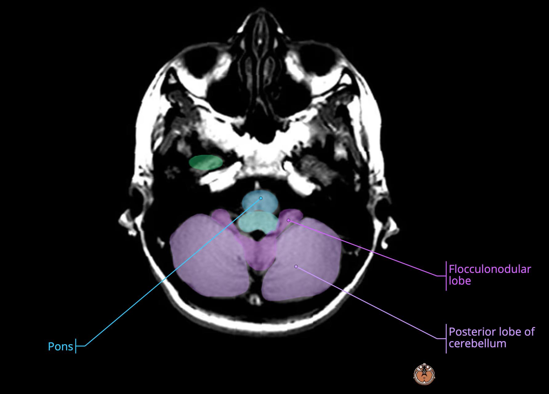
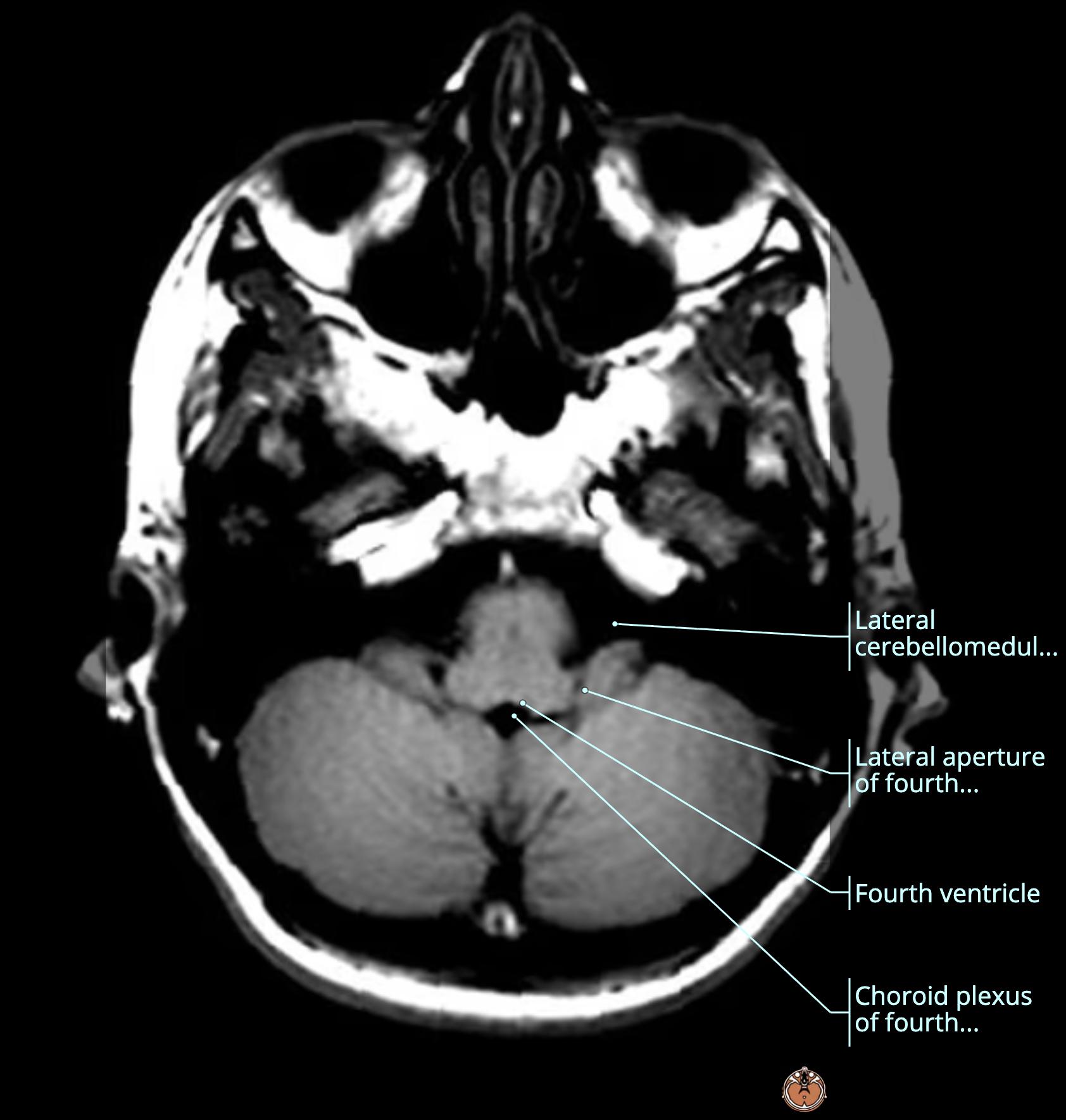
  • this descends further until it turns into the 4th ventricle[…] (light blue) at the level of pons[…]

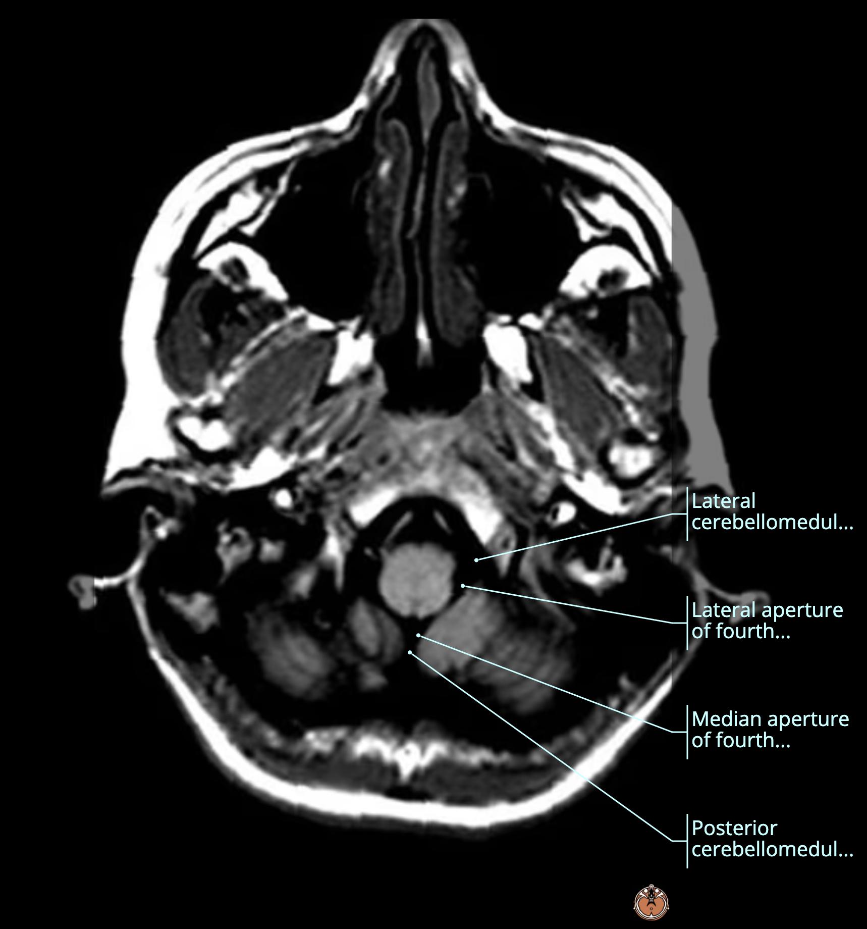
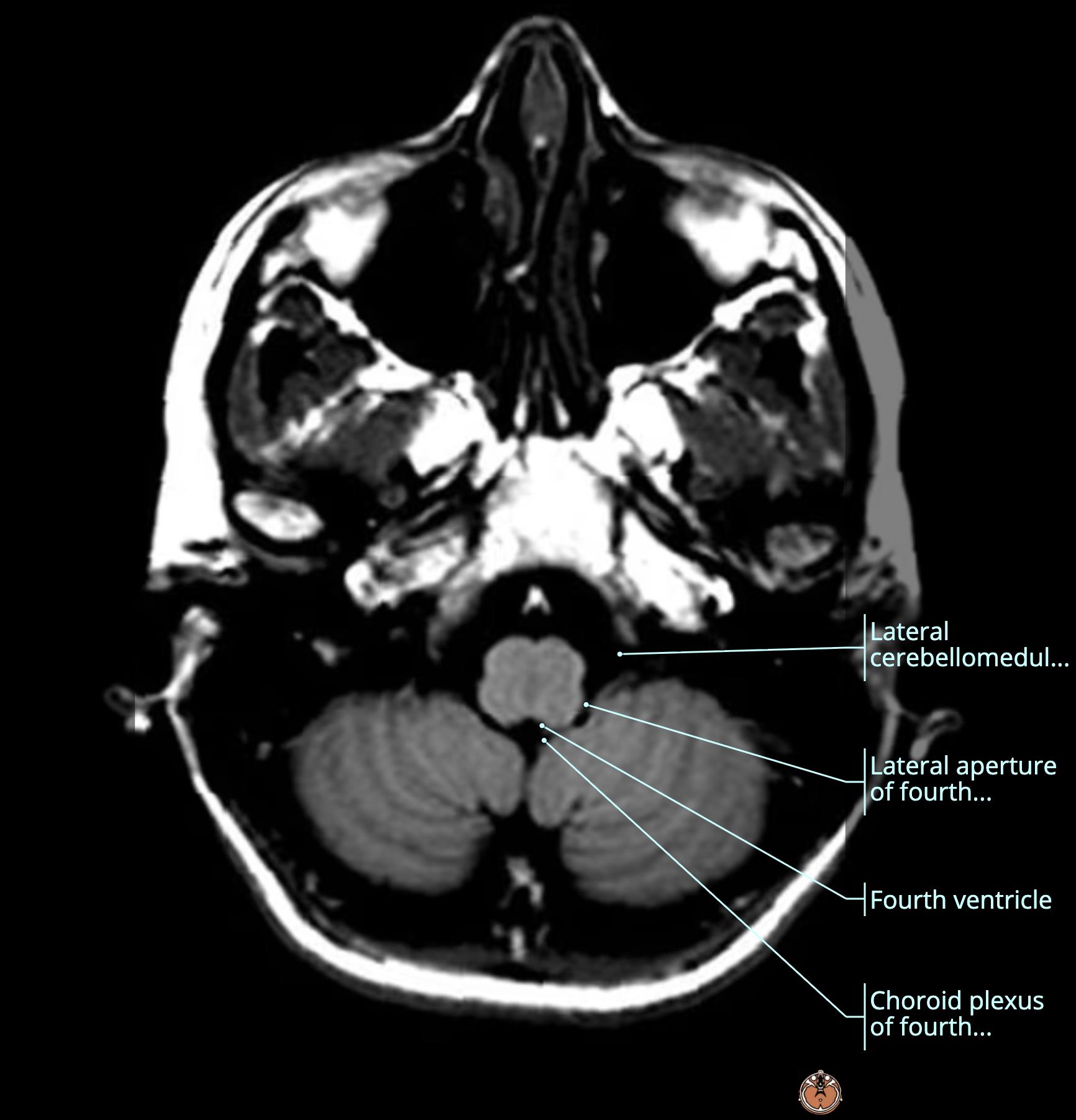
Ventricular System Anatomy Review (Top to Bottom) (14-16)
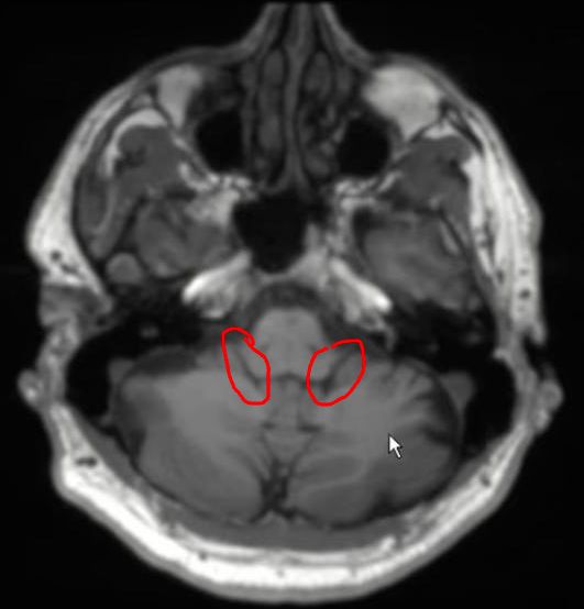
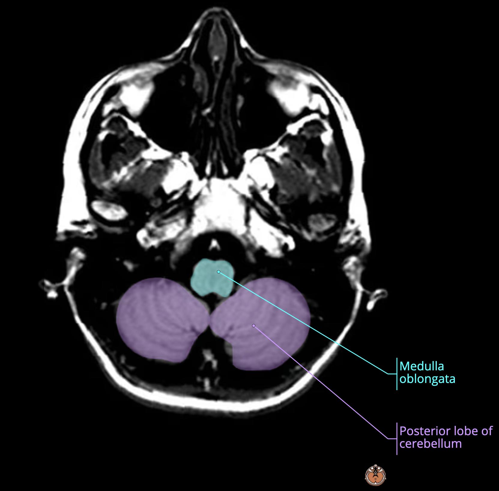
  • these are the lateral apertures[…] (red) aka the foramen of Luschka[…] (red) at the level of pons[…]
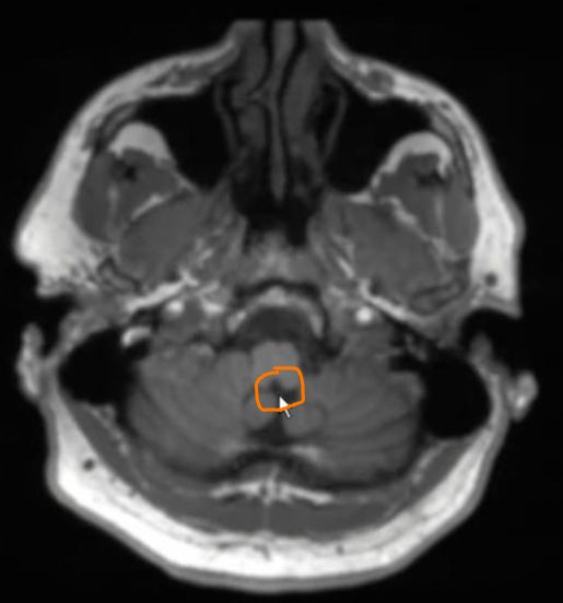
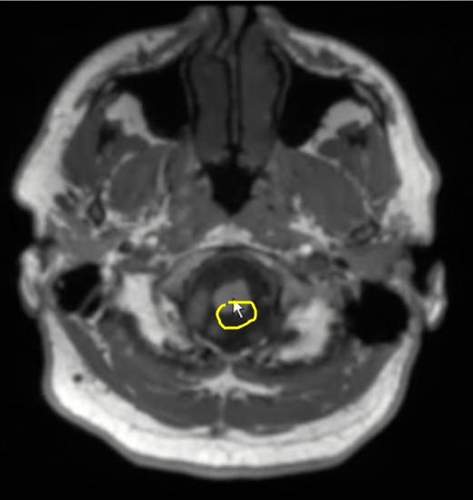
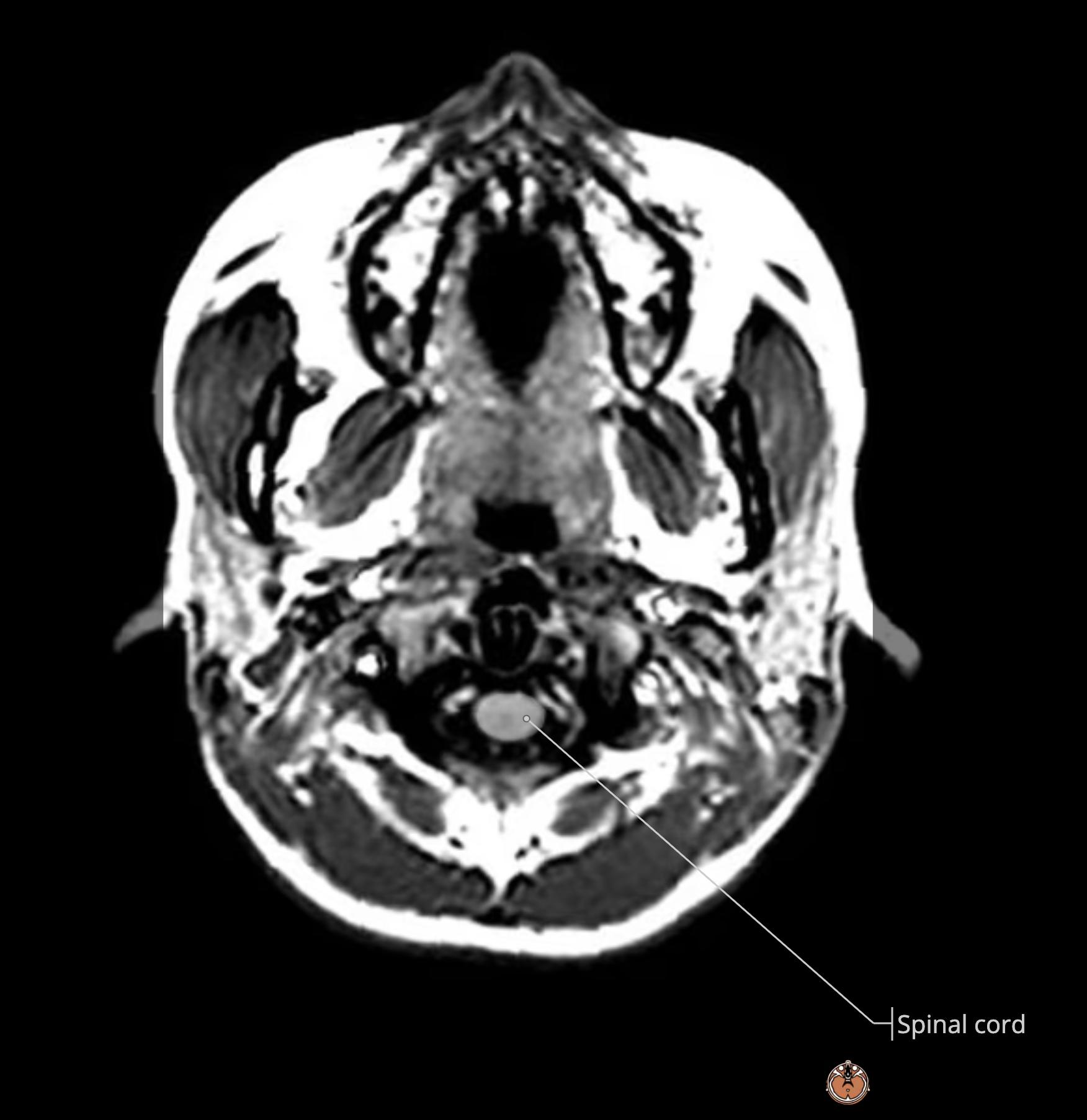
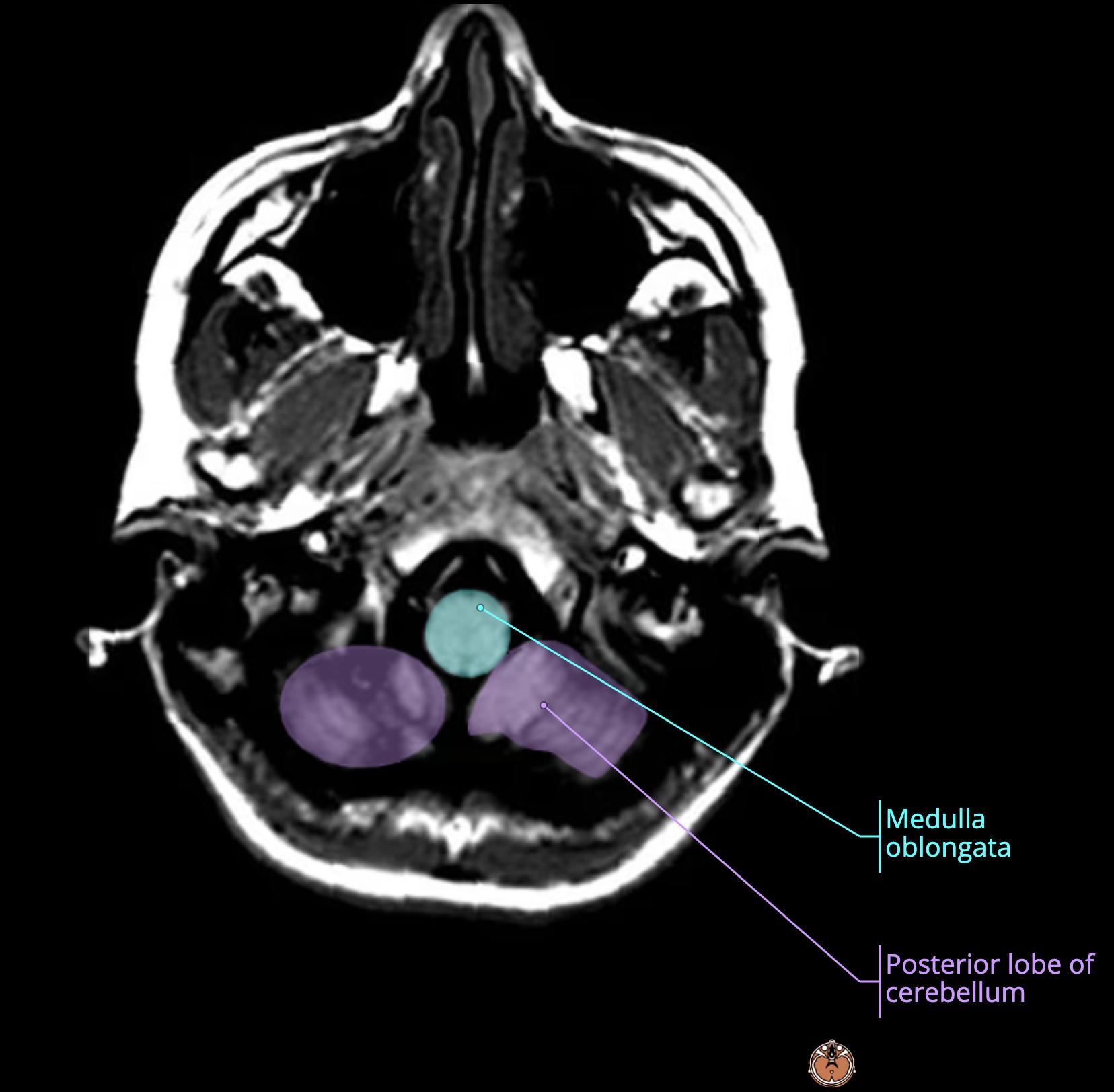
  • descend further and you see the median aperture[…] (orange) aka the foramen of Magendie[…] (orange) which is continuous if you go down a smidge lower with the spinal cord central canal[…] (yellow)

Ventricular System Anatomy Review (L to R) (1-2)
  • this is the temporal horn, lateral ventricle[…] (red, most lateral) which gives off the occipital horn, lateral ventricle[…] (orange) as you move medial

Ventricular System Anatomy Review (L to R) (3)
  • this is the trigone/atra, lateral ventricle[…] (red) 

Ventricular System Anatomy Review (L to R) (4)
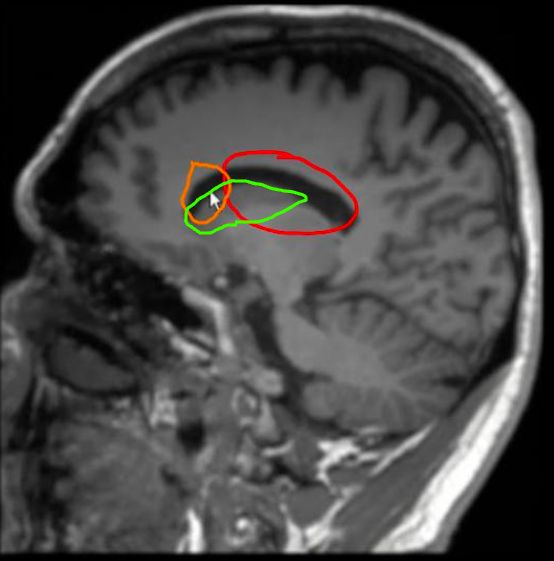
  • this is the body, lateral ventricle[…] (red) and anterior horn, lateral ventricle[…] (orange) with its laterally running caudate nucleus[…] (green)

Ventricular System Anatomy Review (L to R) (7)
  • this is the interventricular foramen / foramen of monroe[…] (red) connecting the lateral ventricle[…] (orange) to the 3rd ventricle[…] (green)

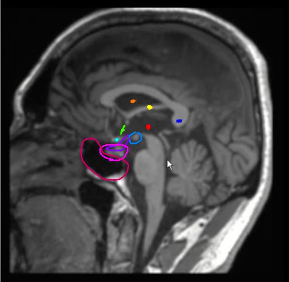
Ventricular System Anatomy Review (L to R) (7)
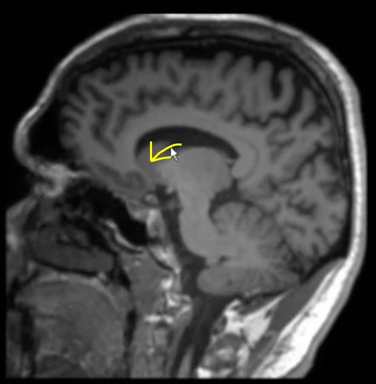
  • 3rd ventricle[…] (red)
  • lateral ventricle[…] (orange)
  • fornix[…] (yellow)
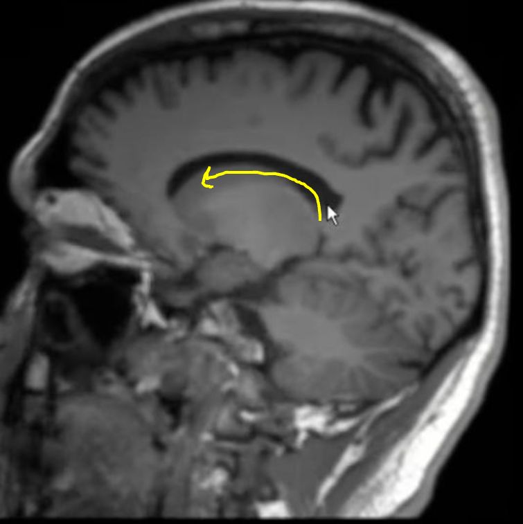
  • lamina terminalis[…] (green)
  • optic chiasm[…] (light blue)
  • mamillary bodies[…] (intermediate blue)
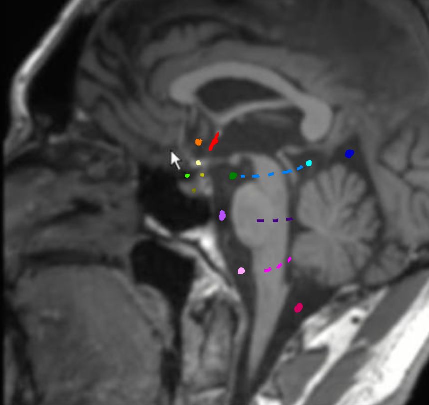
  • pineal gland[…] (dark blue)
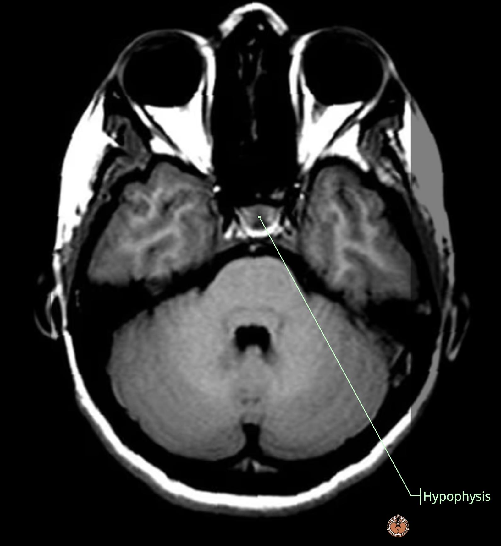
  • infundibular recess[…] (purple) coming down toward the pituitary gland[…] (pink) sitting in the sella turcica[…] (pink)
    • anterior to that is sphenoid sinus[…] (dark pink/red) 

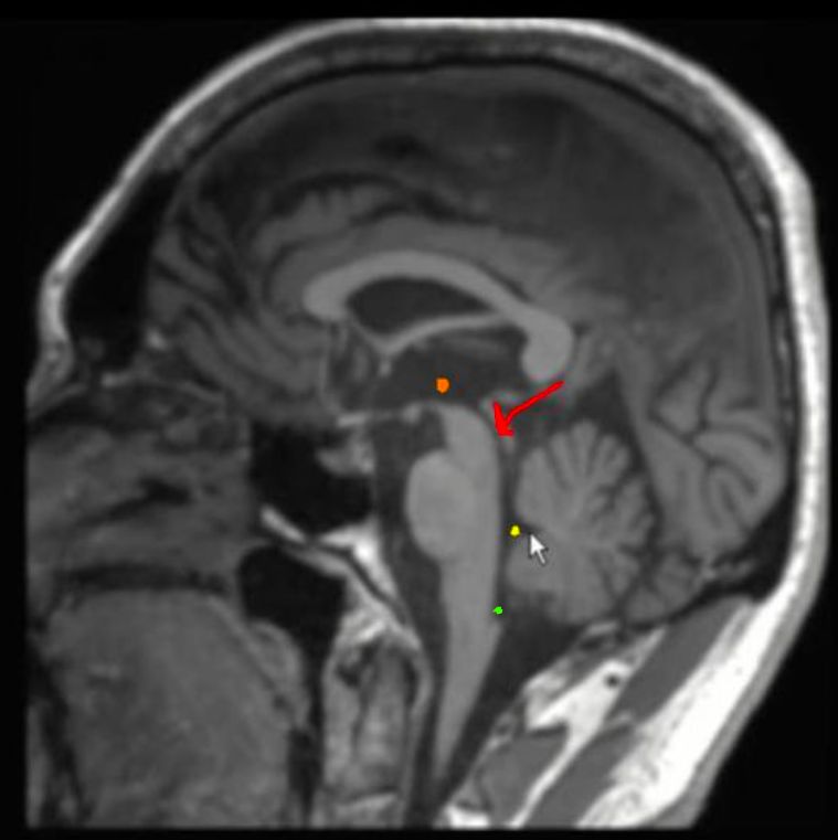
Ventricular System Anatomy Review (L to R) (8)
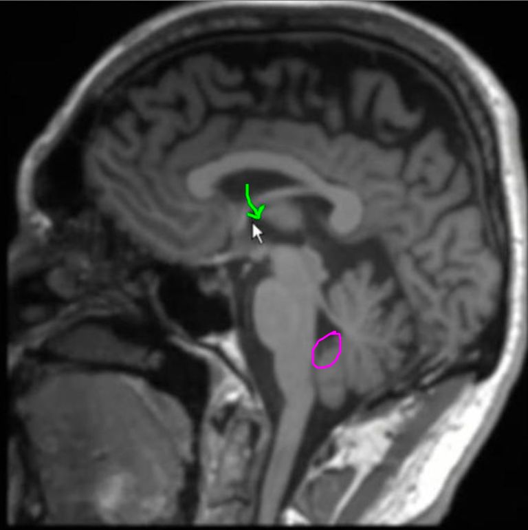
  • orange: 3rd ventricle[…]
  • red arrow: cerebral aqueduct of Sylvius[…]
  • yellow: 4th ventricle[…]
  • green: median aperture (foramen of Magendie)[…]

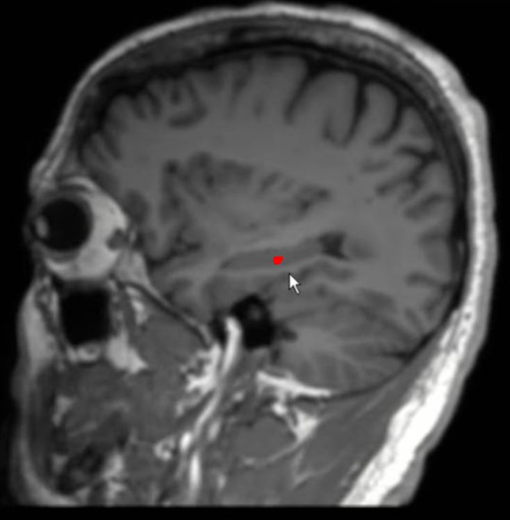
Ventricular System Anatomy Review (L to R) (9)
  • Choroid Plexus[…] Course
    • red: starts in temporal horn, lateral ventricle[…]
    • orange: runs along the roof[…] of this structure all the way around 
    • yellow: runs along the floor[…] of the lateral ventricle (body)[…]
    • green: runs into the 3rd ventricle[…] and runs along the roof[…] of it
    • note 1: there's no choroid plexus in the anterior horns[…] or occipital horns[…] of the lateral ventricles
    • purple - note 2: there's also choroid plexus on the floor[…] of the 4th ventricle[…]

Ventricular System Anatomy Review (L to R) (10)
  • Choroid Plexus Arterial Supply
    • The anterior part of the choroid plexus and ventricles are supplied by the Anterior Choroidal (AChA)[…] Artery which branches off the Communicating ICA (C7 segment)[vessel, segment?] 
    • The posterior part of the choroid plexus and ventricles are supplied by the 
      • (1) Posterior Choroidal[…] Artery which typically branches off the PCA (P2 Segment)[vessel, segment?] 
      • (2) Choroid[…] branch of the PICA[…] which itself branches off the Vert[…]
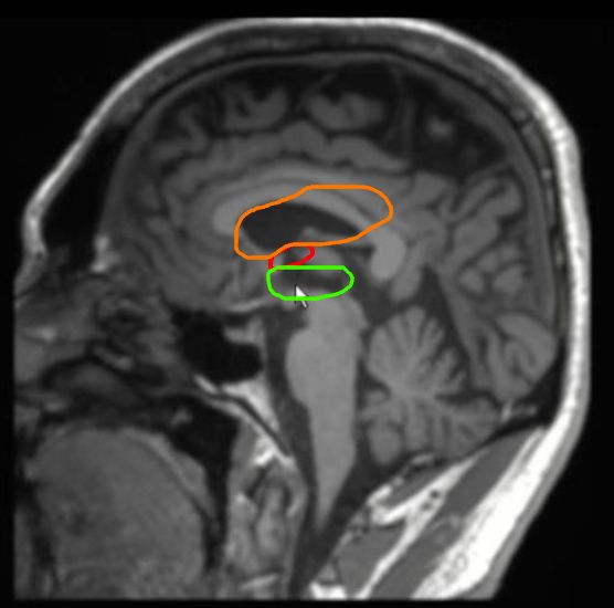
Subarachnoid Cisterns Anatomy Review (Sagittal) 
  • the cisterns are areas where the pia mater[…] is pressed against the brain surface but the arachnoid mater[…] is stretched out and allowing for some space away from the brain surface, generally in the folds[…] of the brain - they generally hold CSF[…] where flow is continuous
  • red (line structure) is the lamina terminalis[…]
  • orange cisterncistern of the lamina terminalis[…]
  • yellowoptic chiasm[…] (light), infindibular stalk[…] (medium), pituitary gland[…] (dark)
  • light greensuprasellar[…] aka chiasmatic[…] cistern
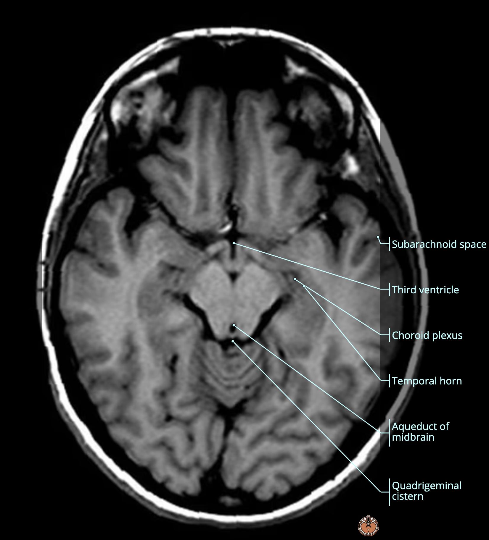
  • dark greeninterpeduncular[…] cistern located anterior to the midbrain[…] and between the cerebral peduncles[…]
  • light bluequadrigeminal[…] cistern posterior to the midbrain tectum[…]
  • medium blue dotted line (seen on axial but imagined here): ambient[…] cisterns wrapping around the midbrain[…] to connect the interpeduncular[…] cistern anteriorly > to the quadrigeminal[…] cistern posteriorly
  • dark bluesuperior cerebellar[…] cistern
  • light purpleprepontine[…] cistern
  • dark purplecerebellopontine angle (CPA)[…] cistern connecting the pons[…] lateral surface > to the cerebellum[…] anterior surface -- into which the lateral apertures (foramen of Luschka)[…] of the 4th ventricle[…] flow into
  • light pinkpremedullary[…] cistern
  • medium pinkcerebellomedullary[…] cistern
  • dark pink/red: the largest cistern at the base of the brain coming through the foramen magnum[structure] which is the cisterna magna[…] - this is often obliterated in situations of high or low ICP[…] with low-lying cerebellar tonsils

Subarachnoid Cisterns Anatomy Review (Axial) 
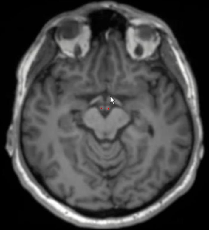
  • red: mamillary bodies[…]

Subarachnoid Cisterns Anatomy Review (Axial) 
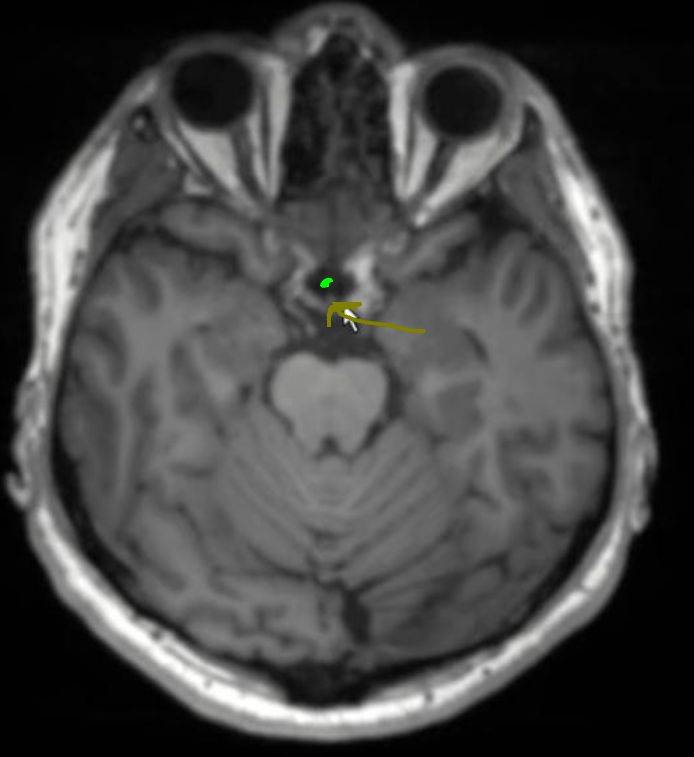
  • light yellow: optic chiasm[…]

Subarachnoid Cisterns Anatomy Review (Axial) 
  • dark yellow: pituitary stalk[…]
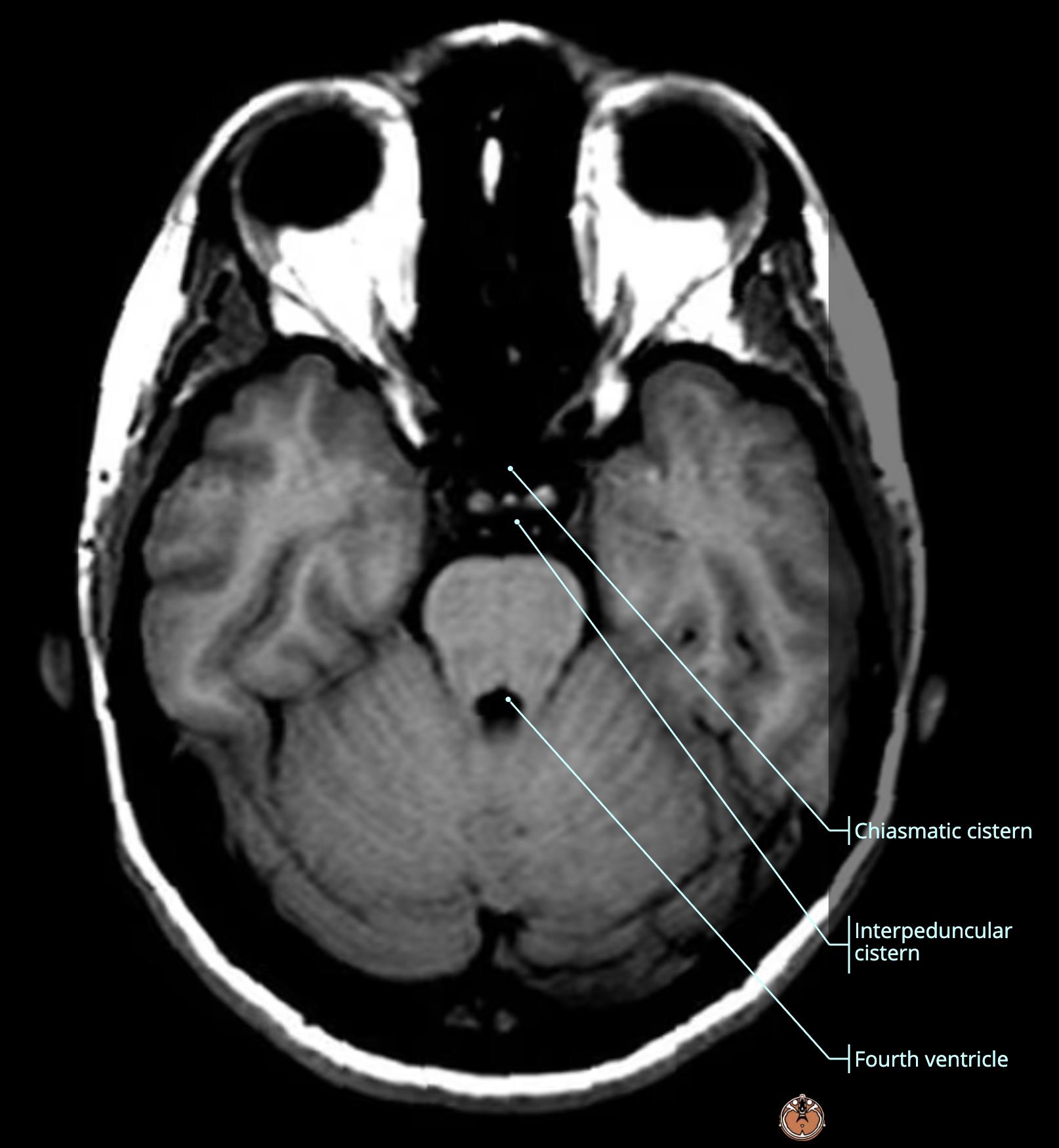
  • light green: supracellar[…] aka chiasmatic[…] cistern

Subarachnoid Cisterns Anatomy Review (Sagittal)
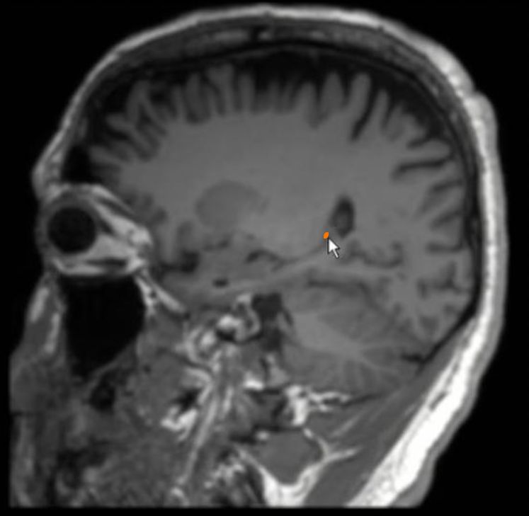
  • dark yellow: pituitary stalk[…]
  • light green: supracellar[…] cistern

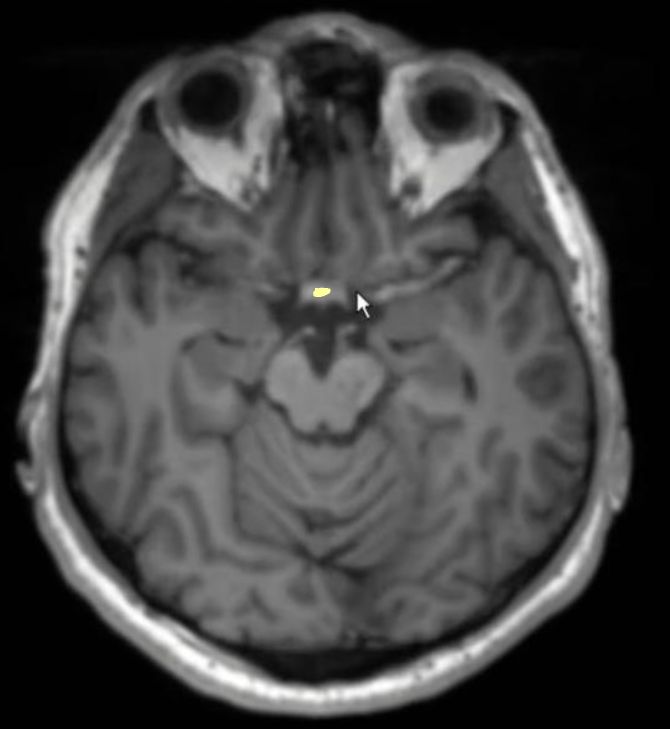
Subarachnoid Cisterns Anatomy Review (Axial)
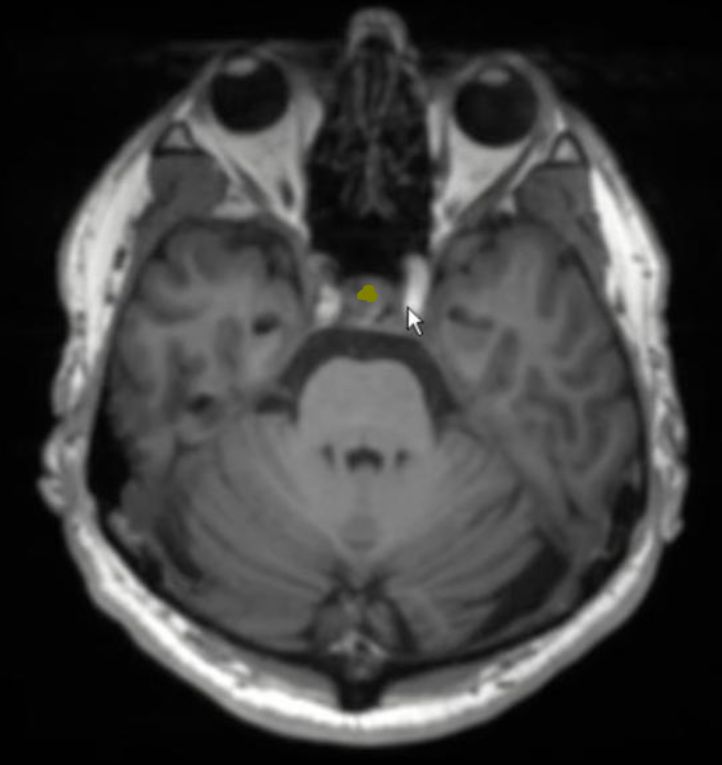
  • dark yellow: pituitary gland[…]

Subarachnoid Cisterns Anatomy Review (Axial)
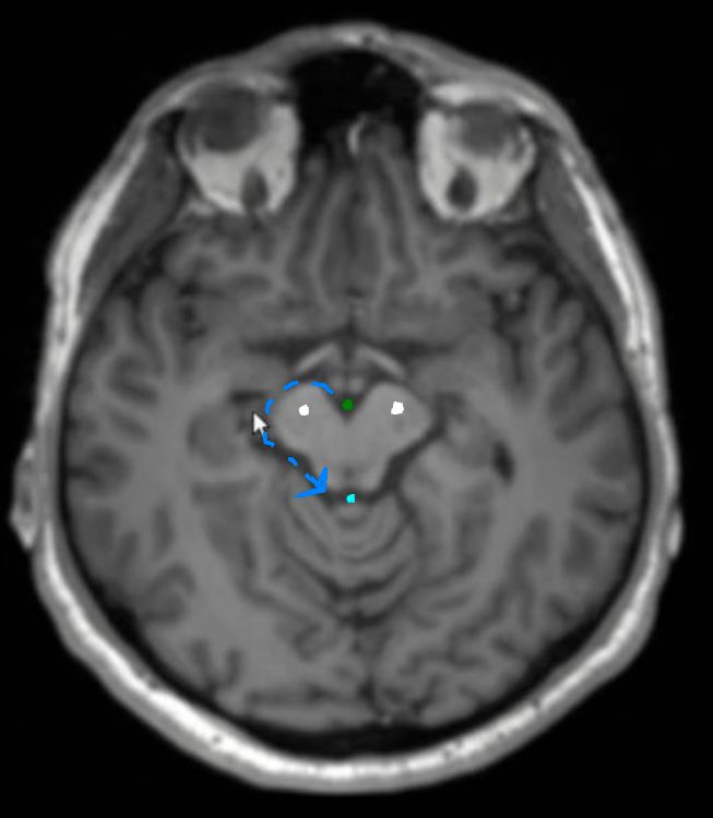
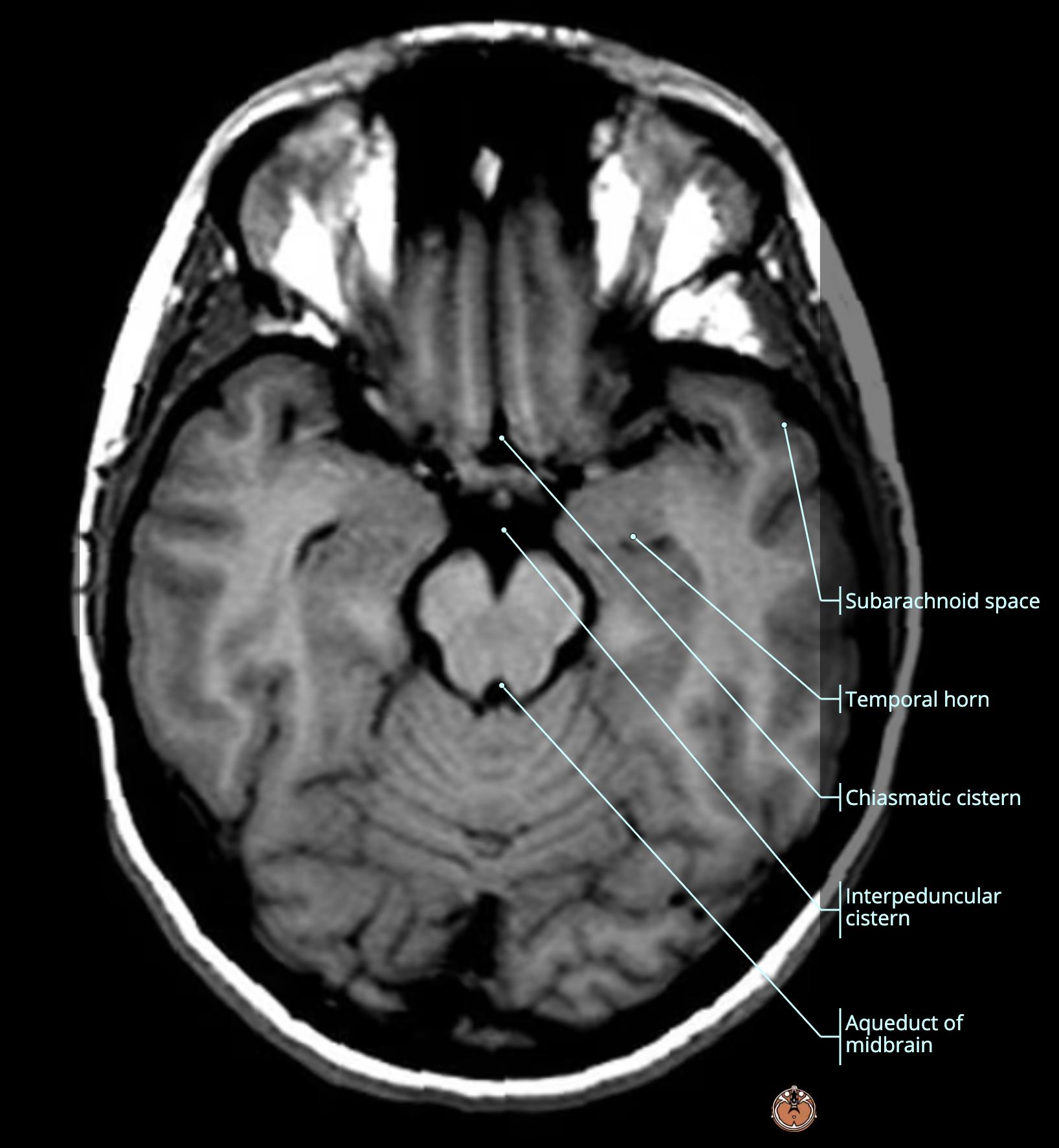
  • dark green: interpeduncular[…] cistern
  • medium blue: ambient[…] cisterns (only one side pictured) > 
    • connecting into the light blue: quadrigeminal[…] cistern posterior to the superior[…] colliculi at this level of the midbrain tectum[structure]
  • white: cerebral peduncles[…]

Subarachnoid Cisterns Anatomy Review (Axial)
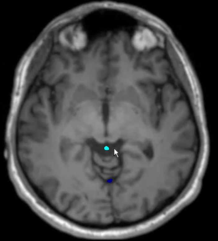
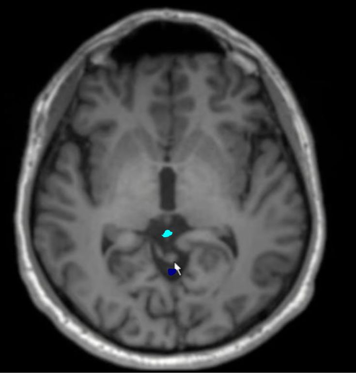
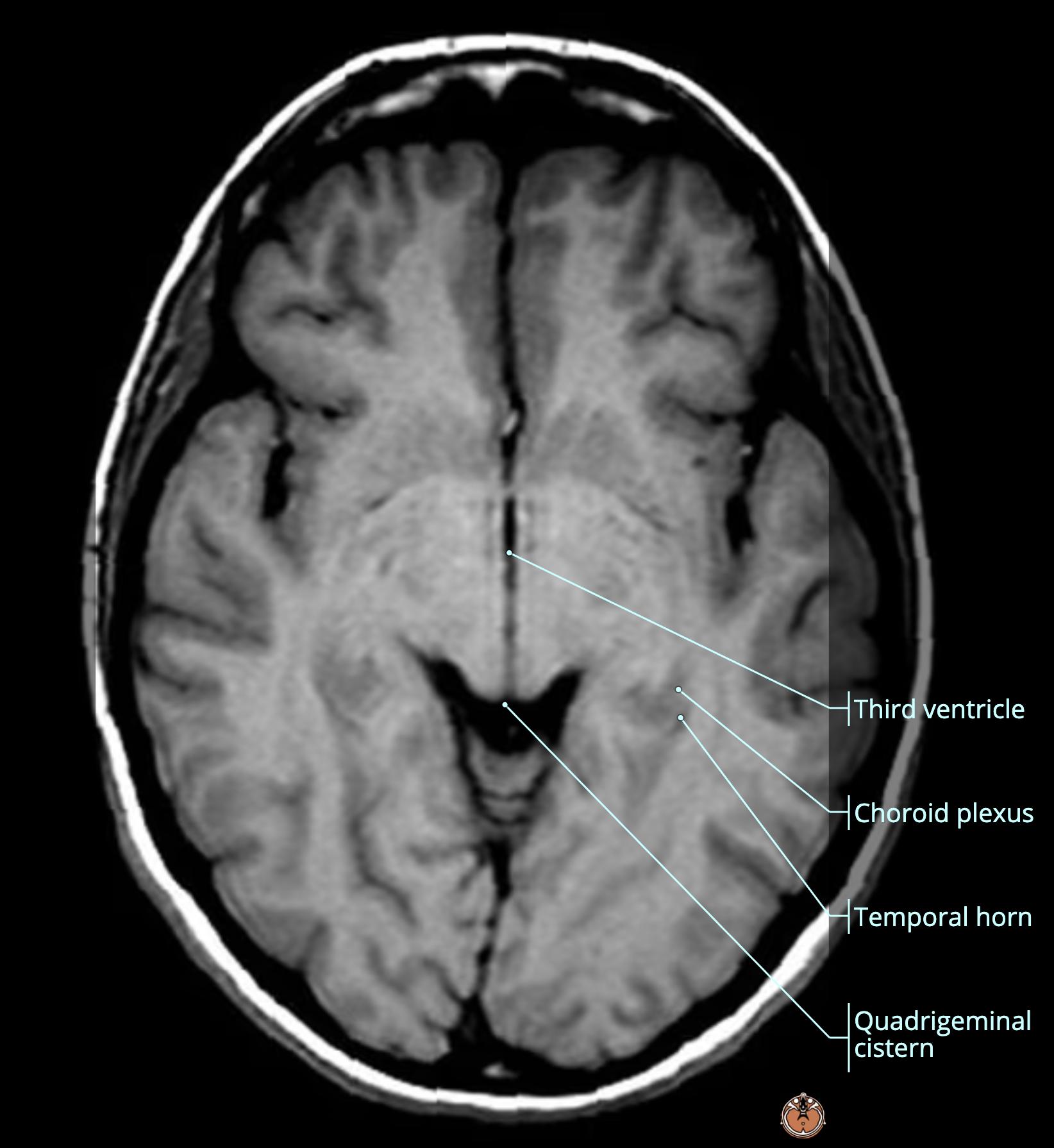
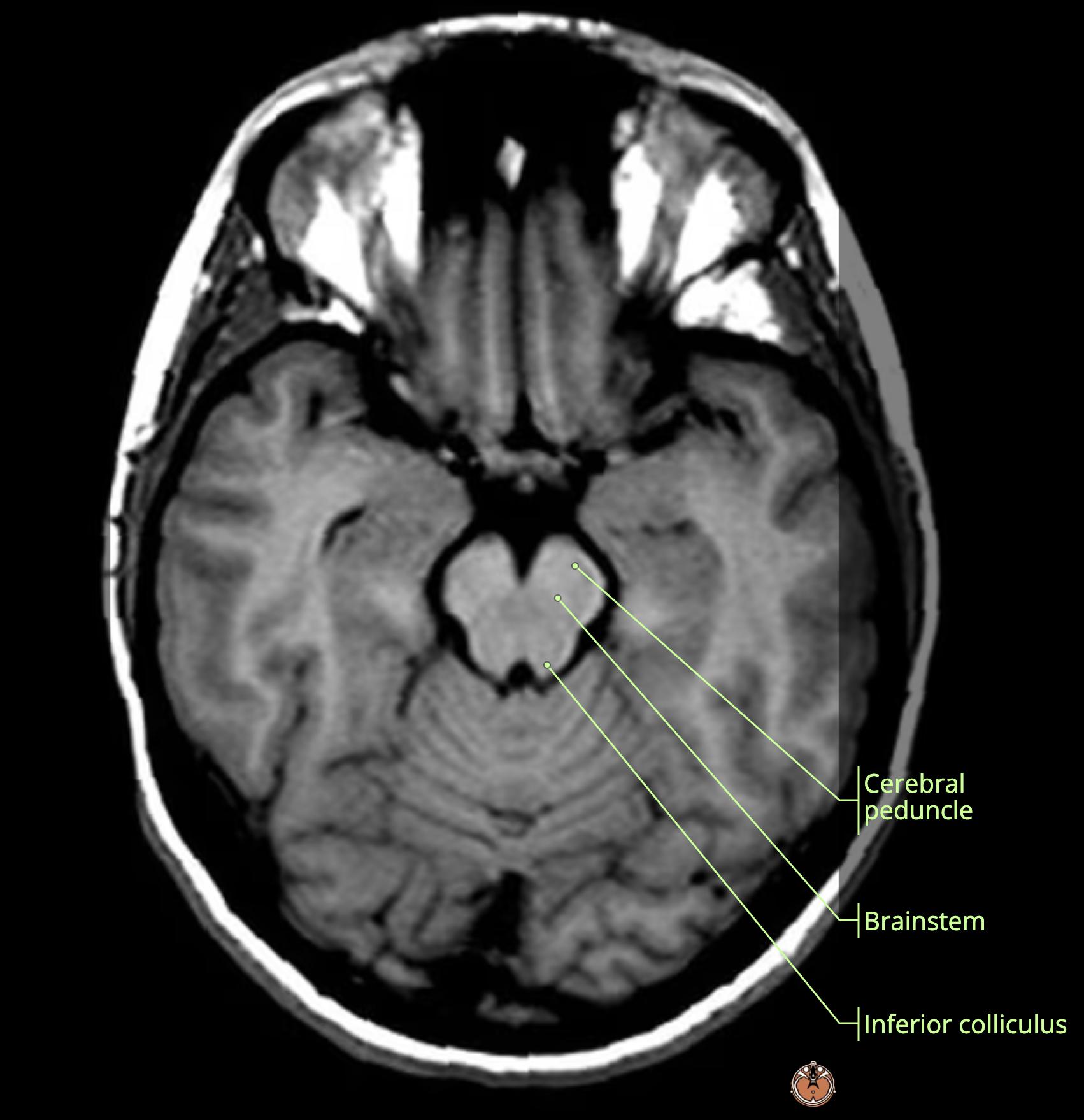
  • light blue: quadrigeminal[…] cistern posterior to the inferior[…] colliculi at this level of the midbrain tectum[structure]
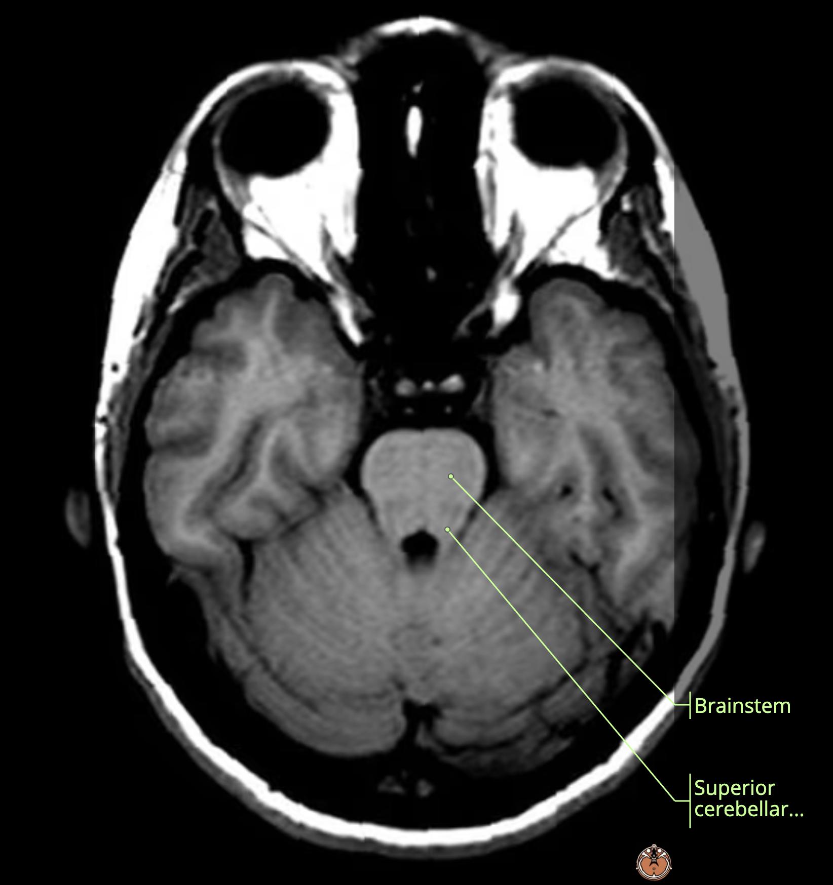
  • dark blue: superior cerebellar[…] cistern

Subarachnoid Cisterns Anatomy Review (Axial)
  • light blue: quadrigeminal[…] cistern
  • dark blue: superior cerebellar[…] cistern

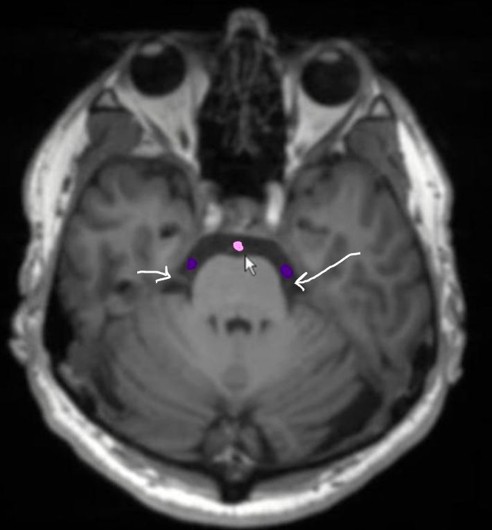
Subarachnoid Cisterns Anatomy Review (Axial)
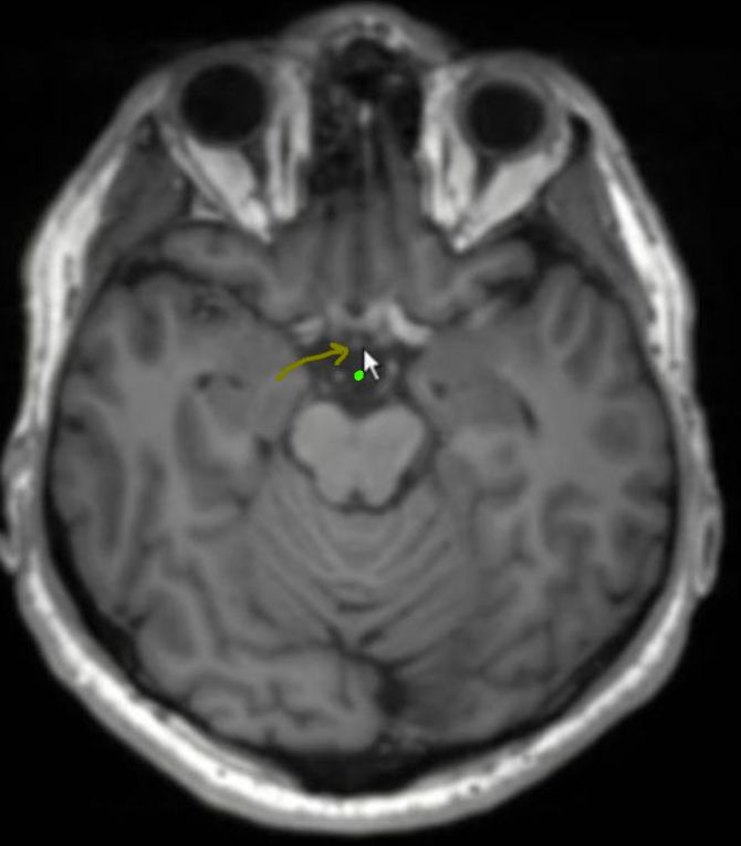
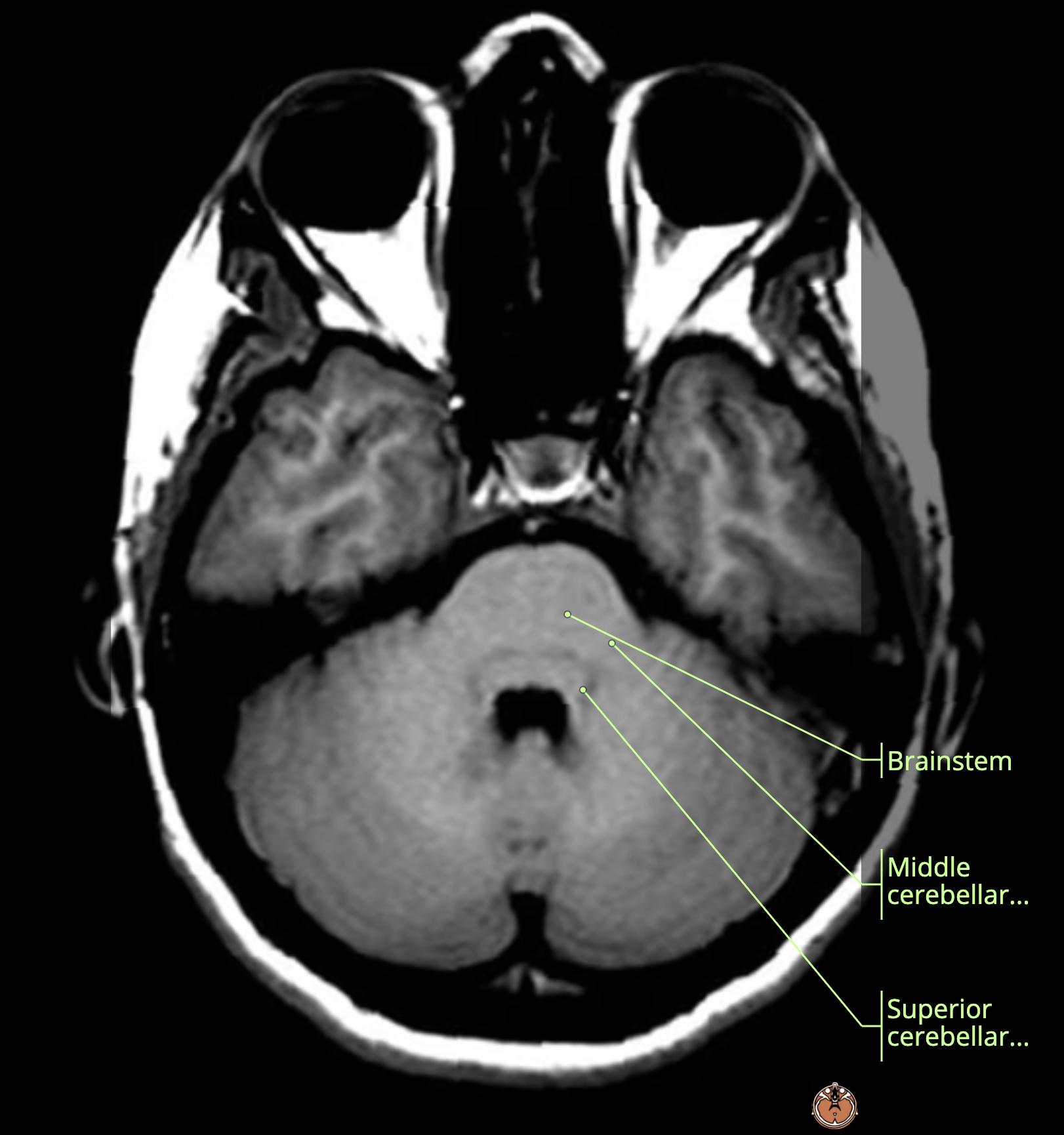
  • light purple: prepontine[…] cistern
  • dark purple: cerebellopontine[…] cisterns through which the trigeminal[…] nerve runs (white arrows)

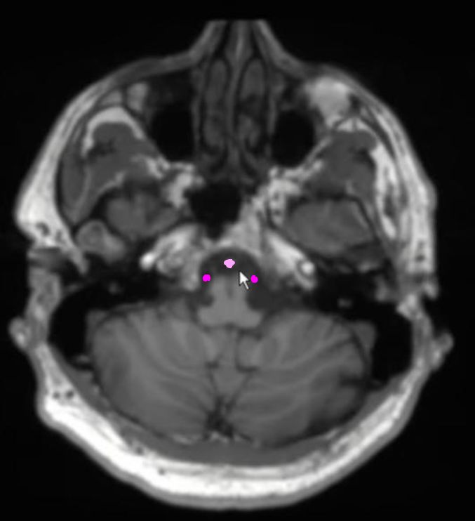
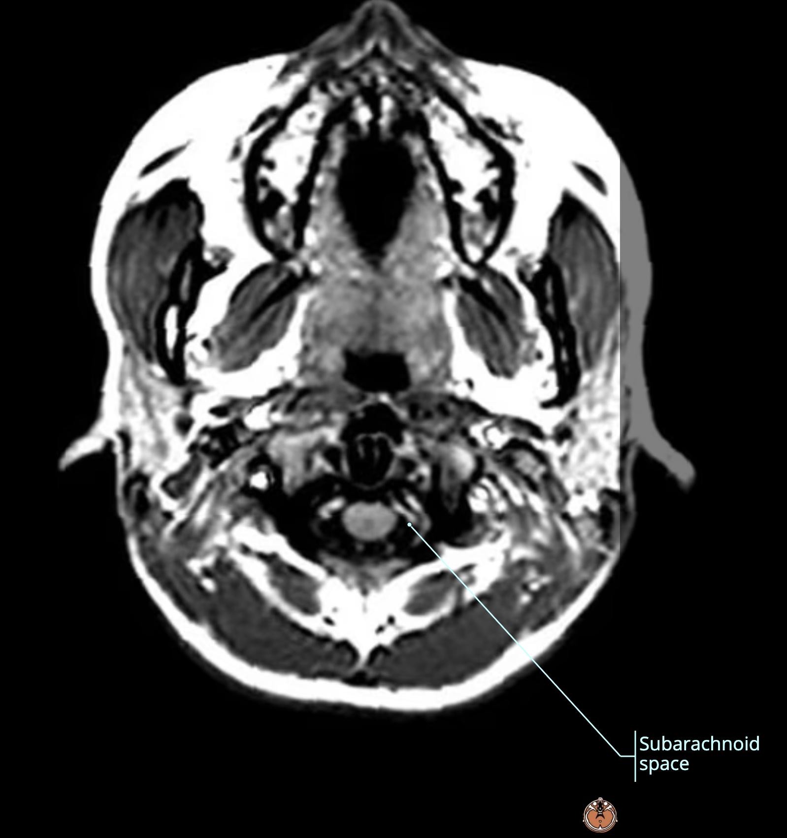
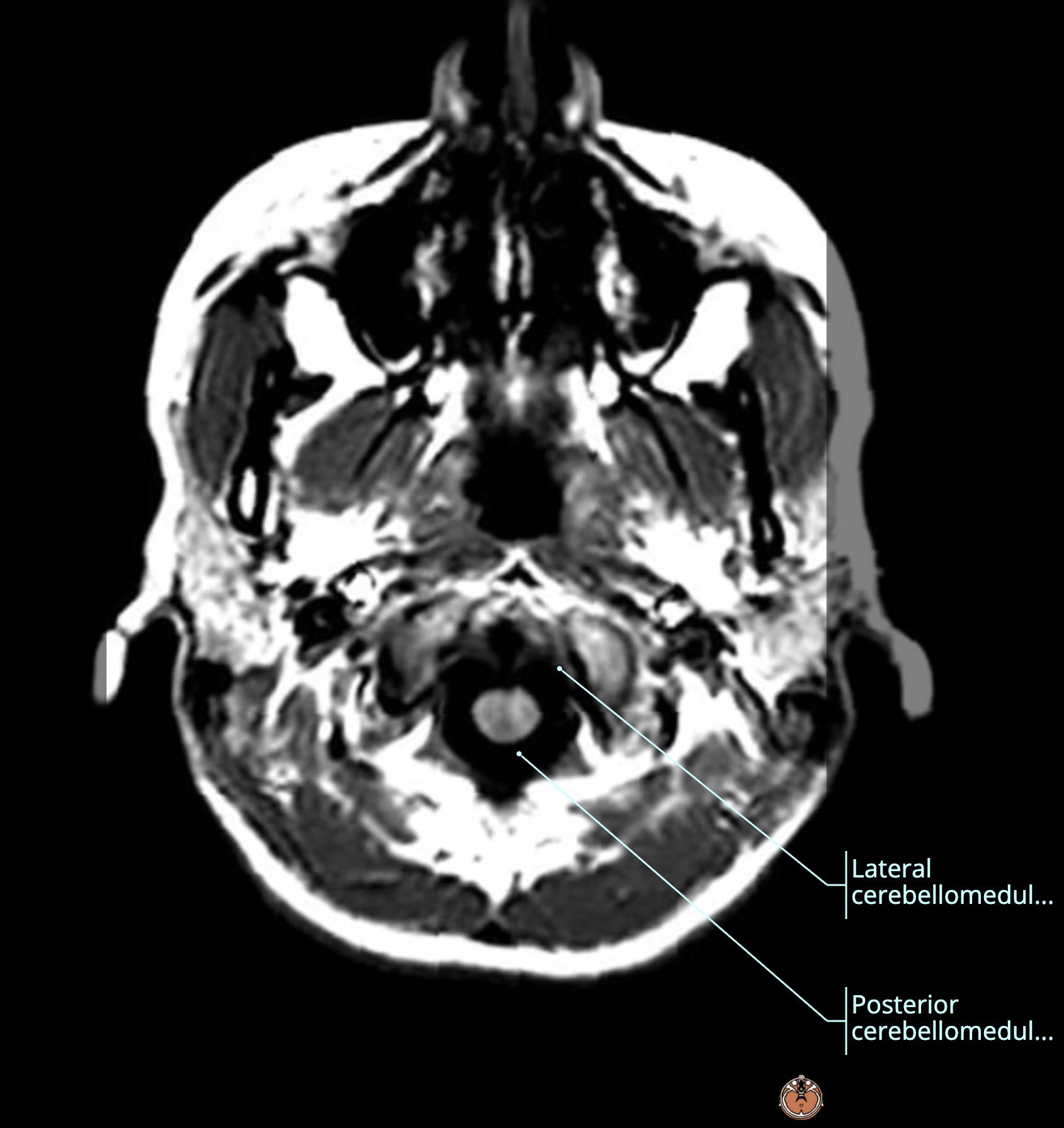
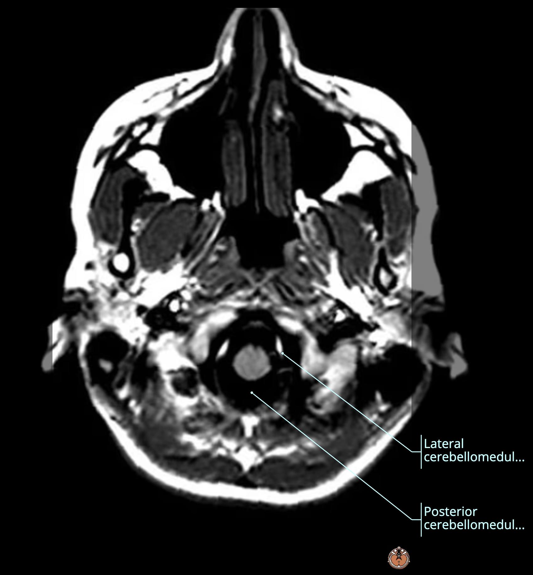
Subarachnoid Cisterns Anatomy Review (Axial)
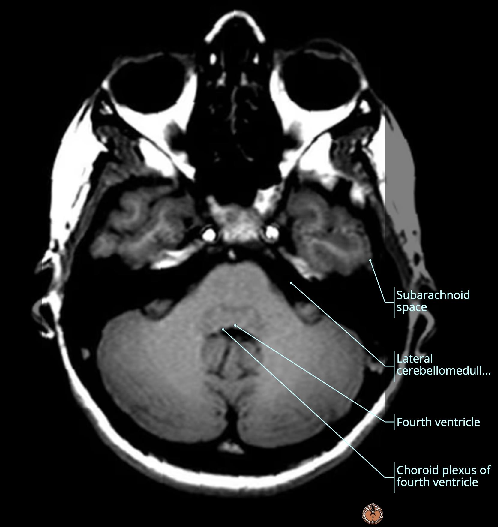
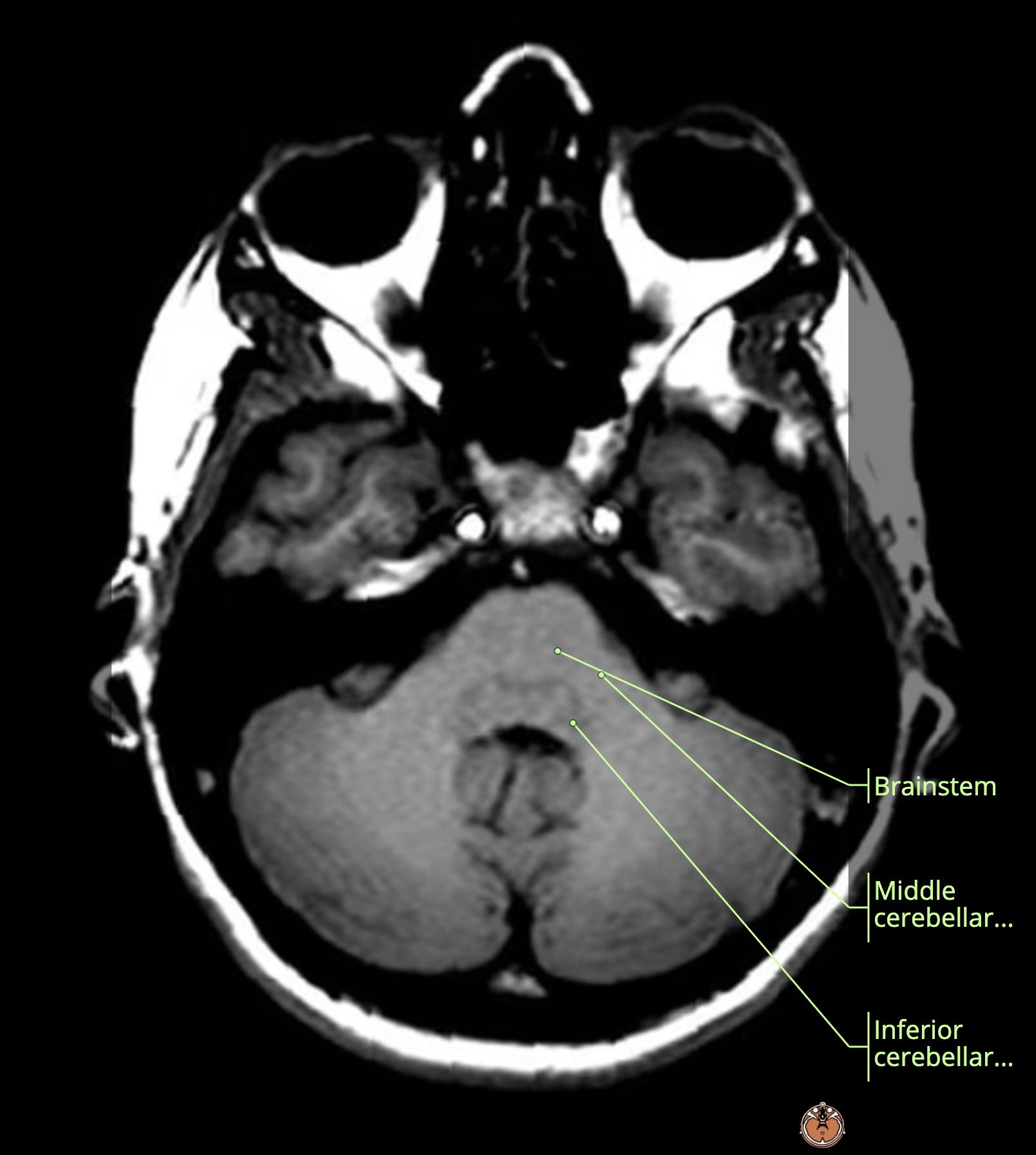
  • light pink: premedullary[…] cistern
  • medium pink: cerebellomedullary[…] cisterns

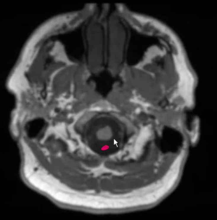
Subarachnoid Cisterns Anatomy Review (Axial)
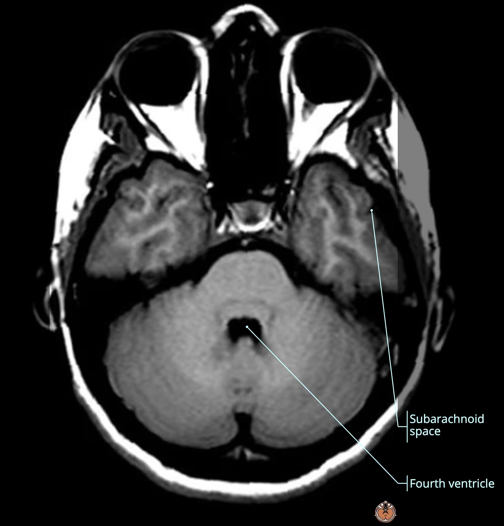
  • dark pink: cisterna magna[…]

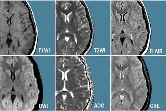
Standard MRIb wo order typically includes the following sequences (6):
  • T1, T2, FLAIR, DWI, ADC, GRE[…]
Edema Pathophysiology and Imaging
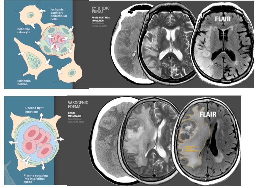
  • normal neurovascular unit aka "BBB[…]": comprised of astrocyte[…] foot processes wrapped around capillary endothelial[…] cells connected by tight[…] junctions with parenchymal neurons[…] located inside and outsode of the capillary endothelial cells; all with capillary lumen[…] carrying RBCs[…] within
  • Cytotoxic[…] Edema: 
    • ischemia[…] to astrocytes / capillary endothelial cells / parenchymal neurons > these cells can't maintain their Na-K[…] gradients > toxic to cells but the BBB[…] remains intact 
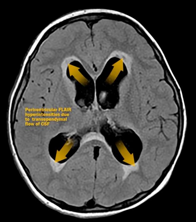
    • you can better see this if you subtract out CSF[…] bright signal - this is aka the T2 FLAIR[…] sequence
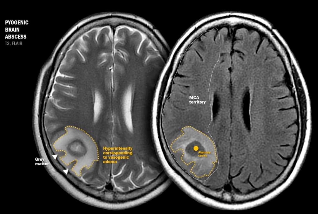
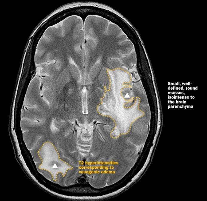
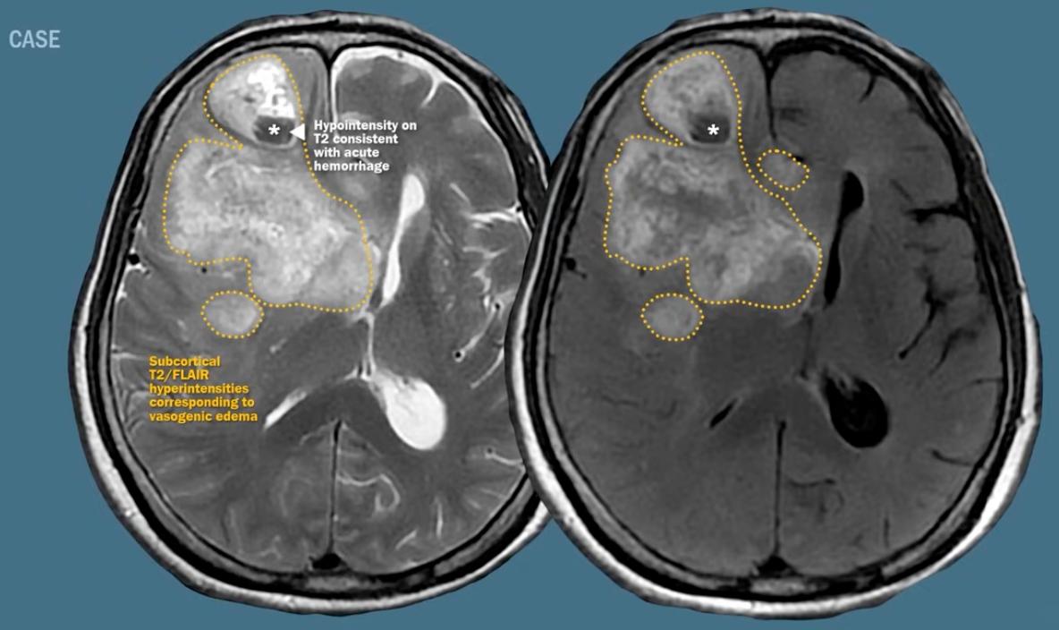
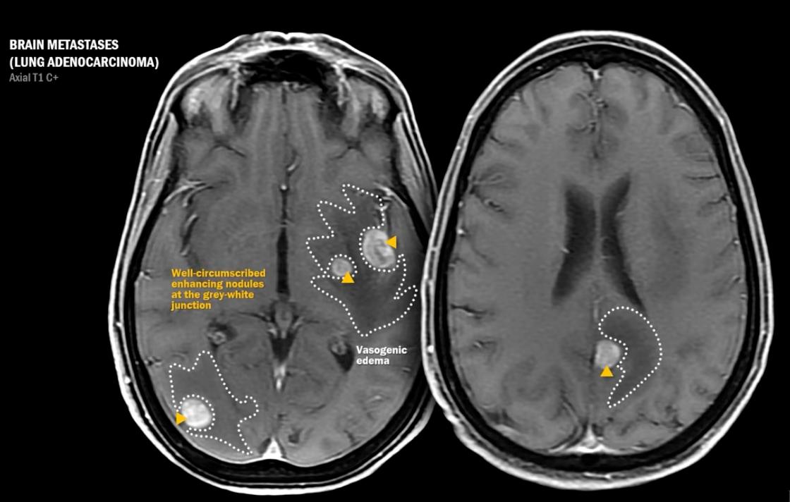
  • Vasogenic[…] Edema: often seen with tumors[…]abscesses[…]PRES[…]/HTNsive emergency, and more
    • tight junctions[…] become leaky > plasma[…] escapes into the interstitial[…] space while preserving the gray-white[…] junction
Stroke Imaging Sequences
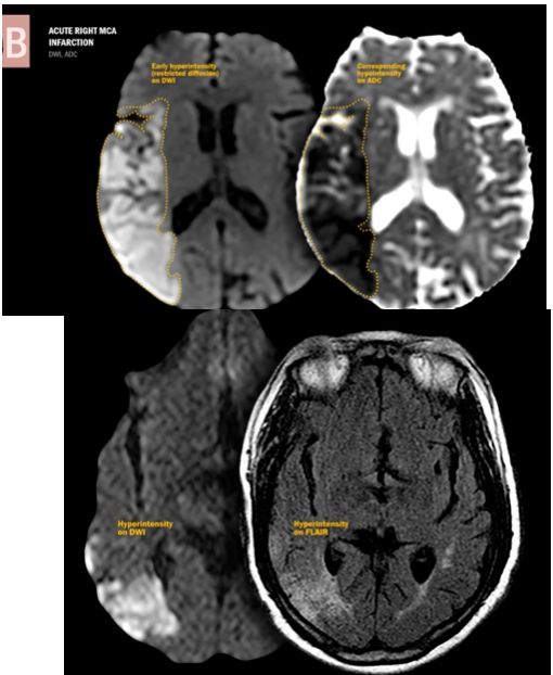
  • It takes several hours[…] for T2-FLAIR[…] to reveal hyperintensity signal change (>6-8)
  • It takes <30mins[…] for DWI/ADC[…] to reveal hyper/hypointense signal change
    • DWI[…] is closely related to T2-FLAIR[…]; both will be bright
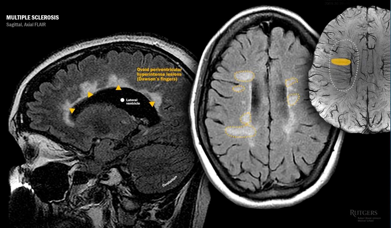
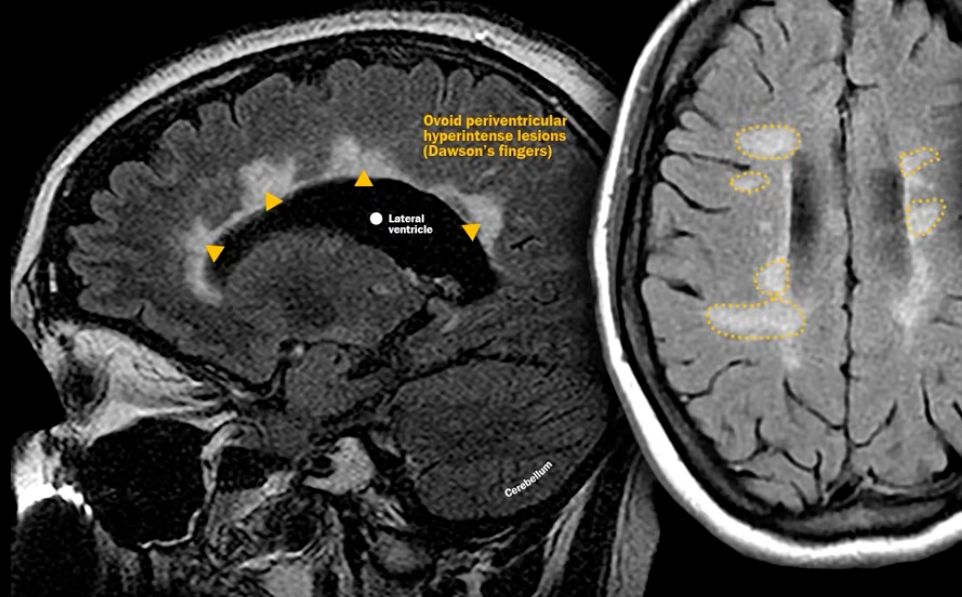
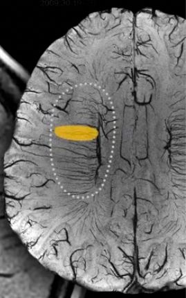
Pathophysiology for why we see ovoid lesions in Dawson's Fingers in MS?
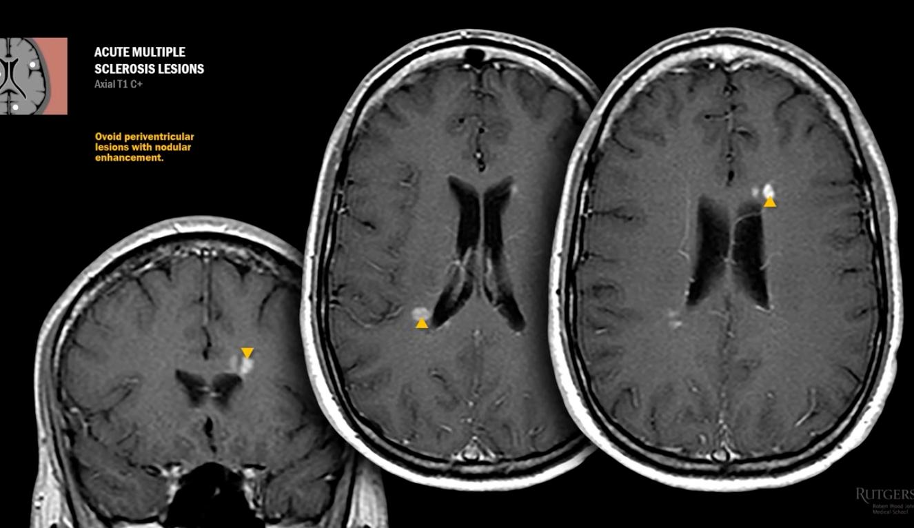
immune cells move through & exit central veins moving perpendicular to lateral ventricles[…]
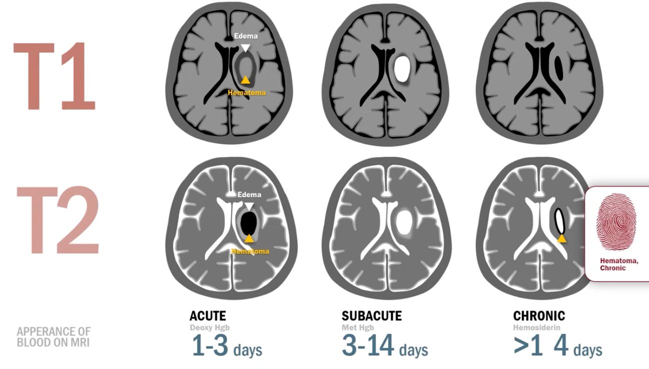
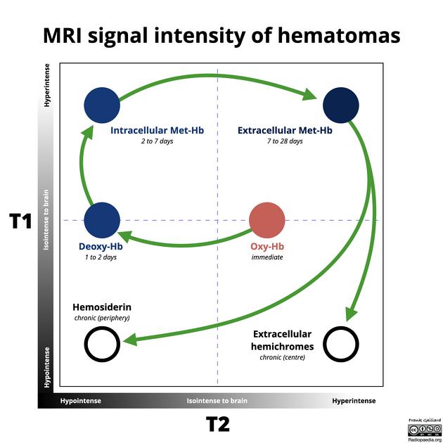
Describe the evolution of a bleed on MRI over time
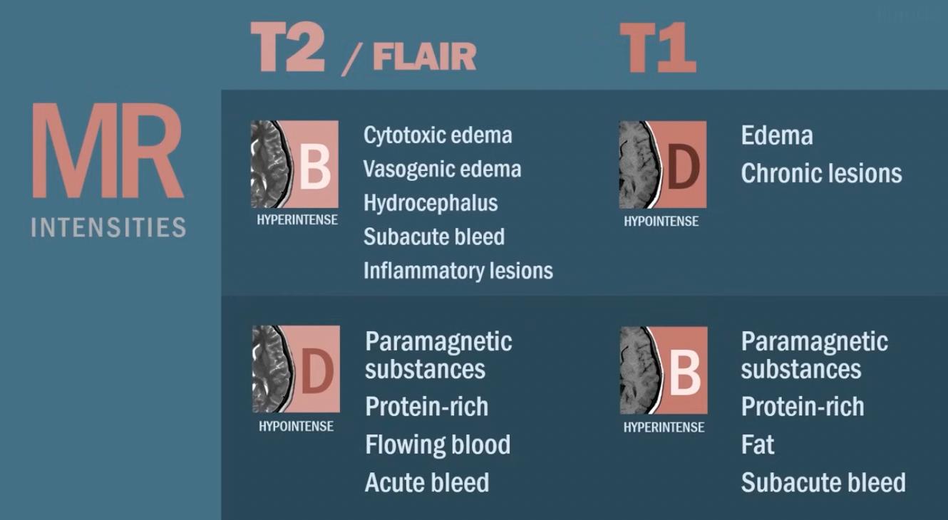
  • Acute[…] (1[…]-3[…] days)
    • iso[…]intense on T1[…]
    • hypo[…]intense on T2[…]
    • contains deoxyHgb[…]
  • Subacute[…] (3[…]-14[…] days)
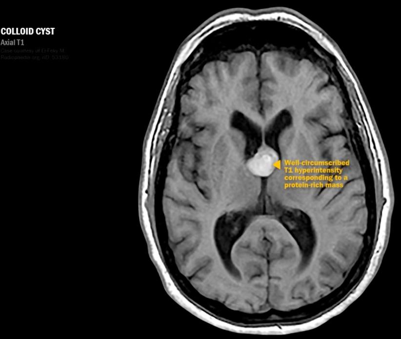
    • hyper[…]intense on T1[…]
    • hyper[…]intense on T2[…]
    • contains MetHgb[…]
  • Chronic[…] (>14[…] days)
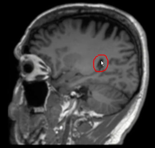
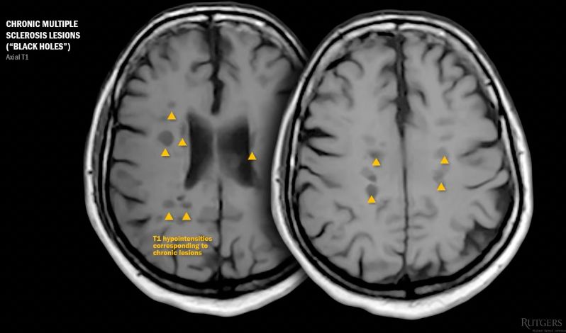
    • hypo[…]intense on T1[…] as the site of hemorrhage is replaced by CSF[…]-filled slit-like hole as if the brain were "stabbed[…]"
    • hyper[…]intense on T2[…] (think of it like a "fingerprint[…]" left by a chronic hemorrhage
    • contains Hemosiderin[…] 
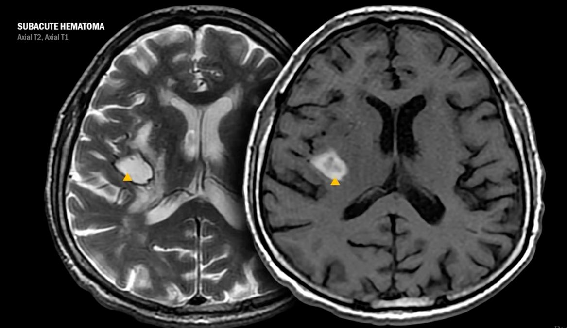
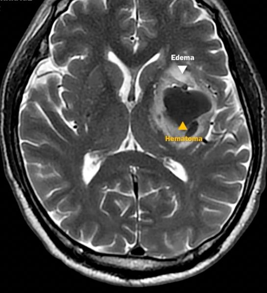
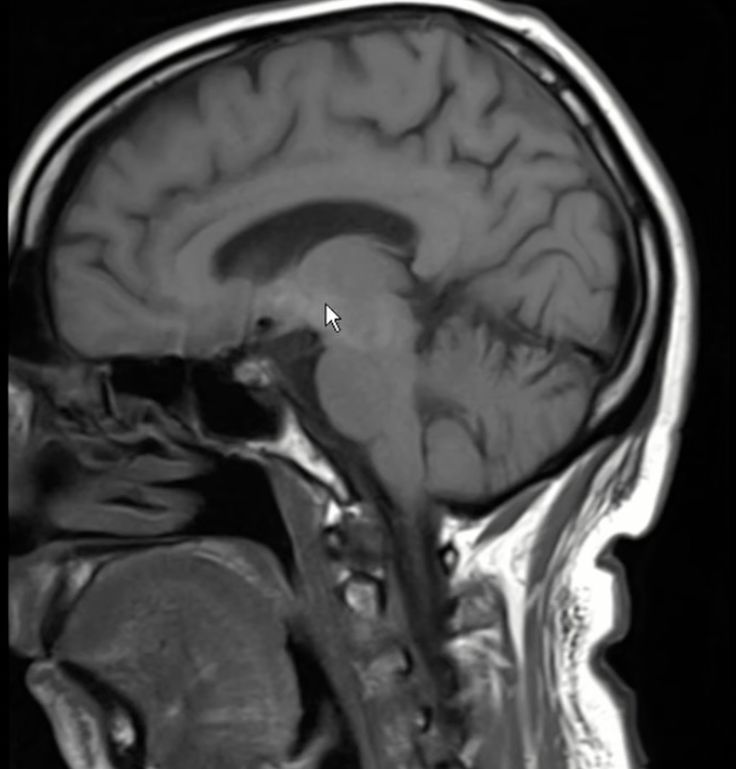
What's the stage of this hematoma?

late subacute: both T1 T2 bright[…]
Rapid Review
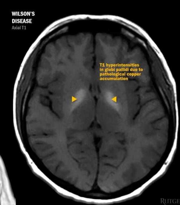
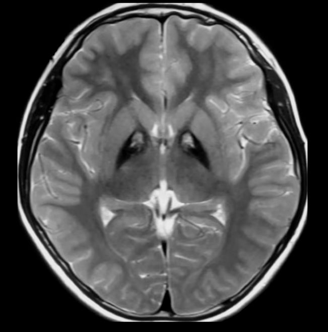
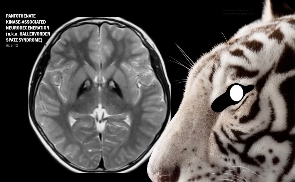
  • "eye of the tiger[…]" sign
  • c/f PKAN (Pantothenate Kinase-Associated Neurodegeneration)[…] aka Hallervorden Spatz[…] Syndrome

Pantothenate = Vitamin B5
Stage this bleed time-course
  • acute[…] (1-3[…] days) hemorrhage containing deoxyHgb[what substance?]

On T1[T1 or T2?] imaging, chronic[acute or chronic] MS lesions appear as hypointensities ("black holes")[…]
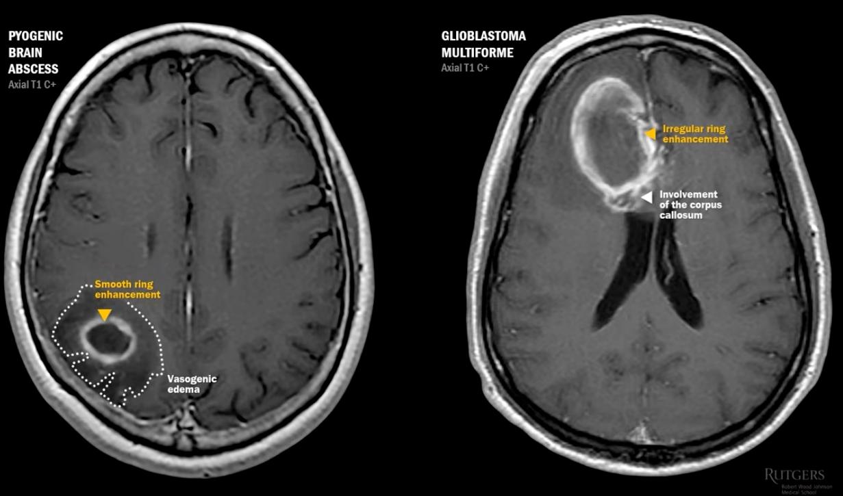
Common Patterns of Enhancement - Ring
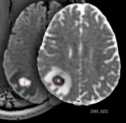
  • if confused between say brain abscess and GBM / other ring enhancing lesions, use DWI/ADC[sequences?] to differentiate
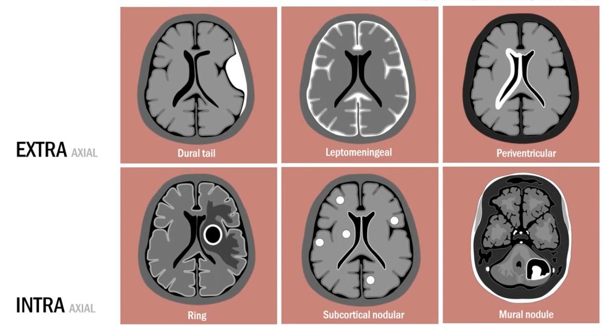
Common Patterns of Enhancement 
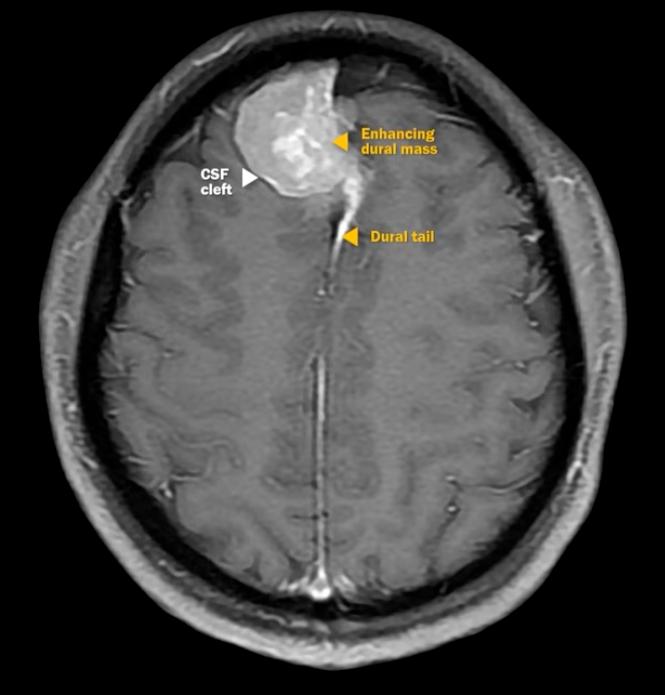
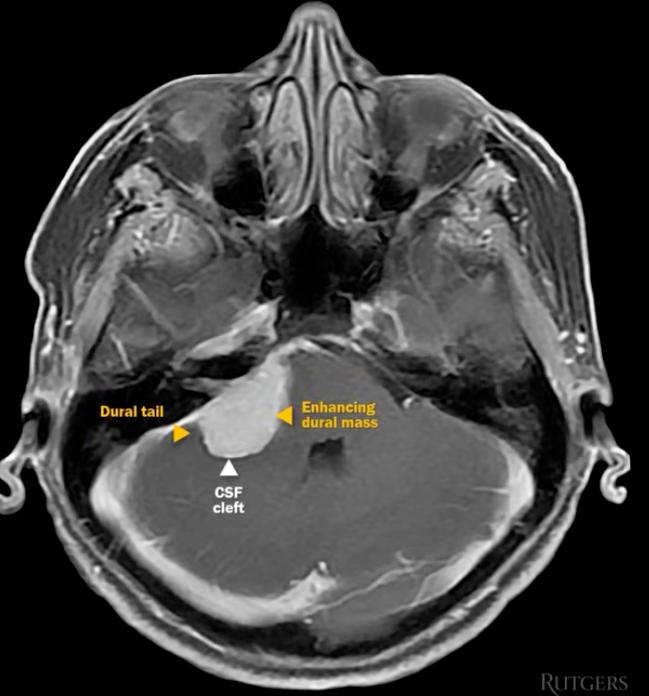
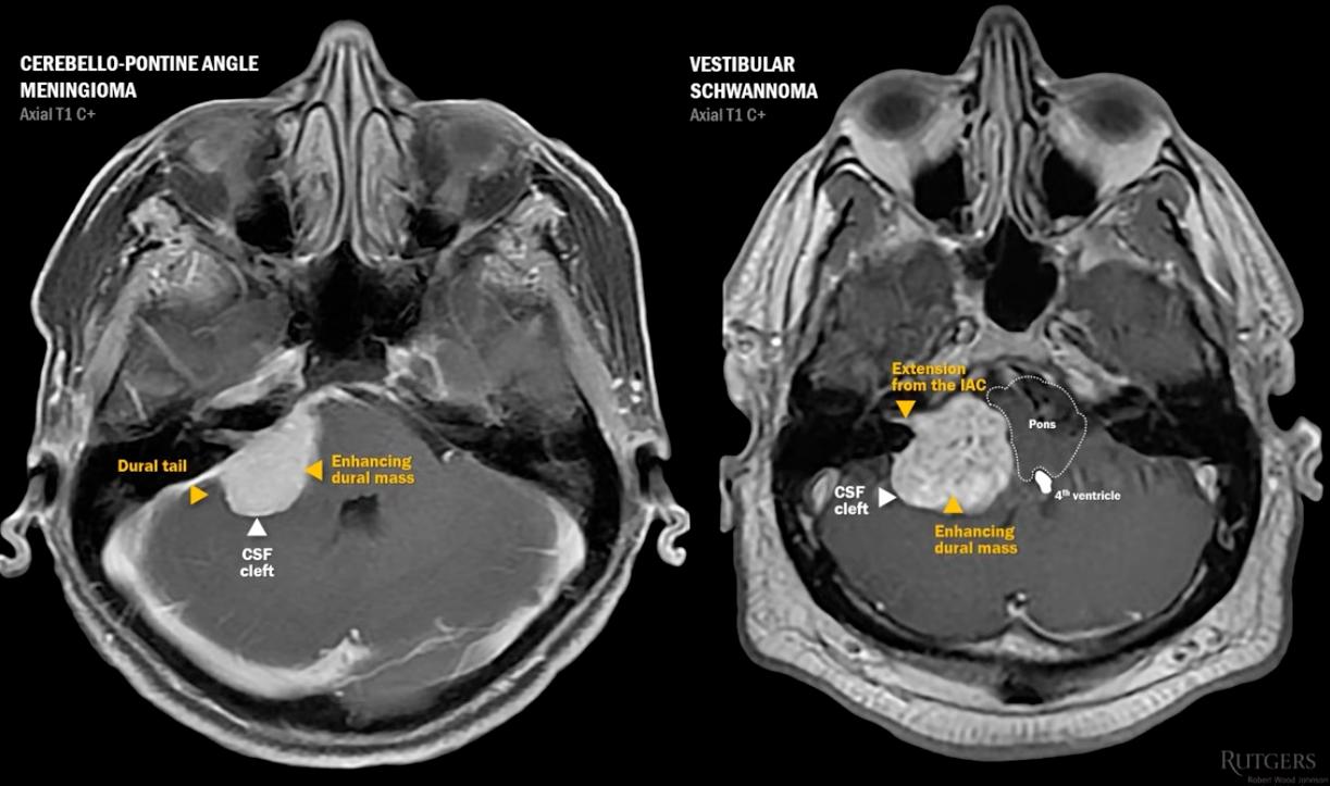
  • dural: meningioma[…] & schwannoma[…]
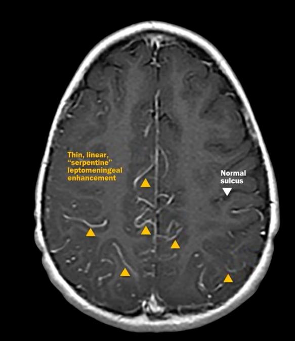
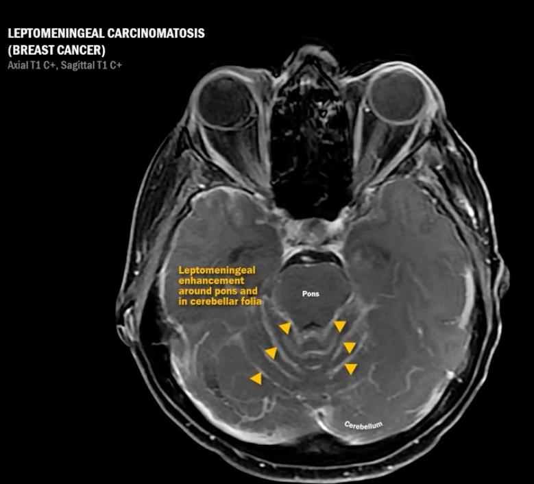
  • leptomeningeal: meningitis[…] & LMM[…]
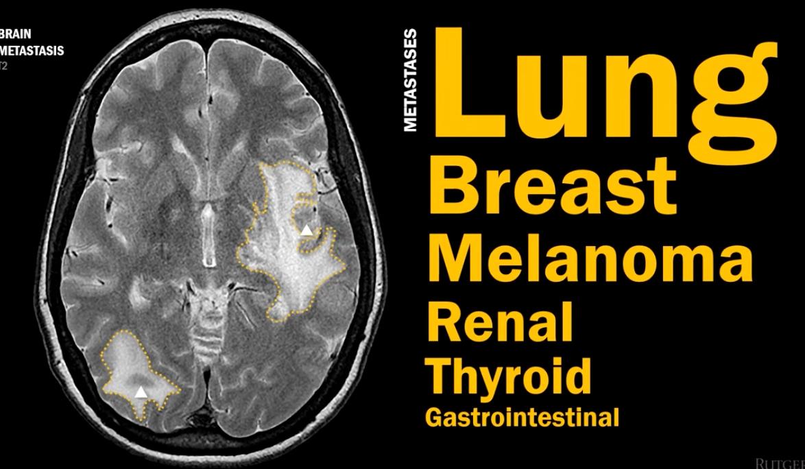
  • subcortical nodular: mets[…] & acute MS ovoid lesions[…]
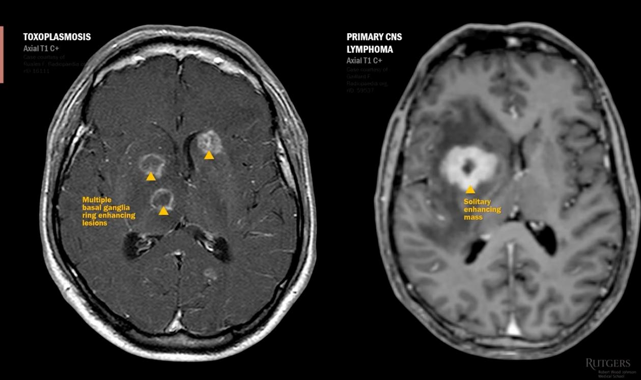
  • ring enhancing: abscess[…], toxo[…], PCNSL[…]
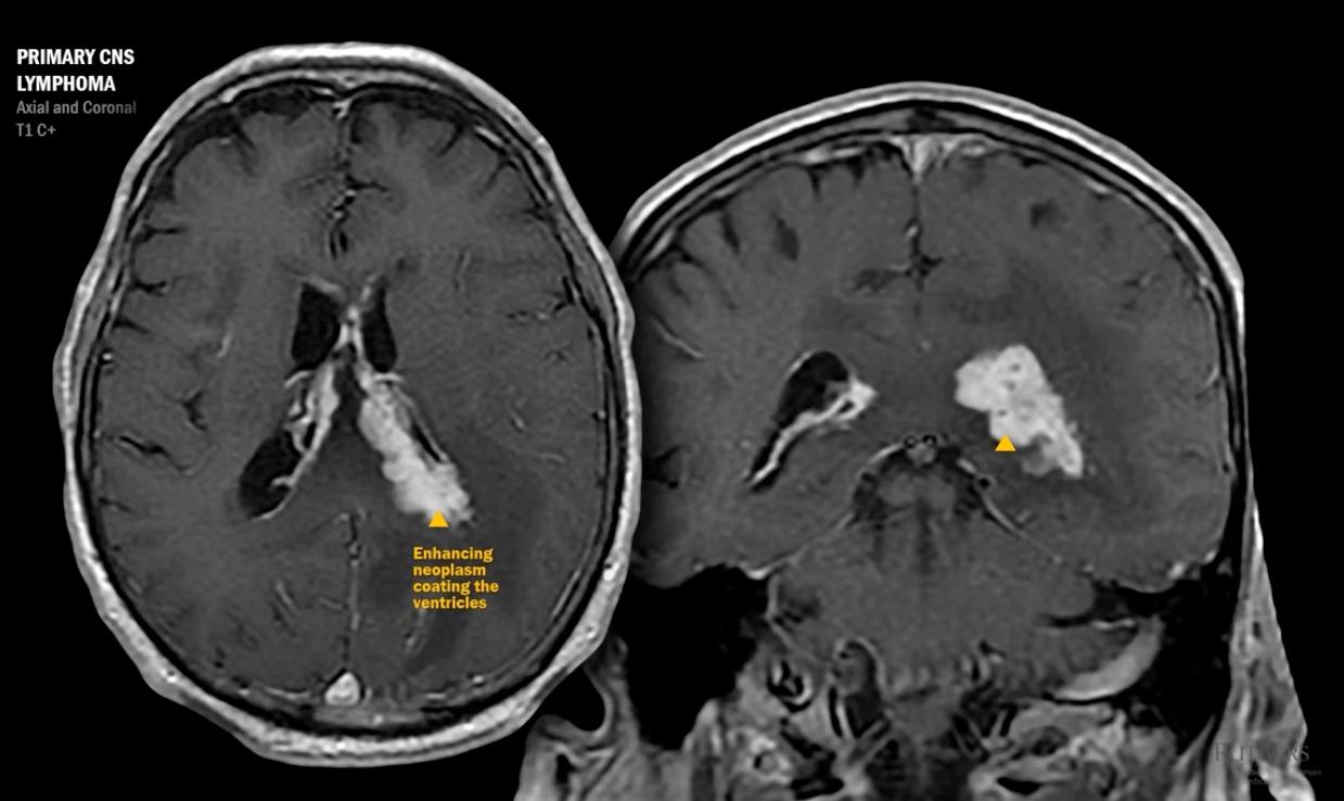
  • paraventricular: PCNSL[…]
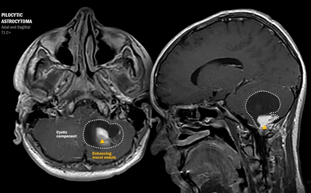
  • mural nodule: pilocytic astrocytoma[…]
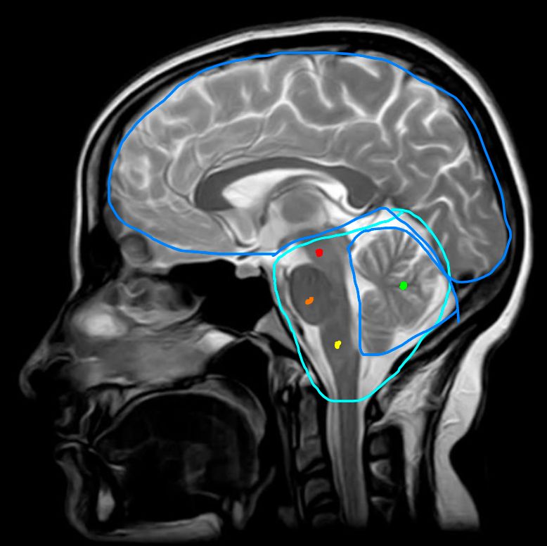
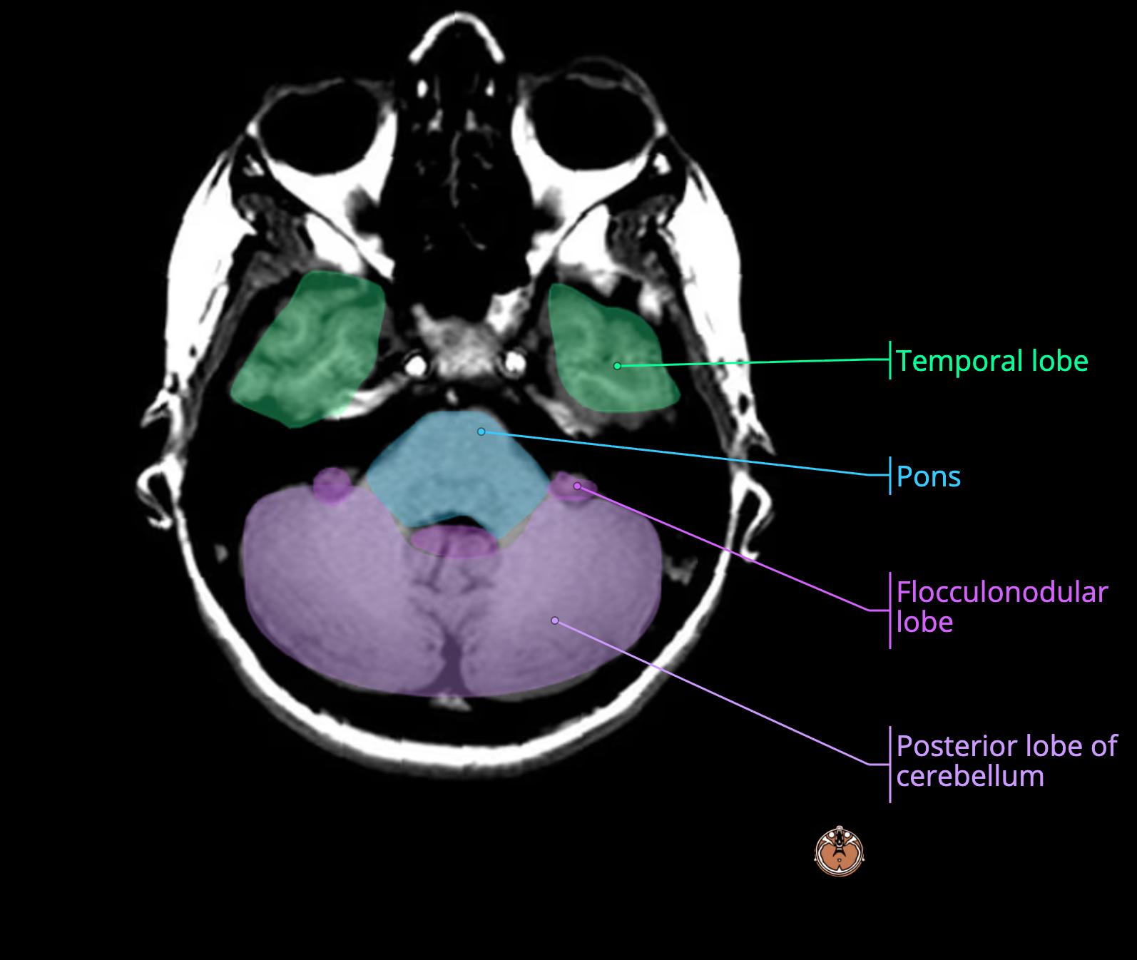
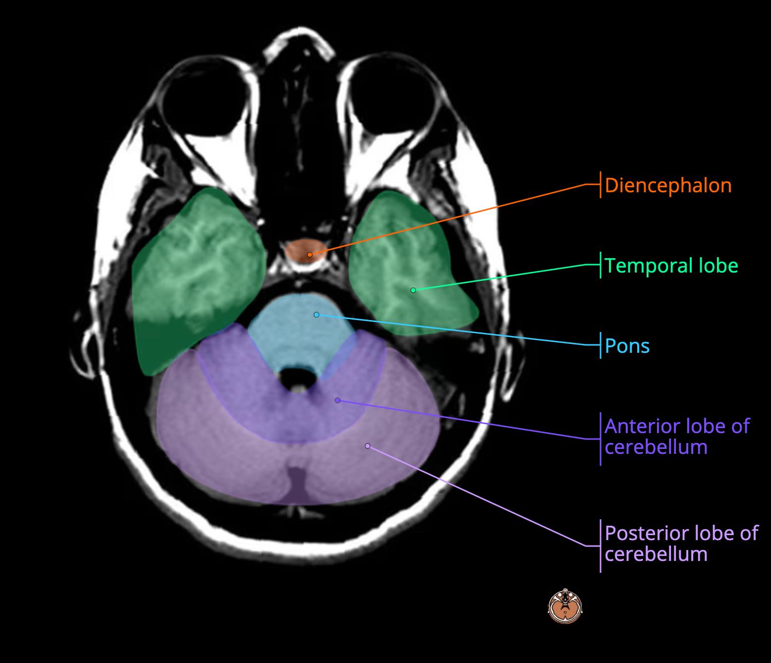
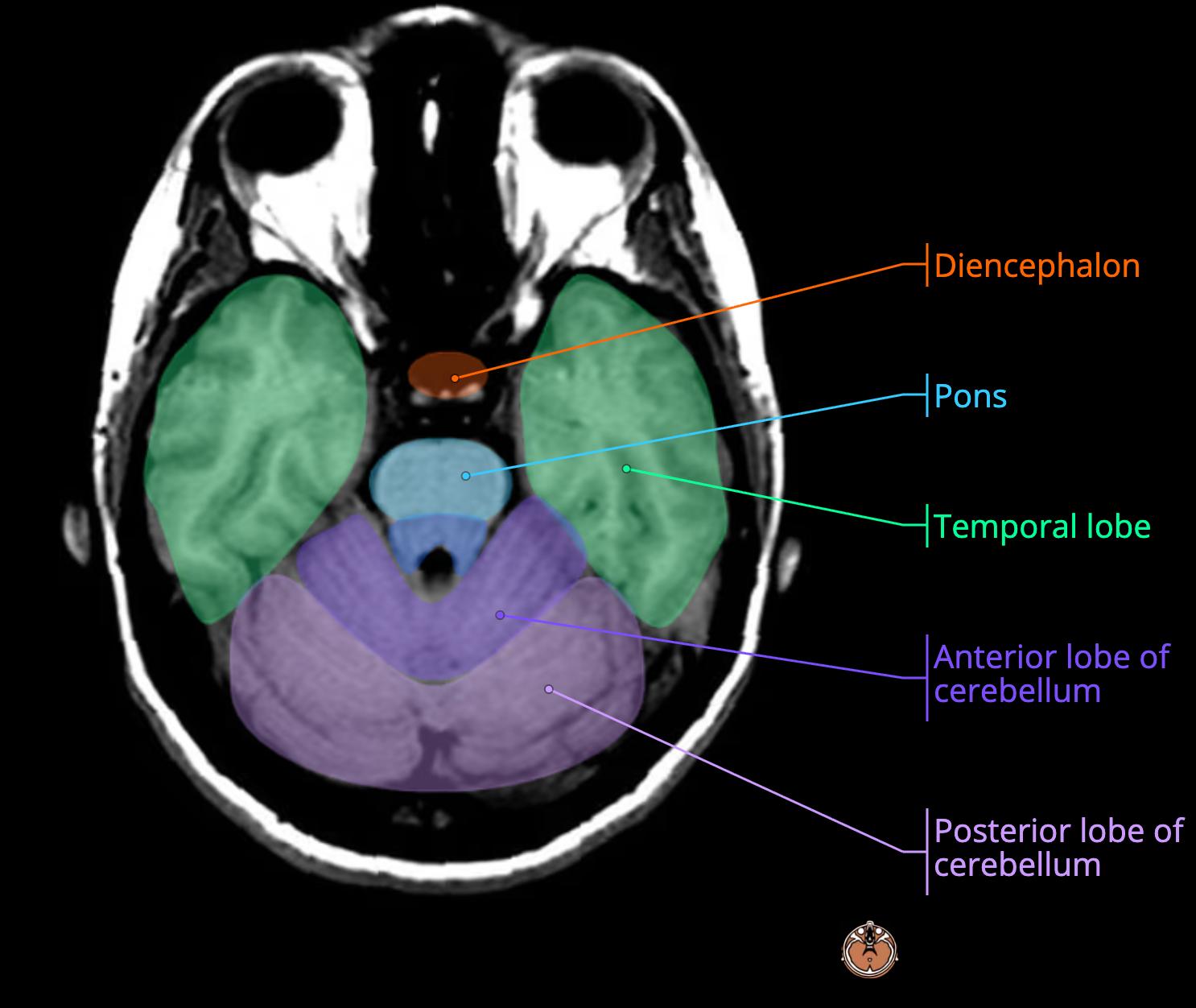
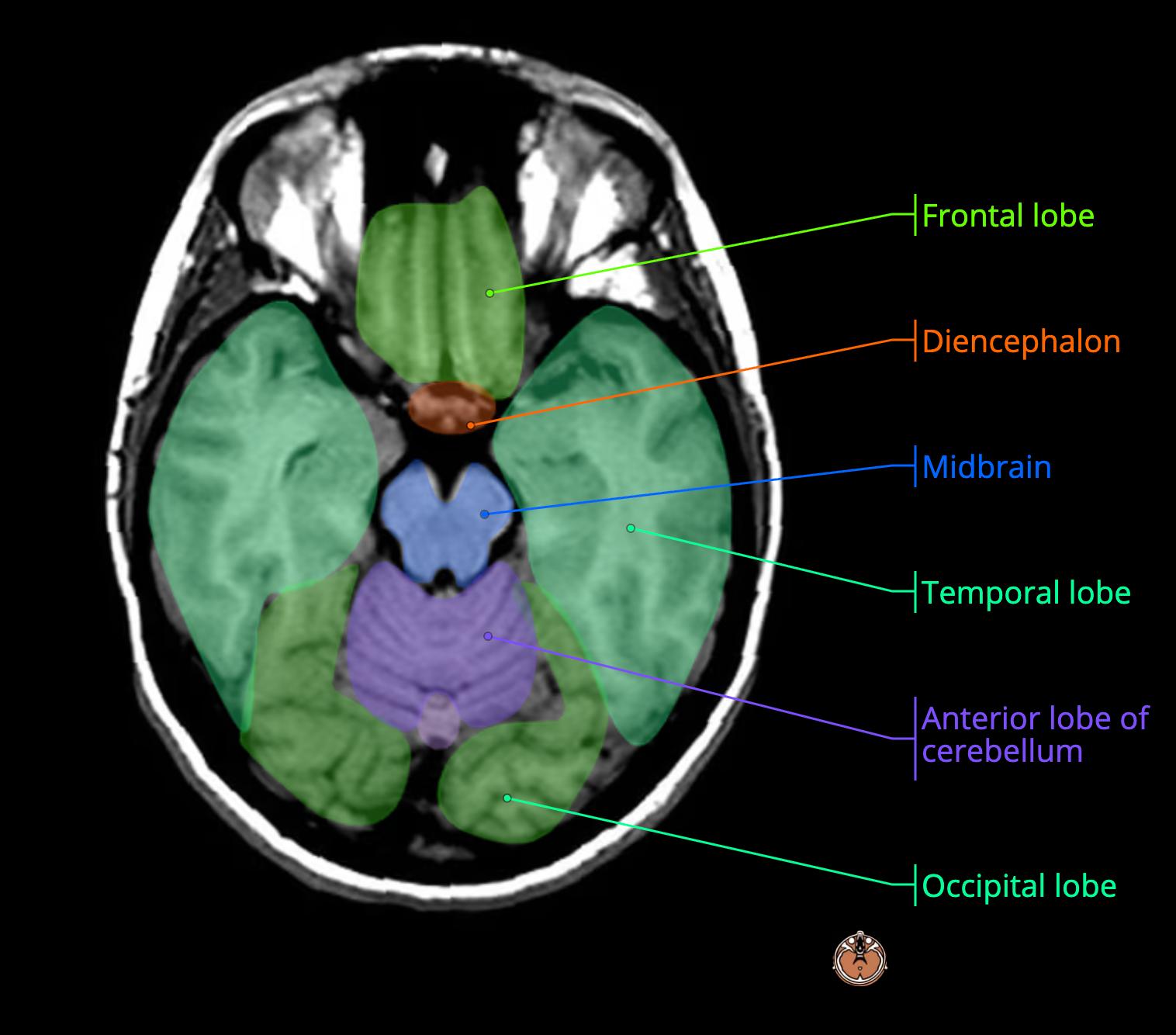
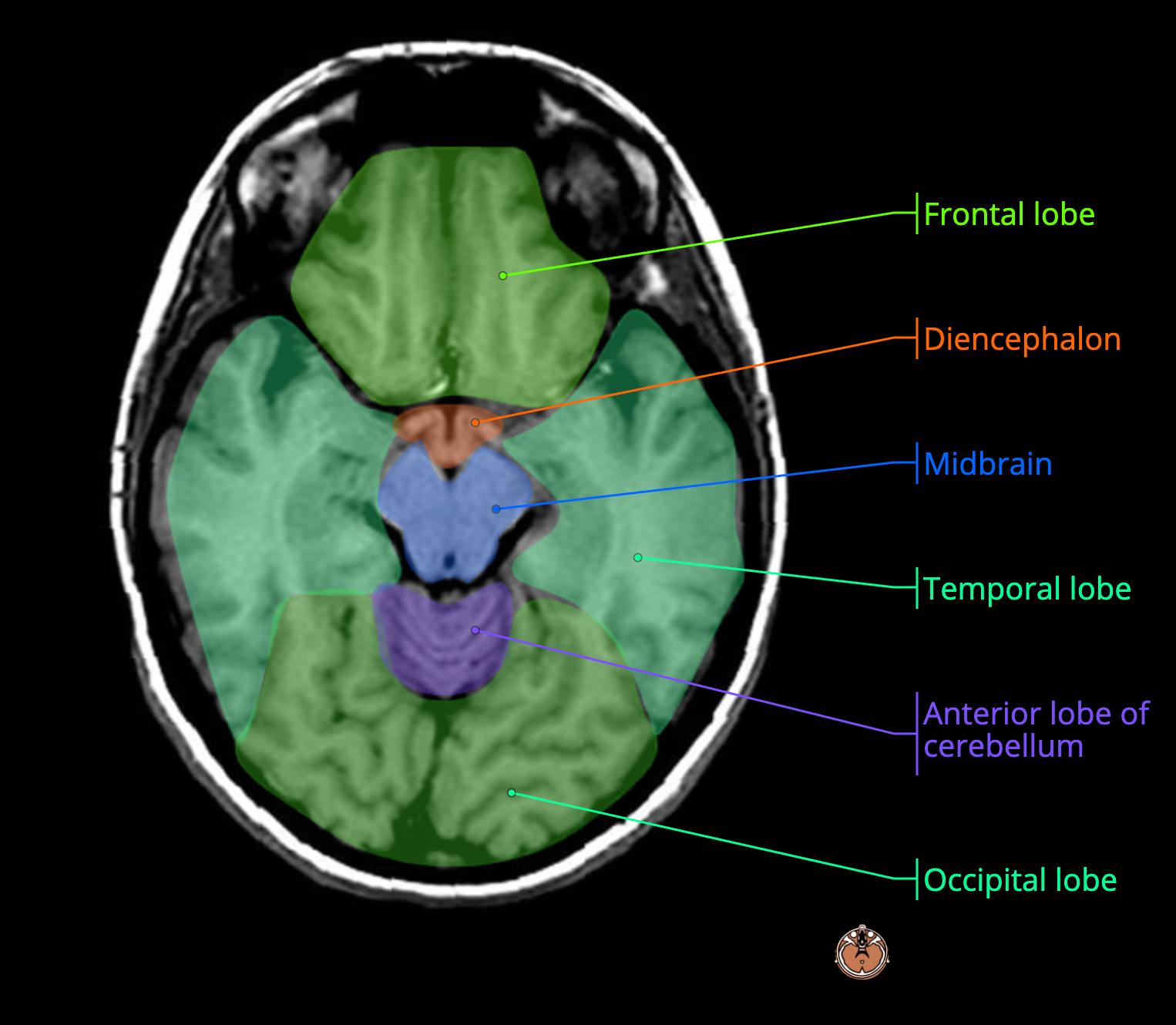
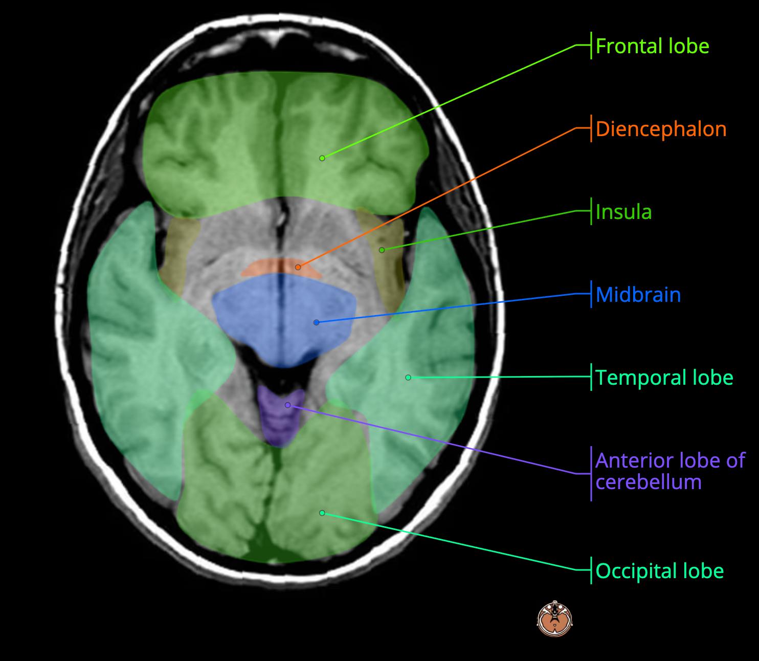
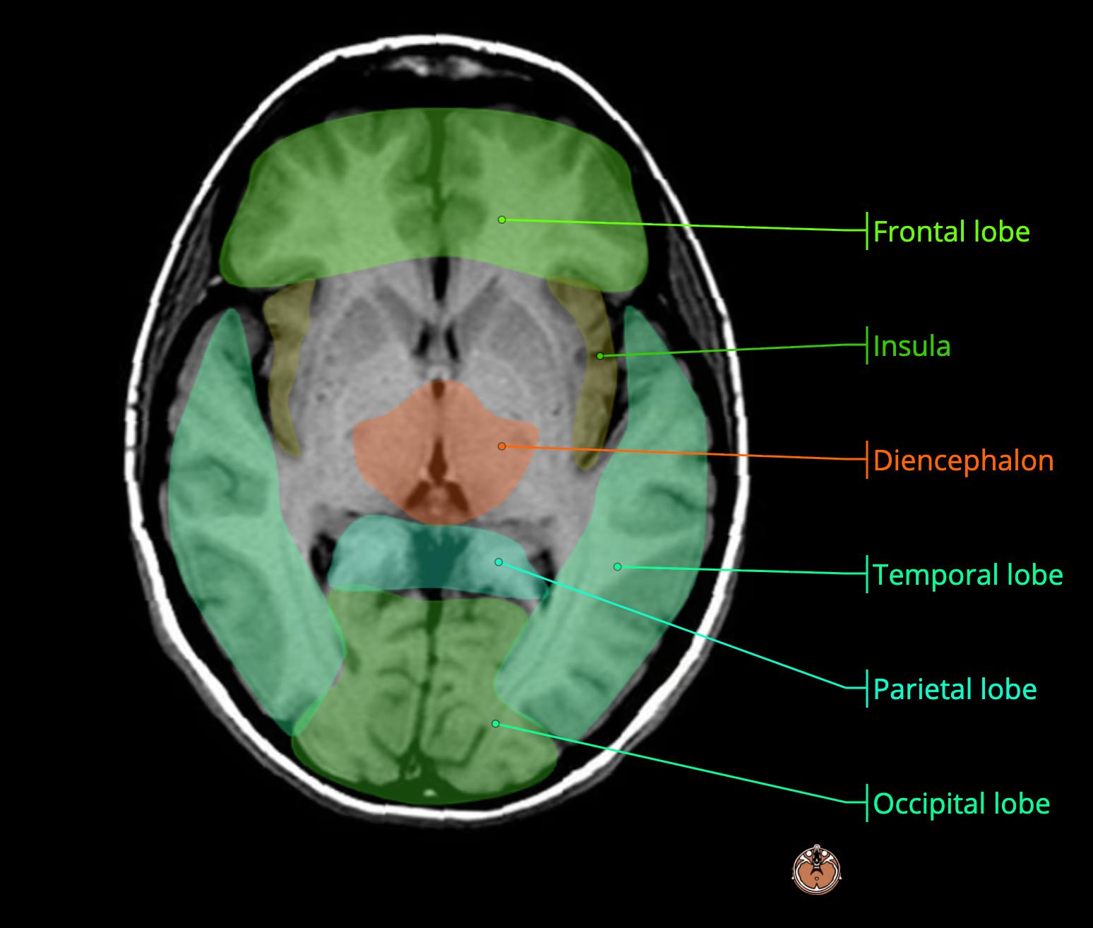
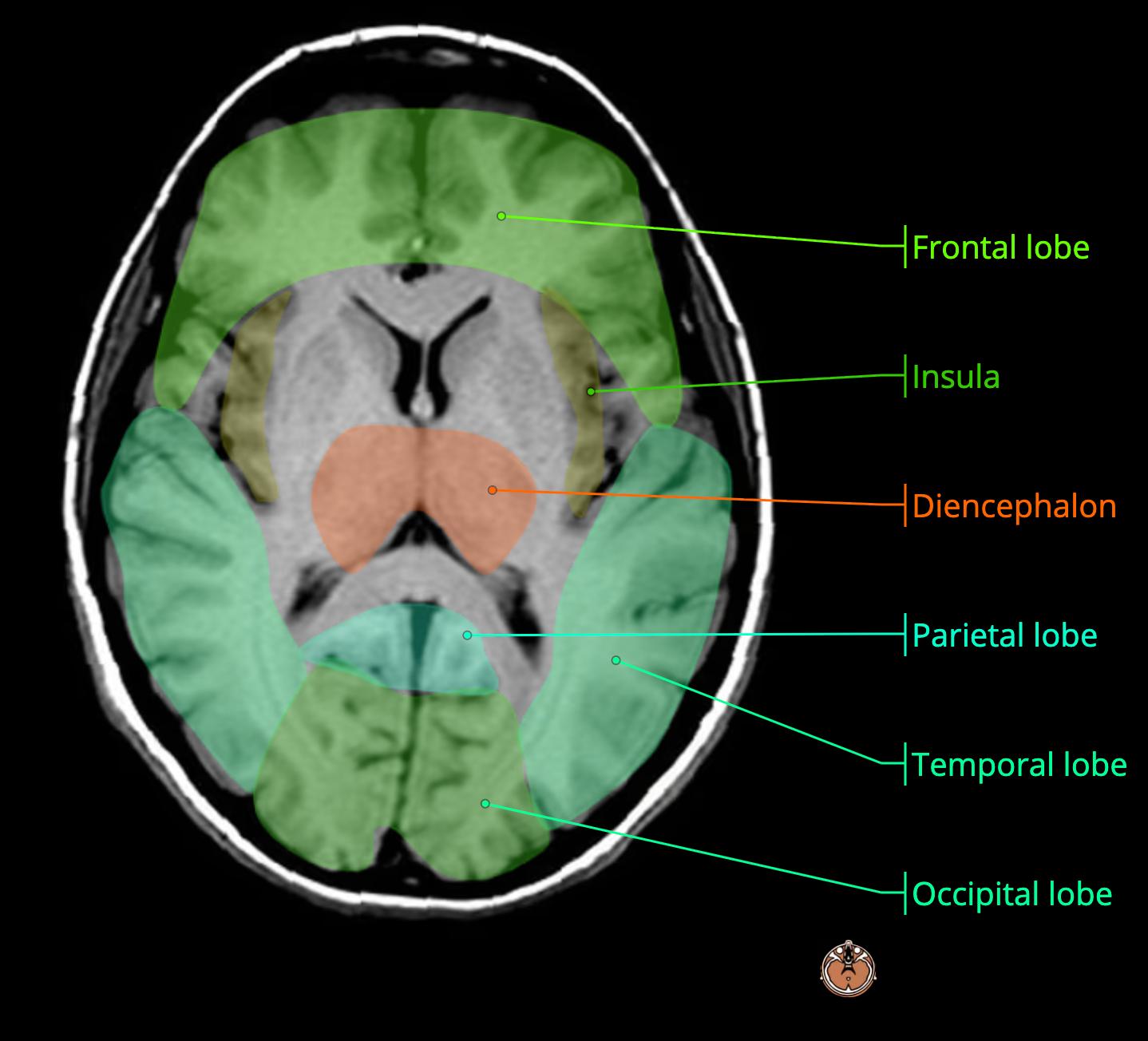
    MRI Brain Anatomy
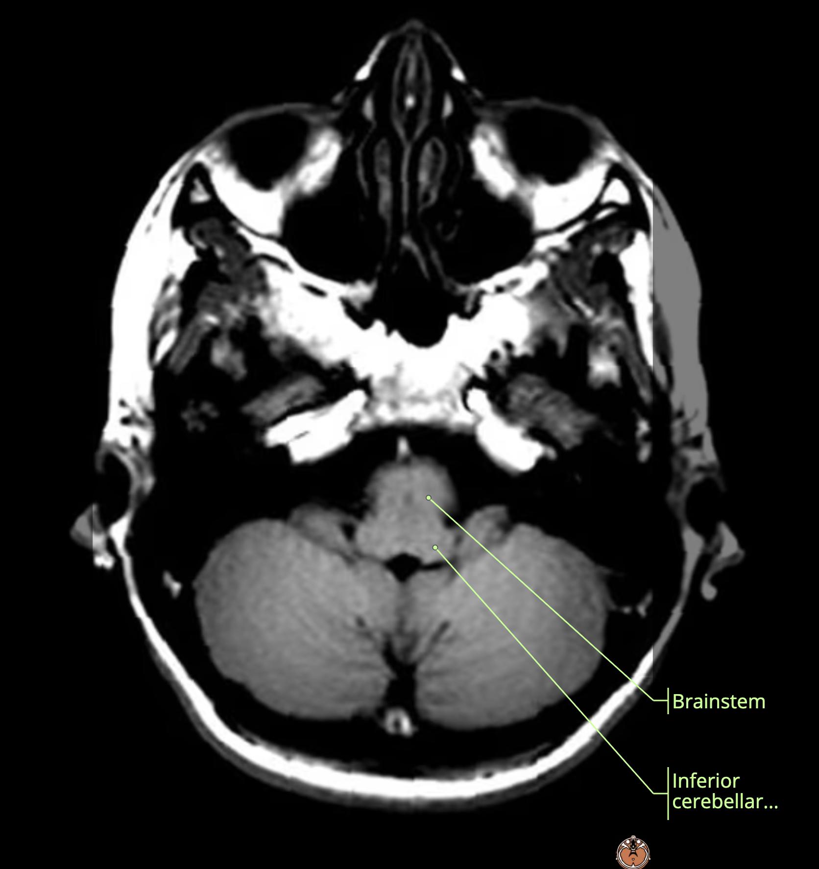
    • red: midbrain[…]
    • orange: pons[…]
    • yellow: medulla[…]
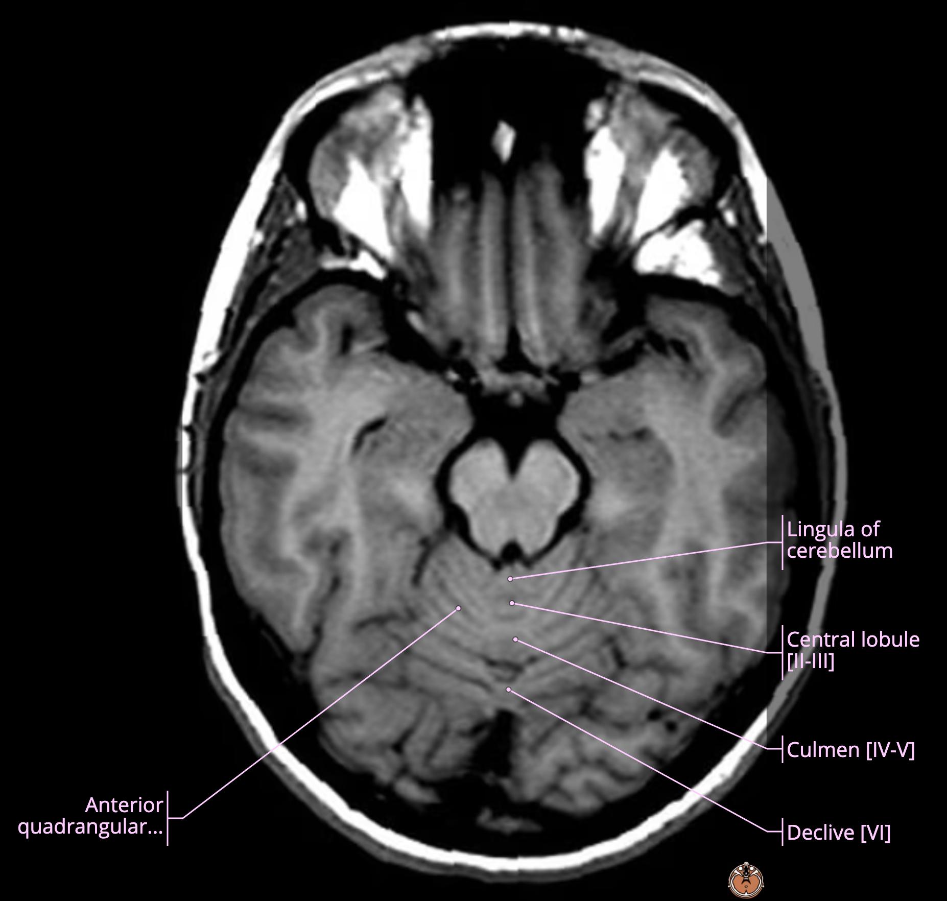
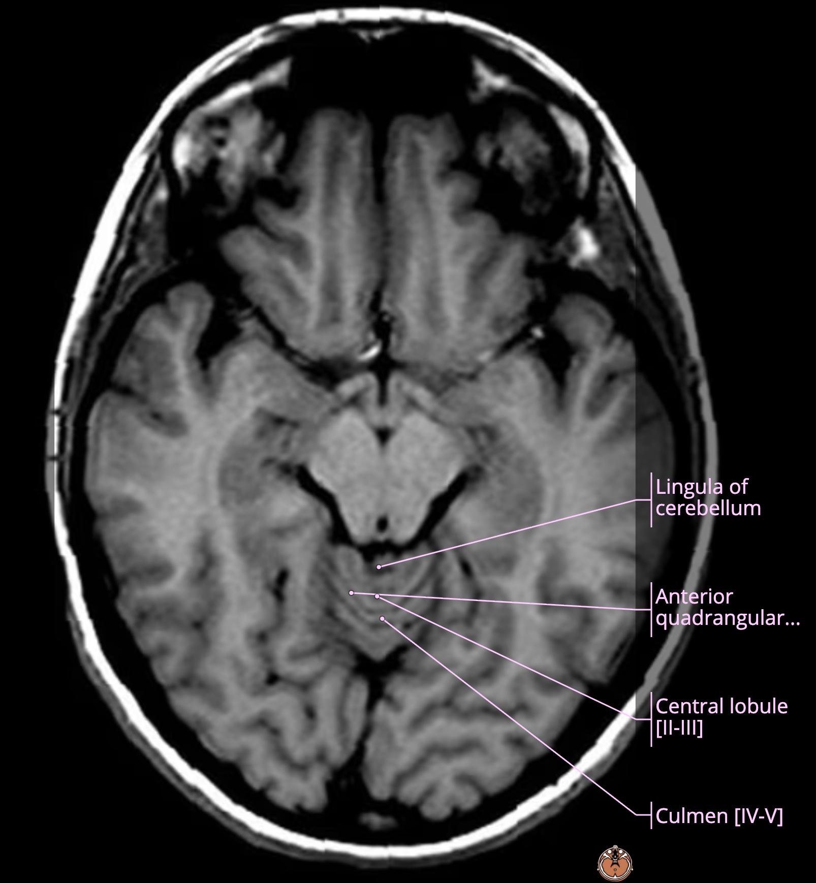
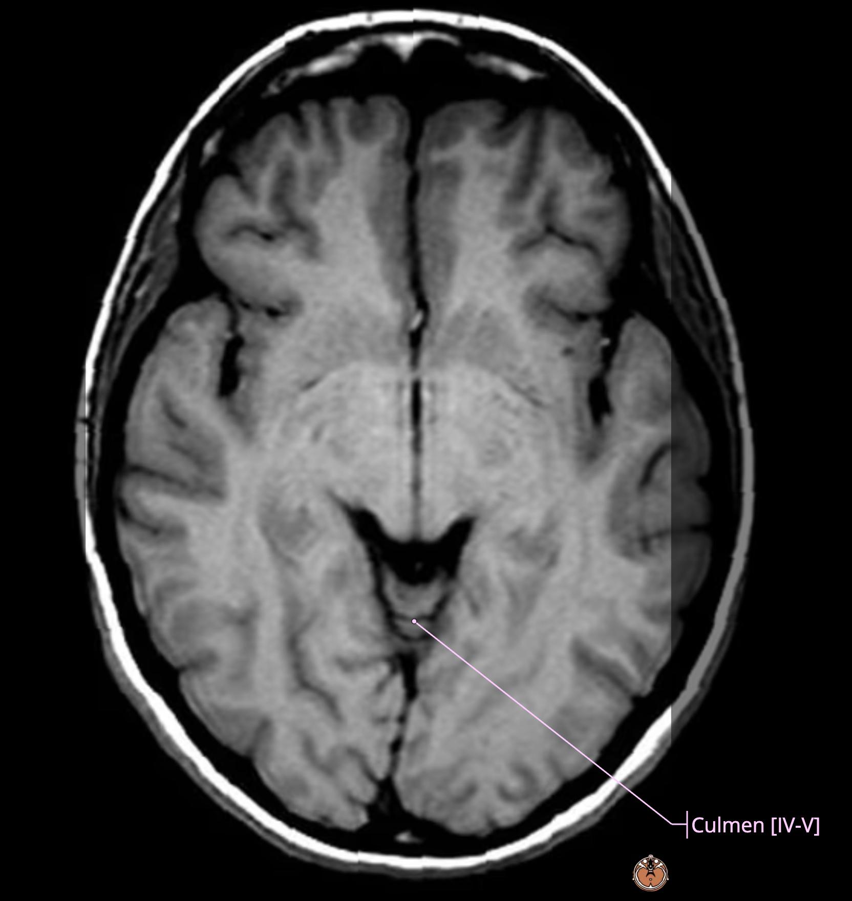
    • green: cerebellum[…]
    • light blue: posterior fossa[…]
    • medium blue: cerebrum[…]cerebellum[…]


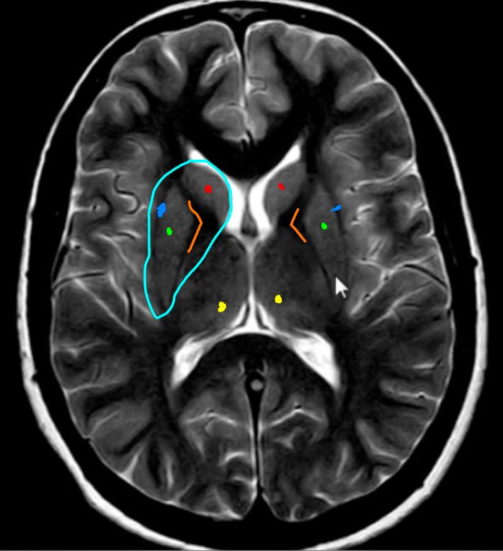
    MRI Brain Anatomy
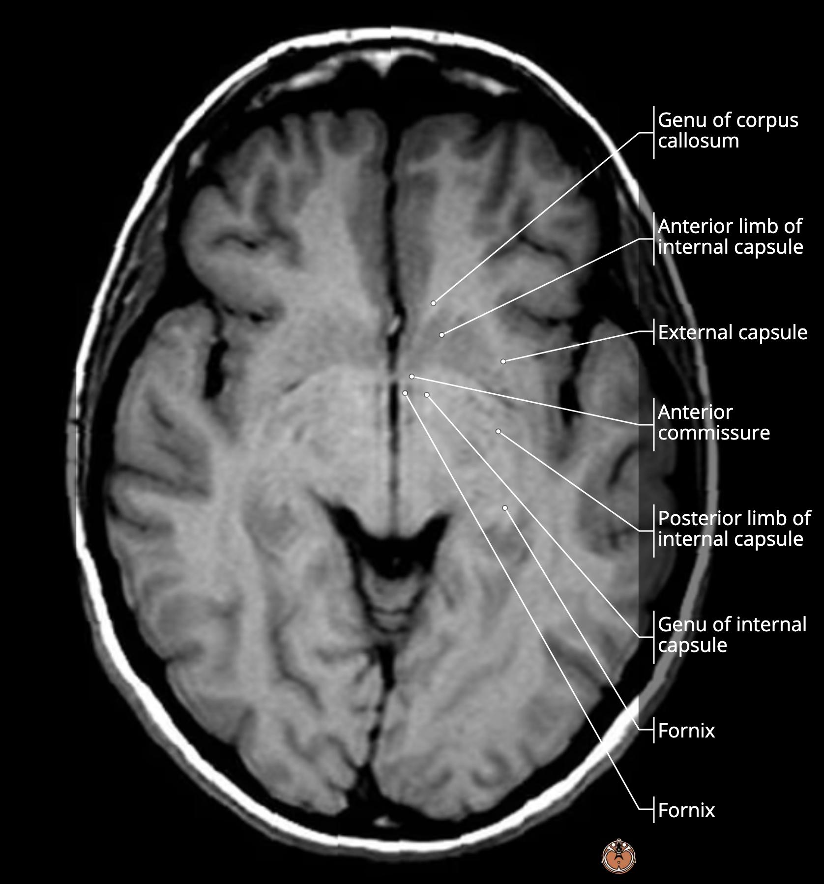
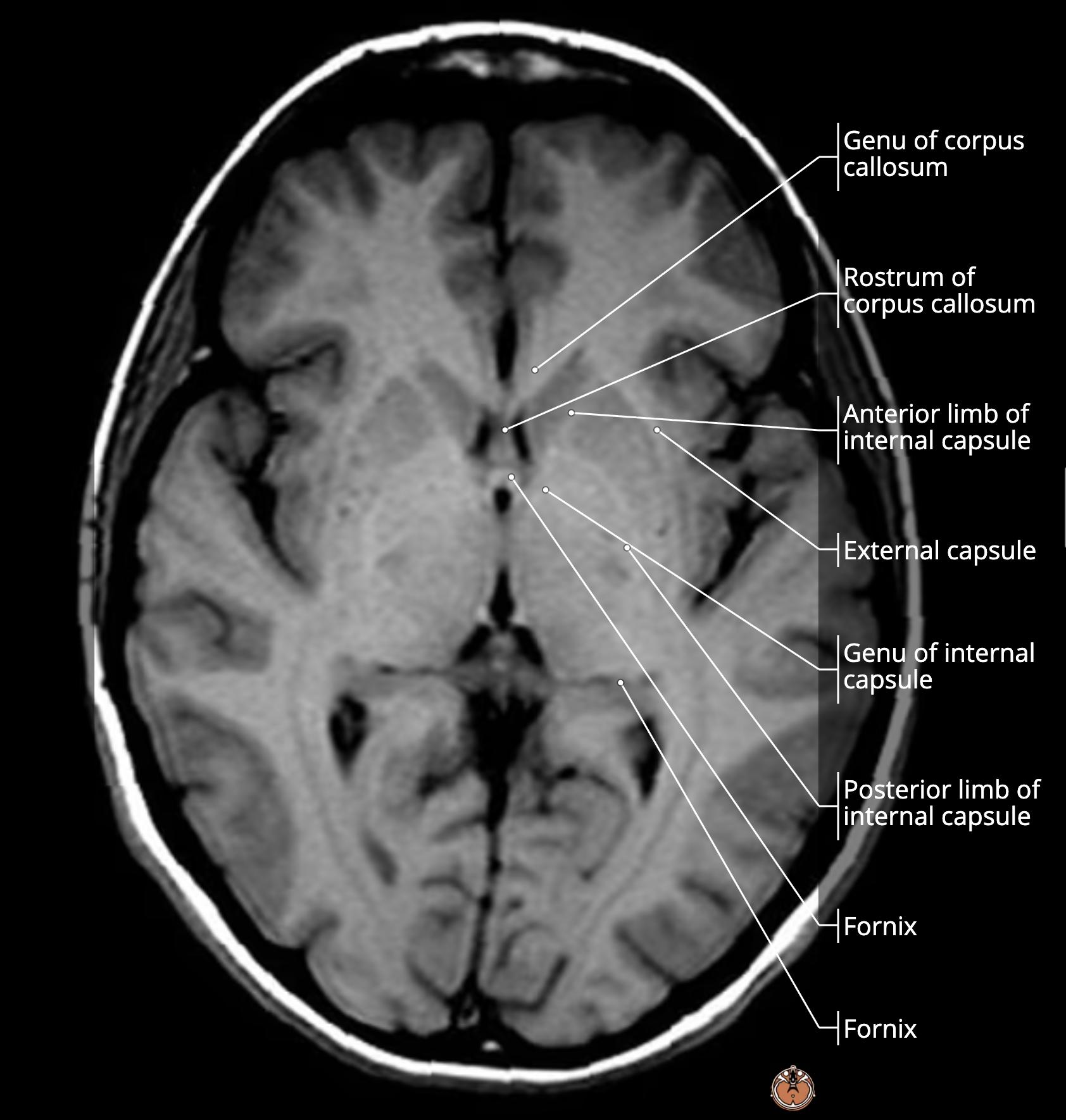
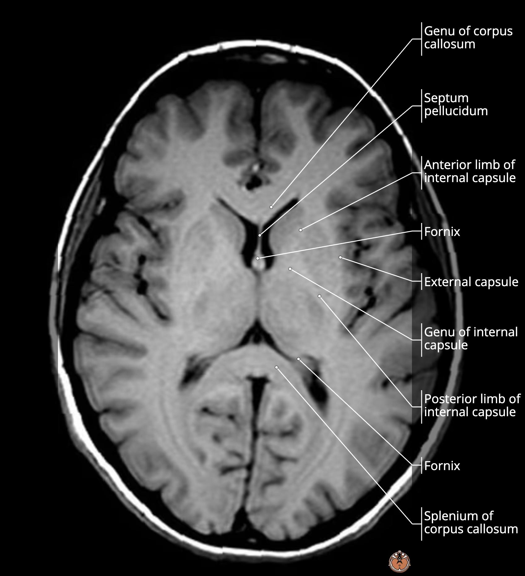
    • red deep gray matter: head of caudate[…]
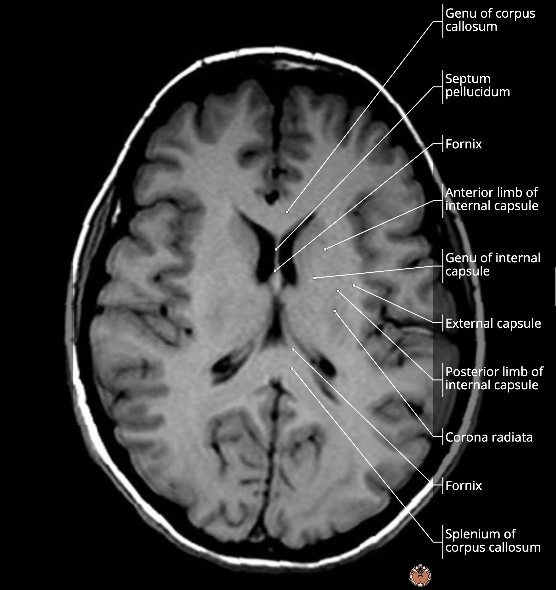
    • orange white matter: anterior limb, genu, posterior limb of IC[…]
    • green deep gray matter: lentiform nucleus (Gpi/e + putamen)[…]
    • light blue: corpus striatum (lentiform nucleus + head of caudate)[…]
    • yellow: thalamus[…]
    • medium blue: external capsule[…]

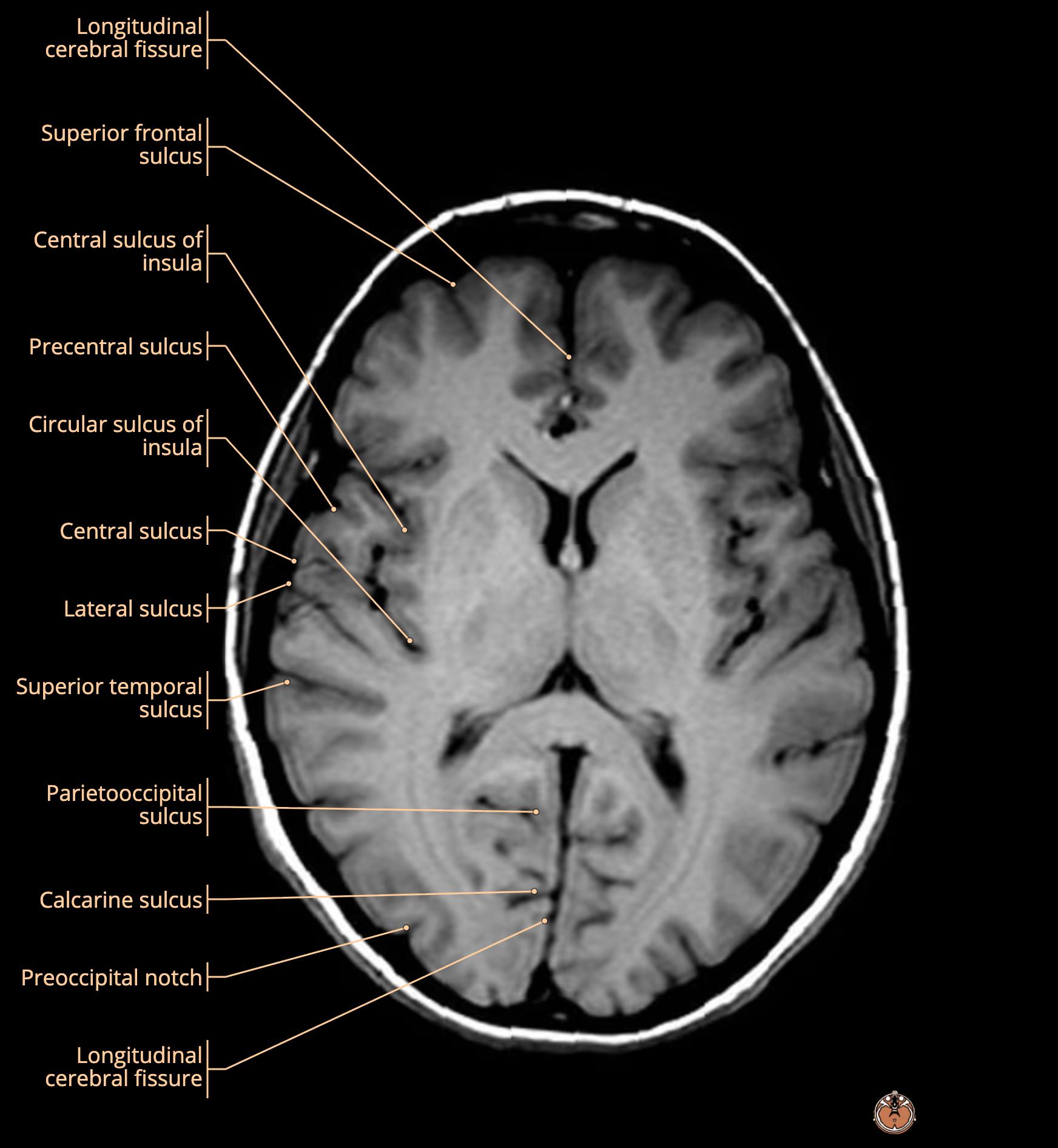
    MRI Brain Anatomy
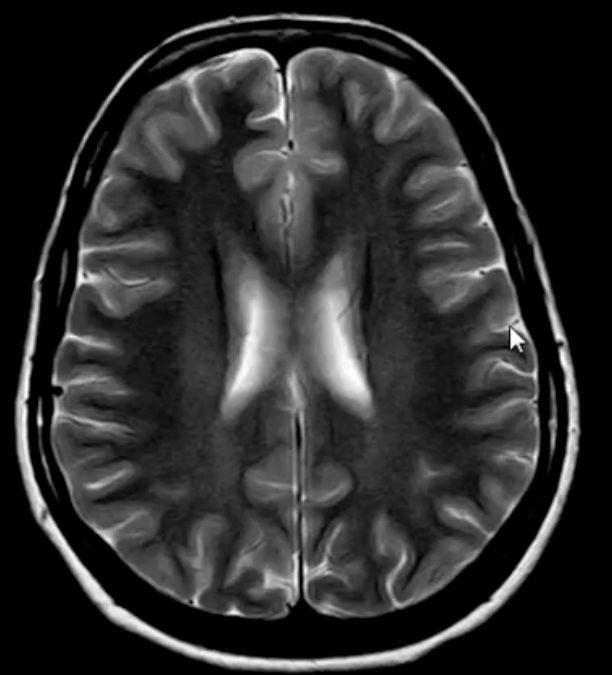
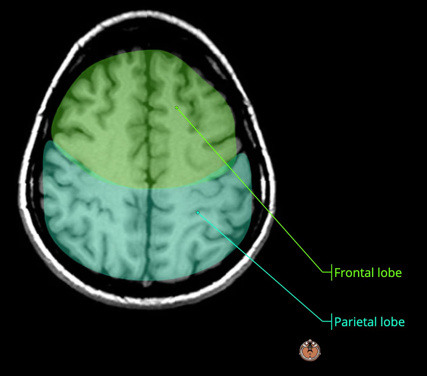
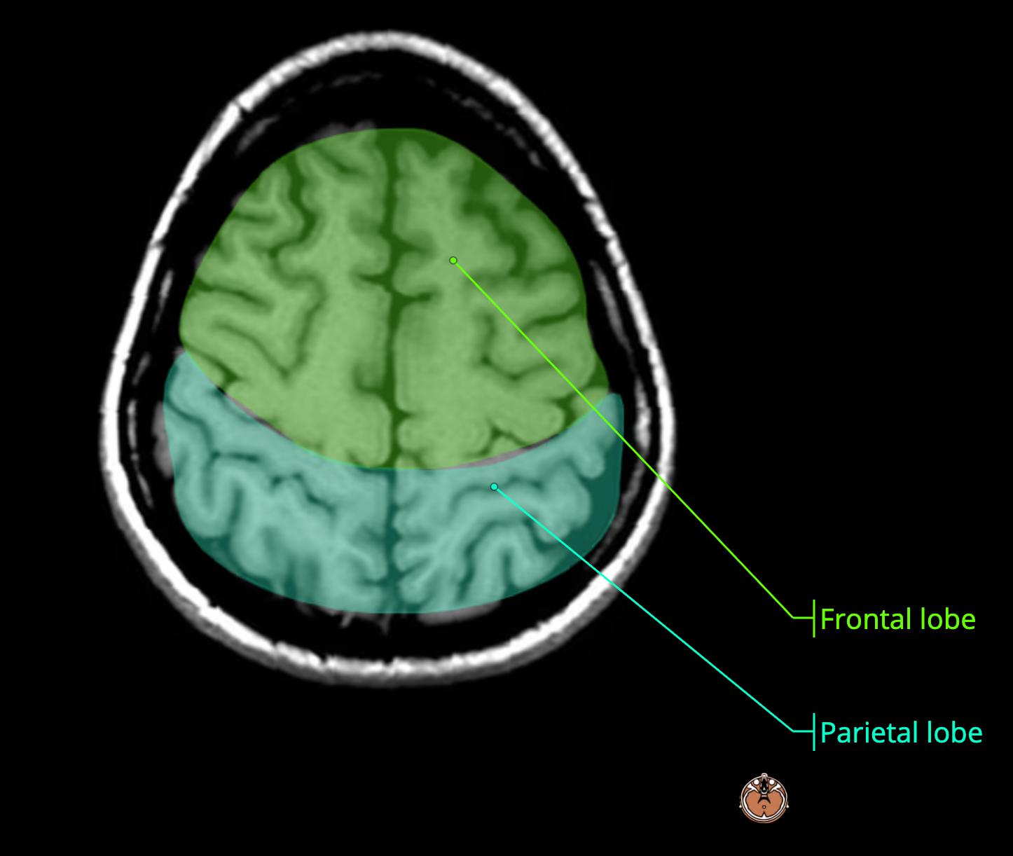
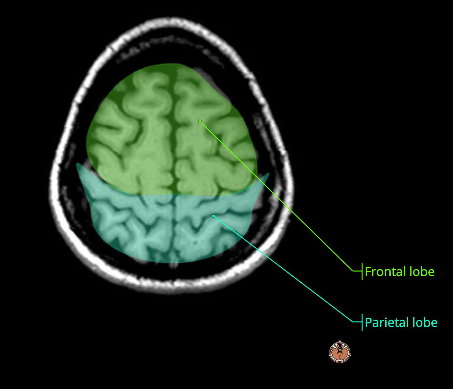
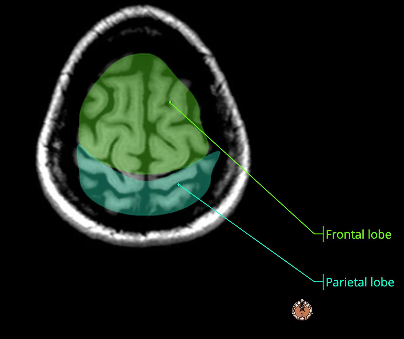
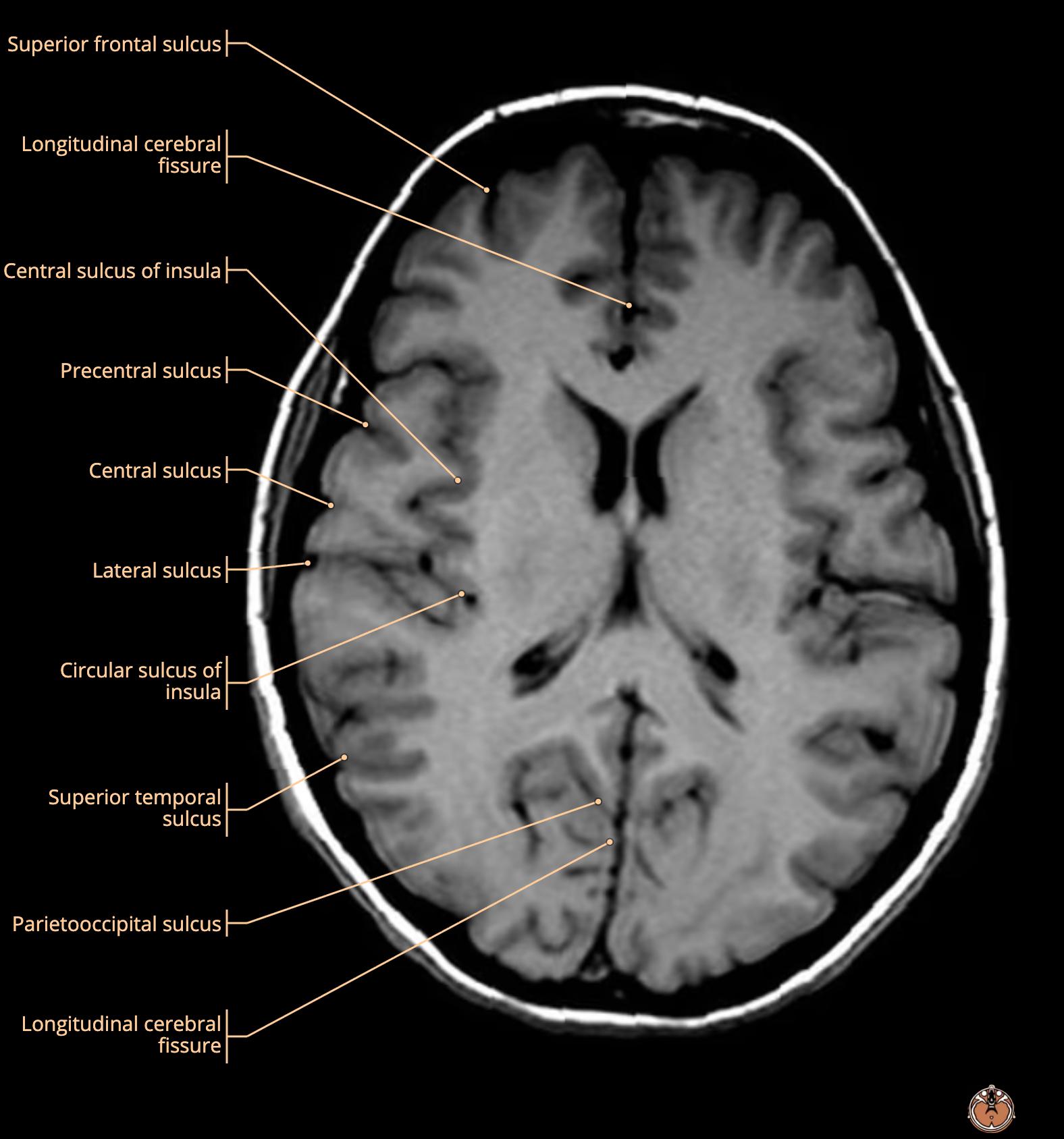
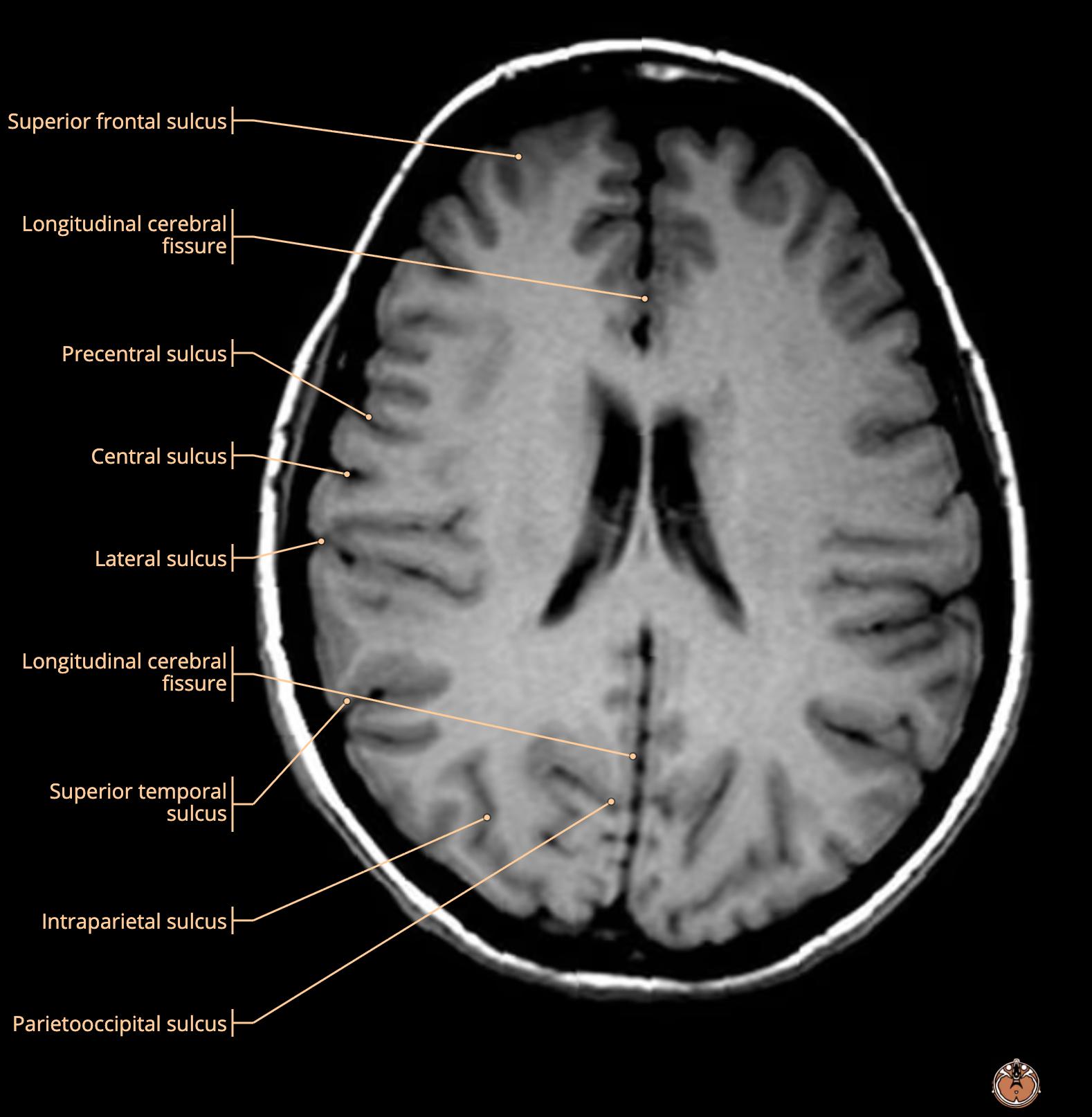
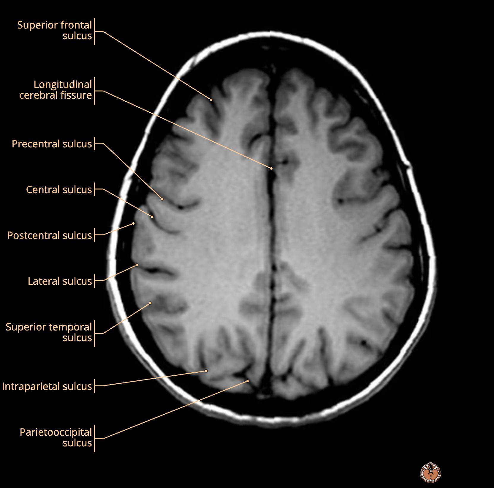
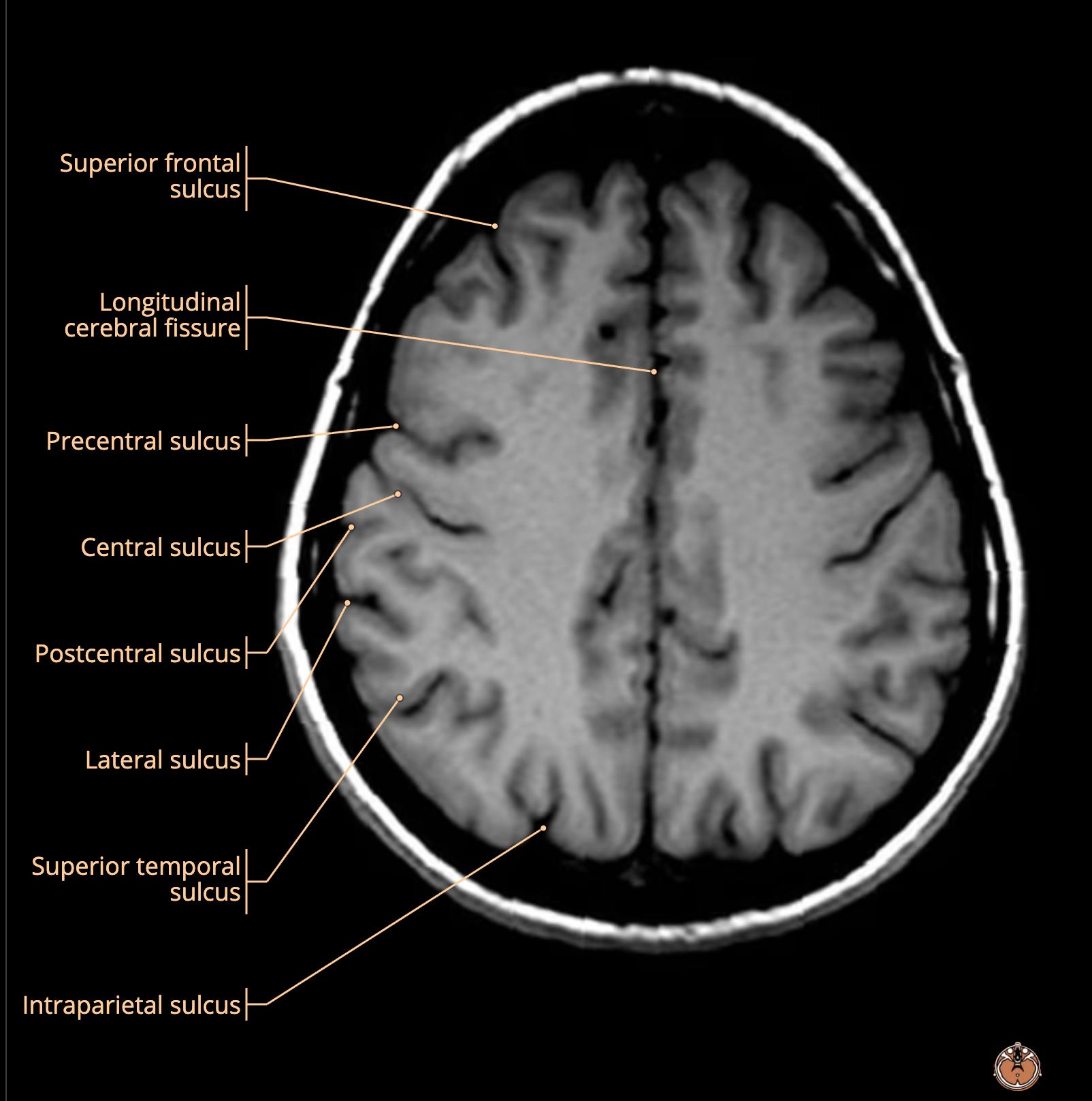
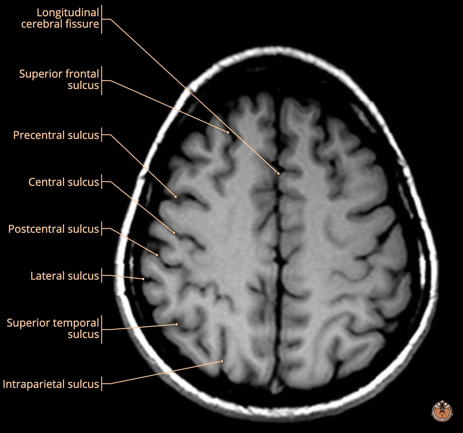
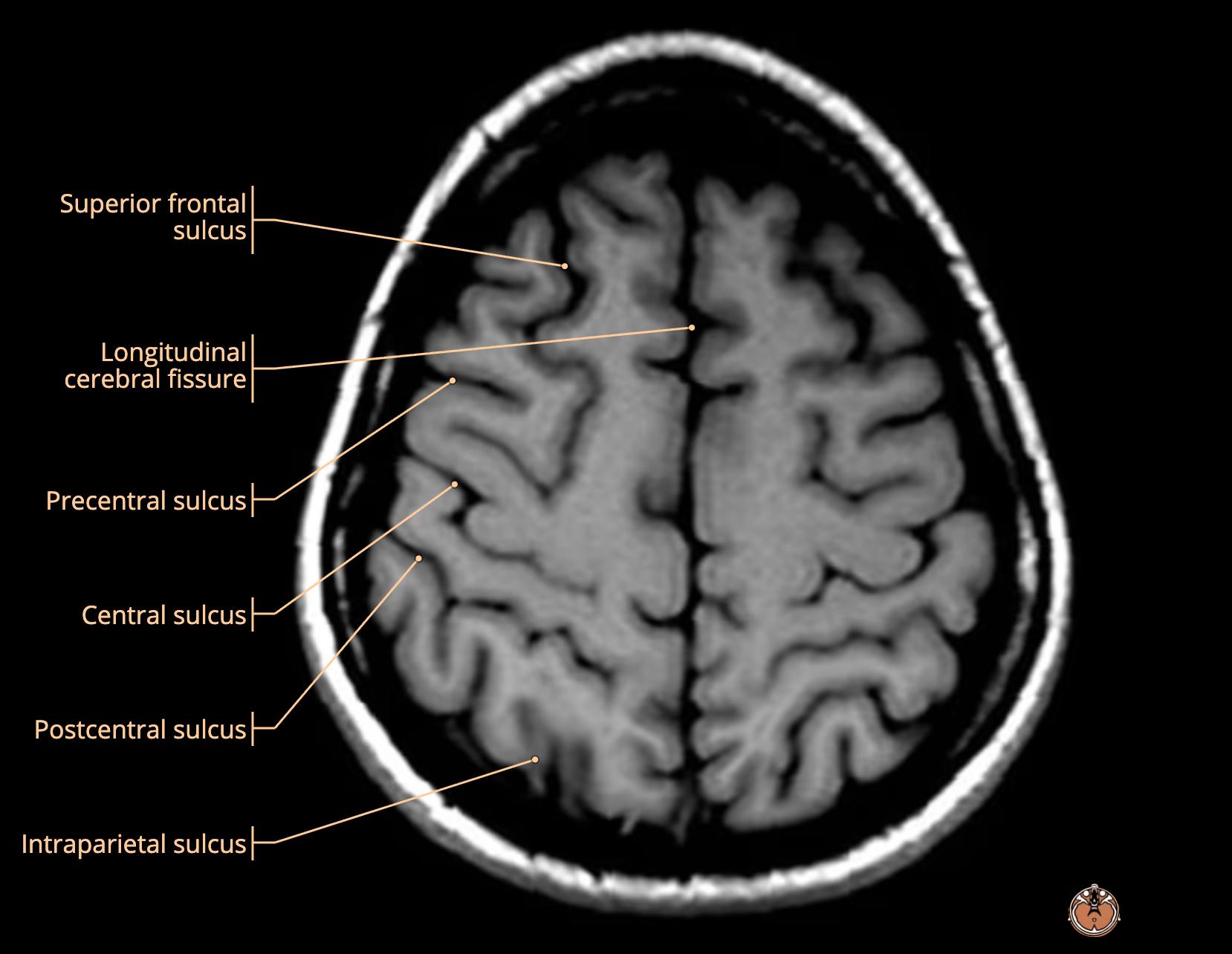
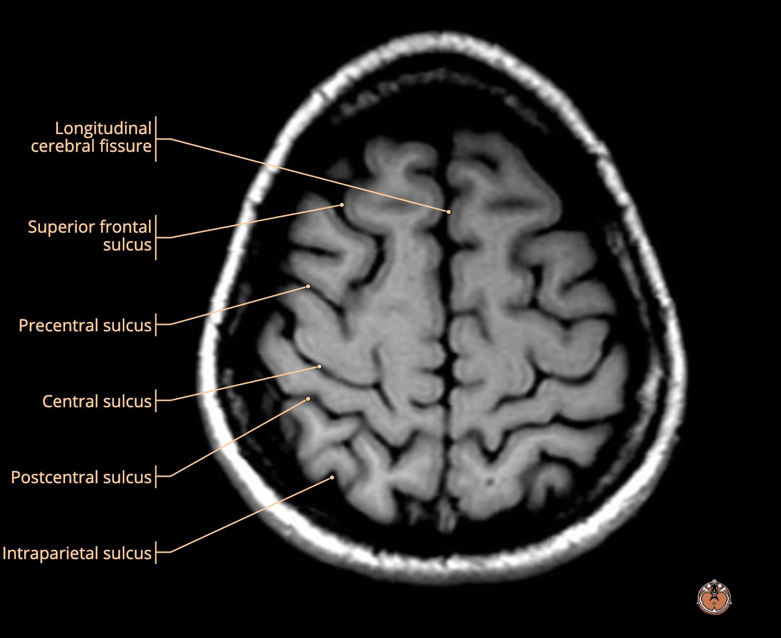
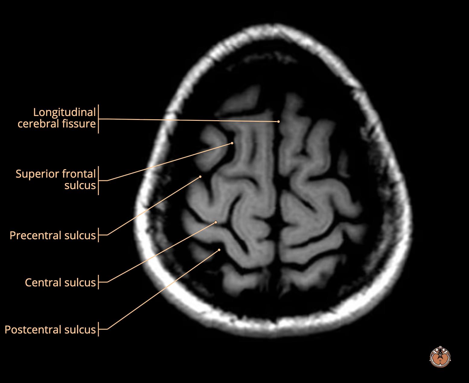
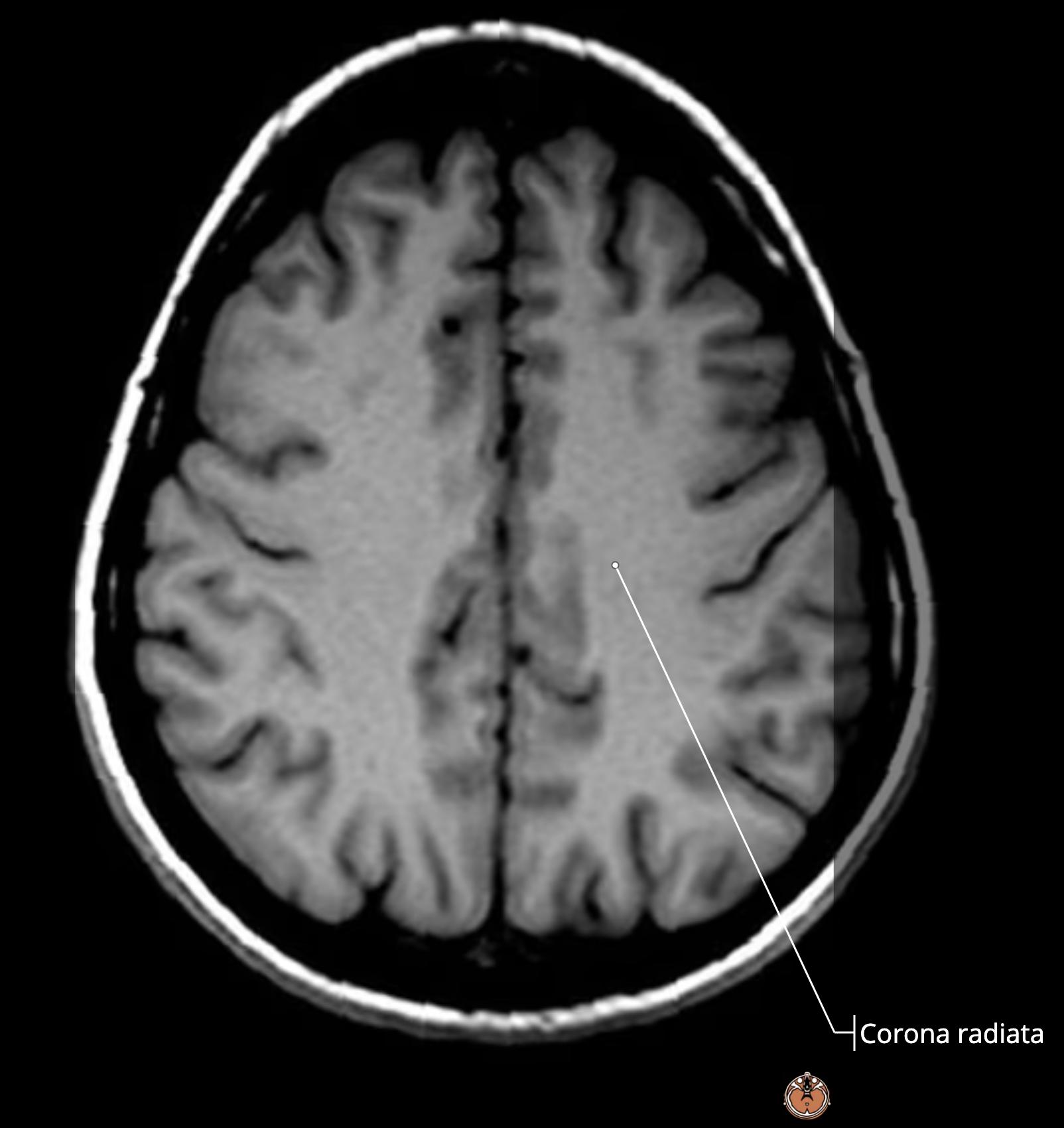
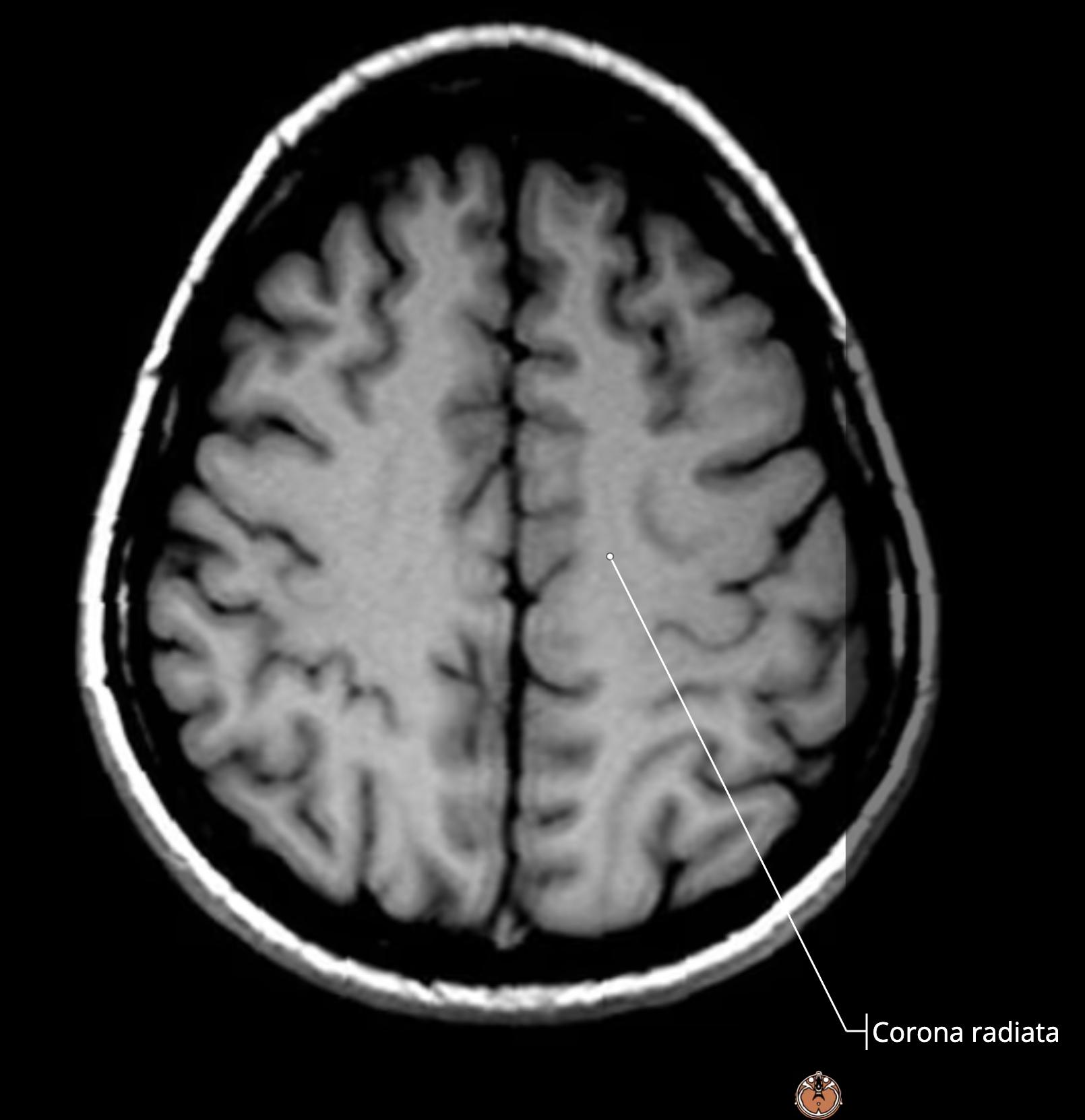
    2 ways to ID the central sulcus
    • look for the sulcus that reaches the midline[…]
    • look for the omega[…] sign
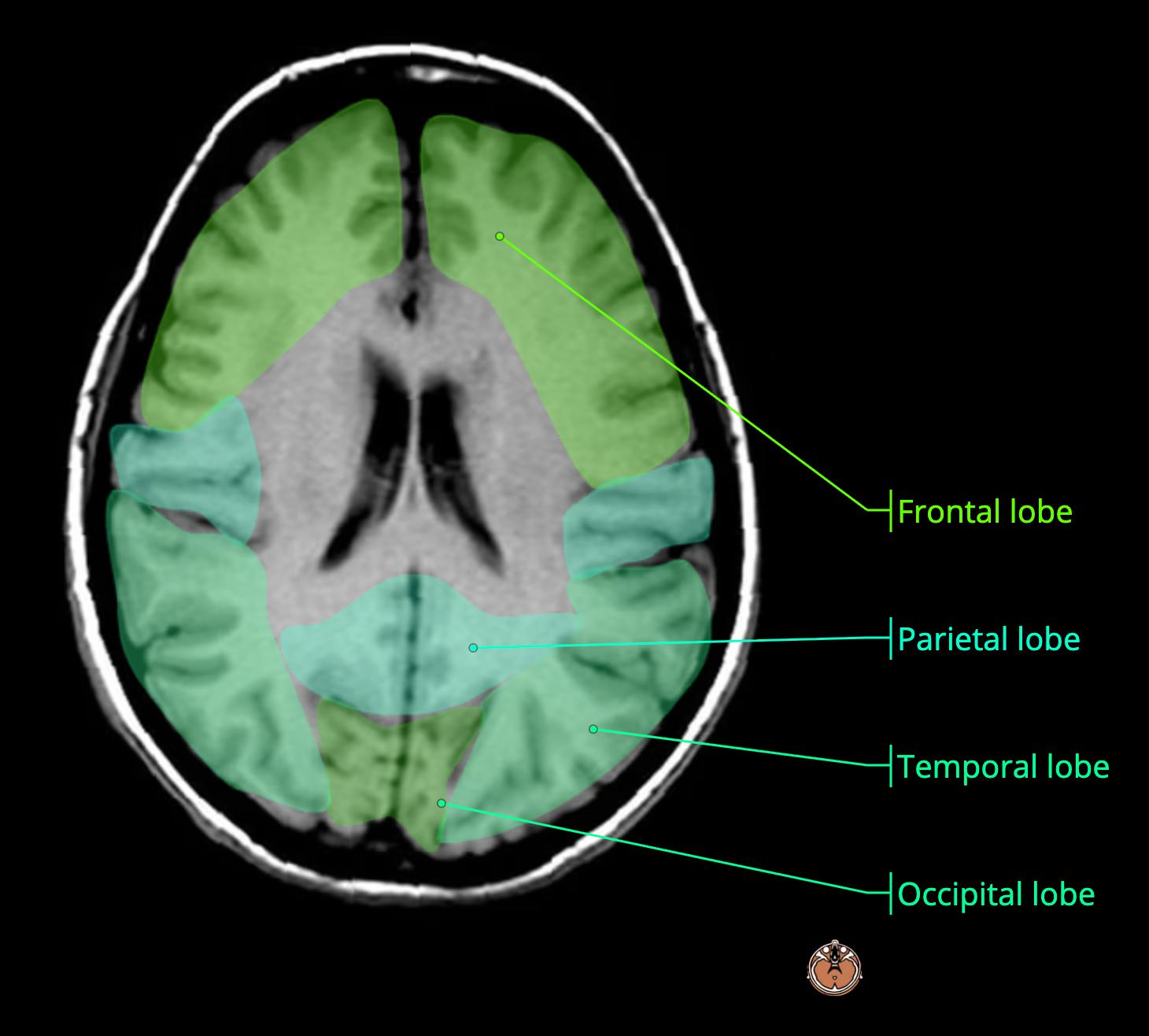
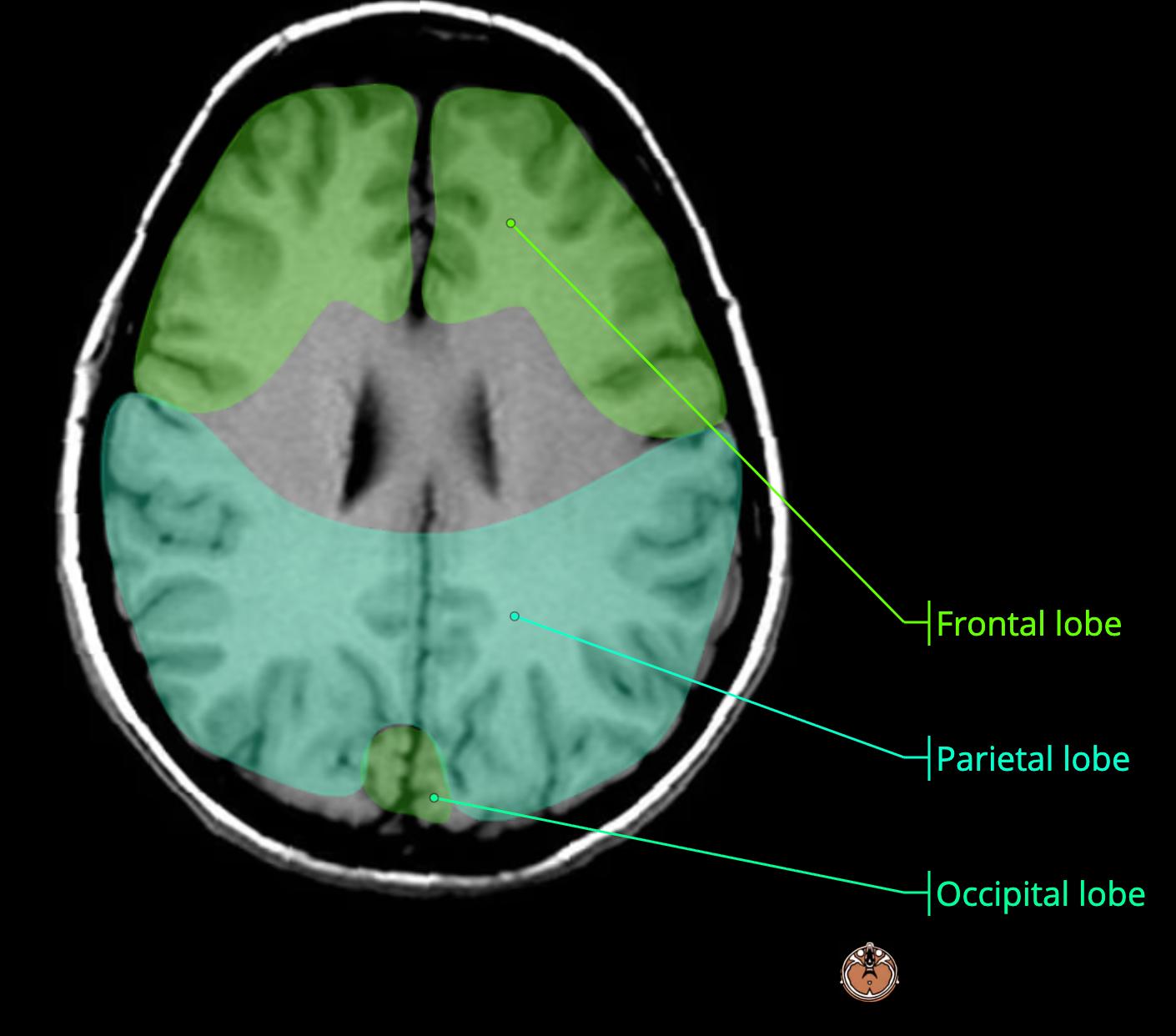
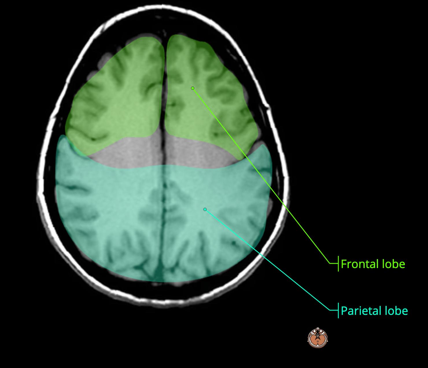
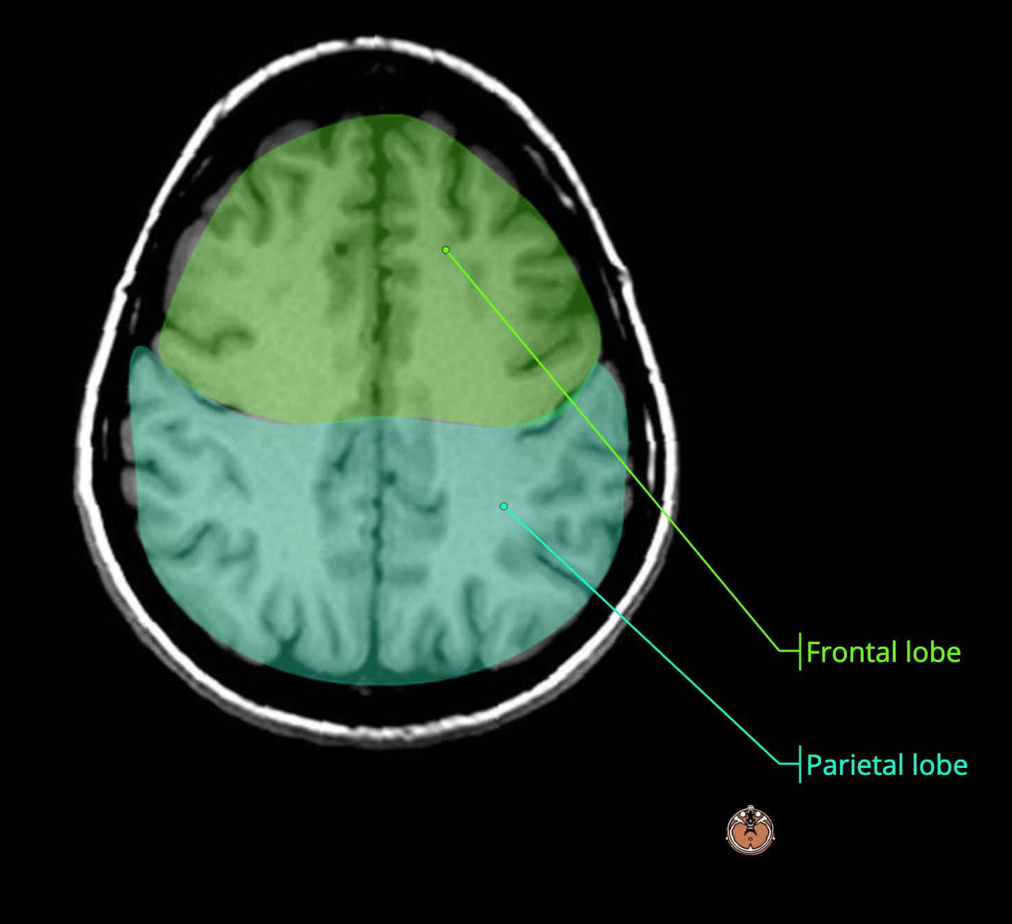
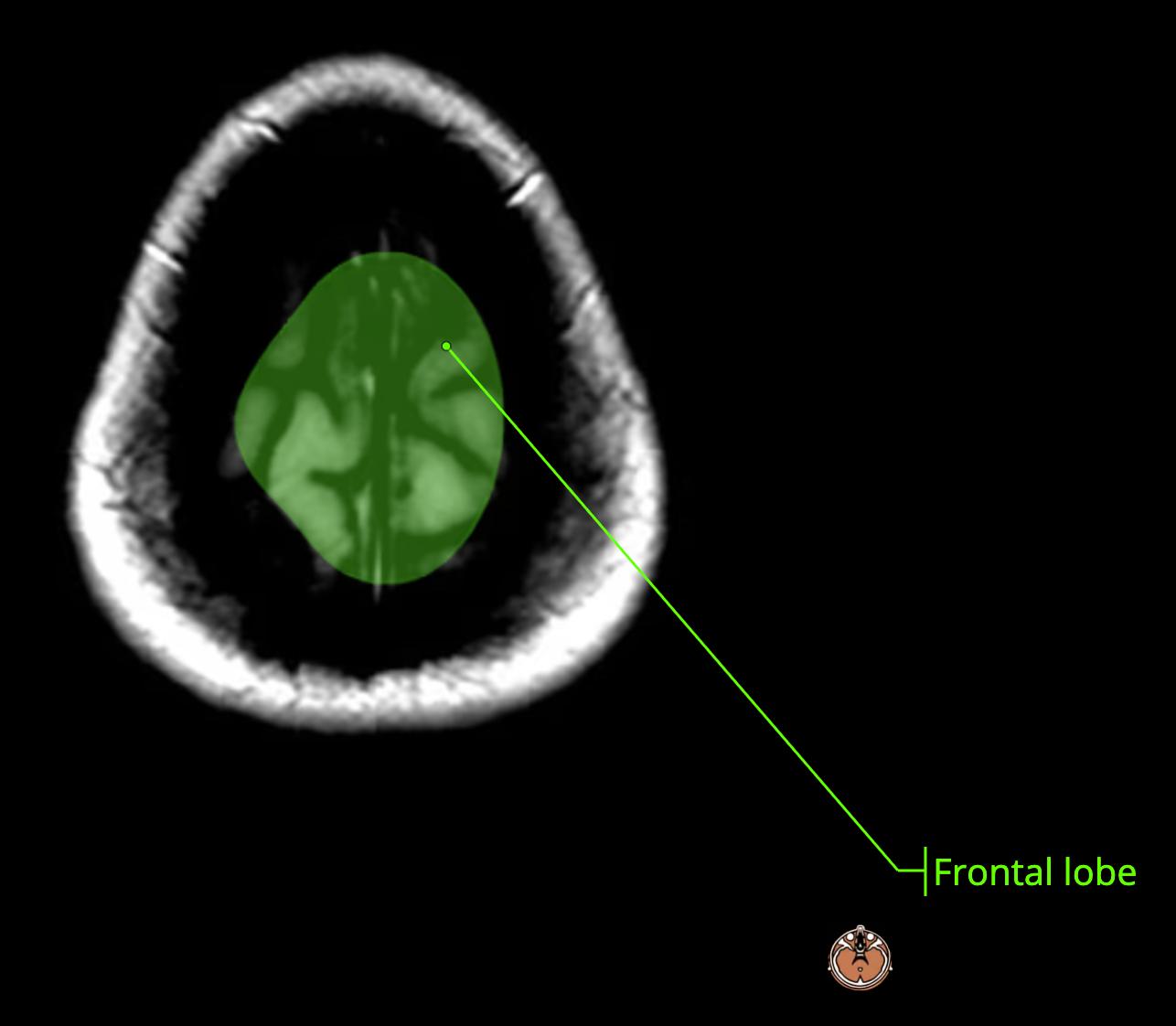
    • everything anterior to it is frontal[…] lobe; posterior is parietal[…] lobe

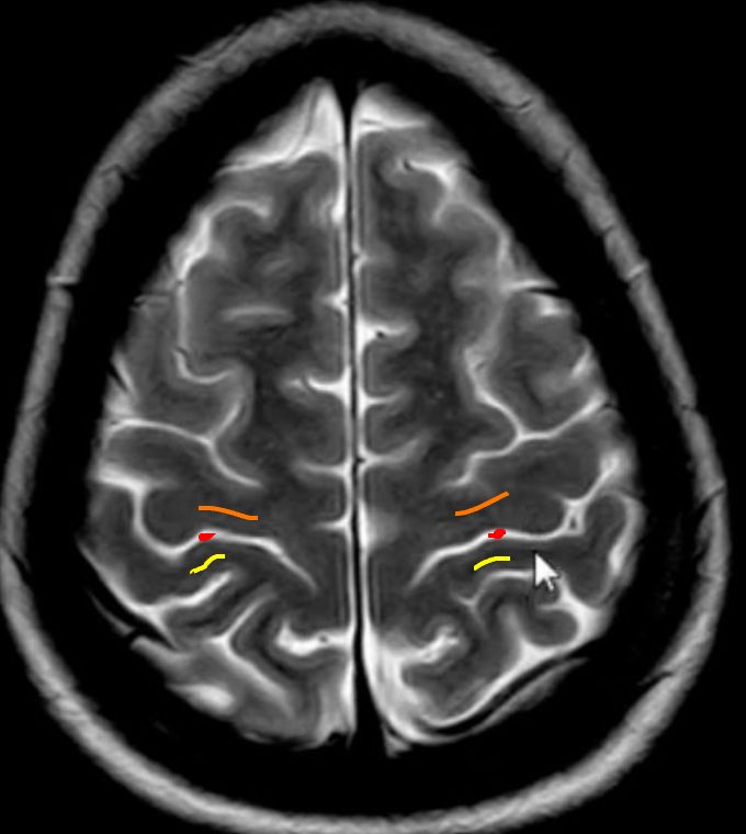
    MRI Brain Anatomy
    • red: central sulcus[…]
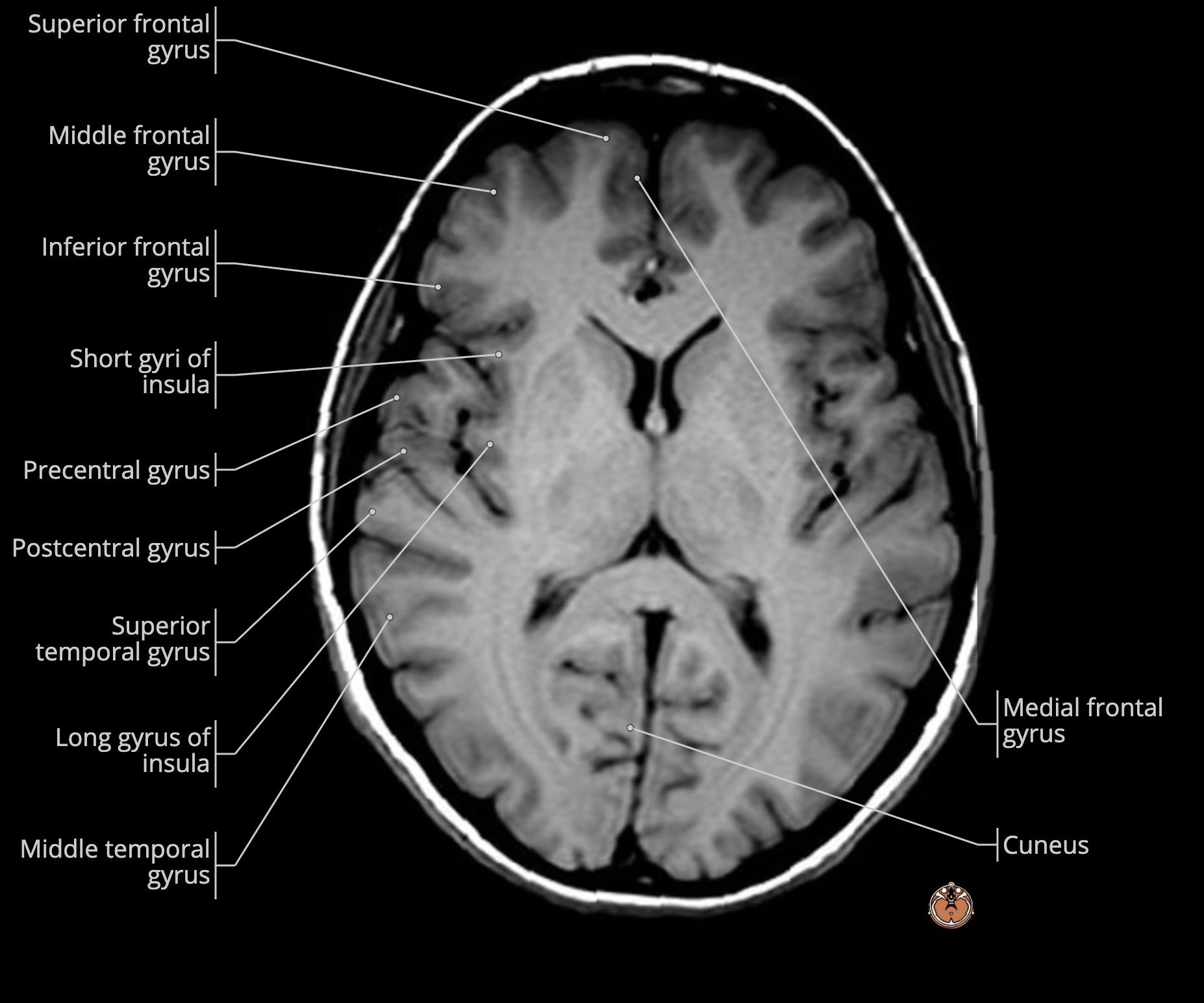
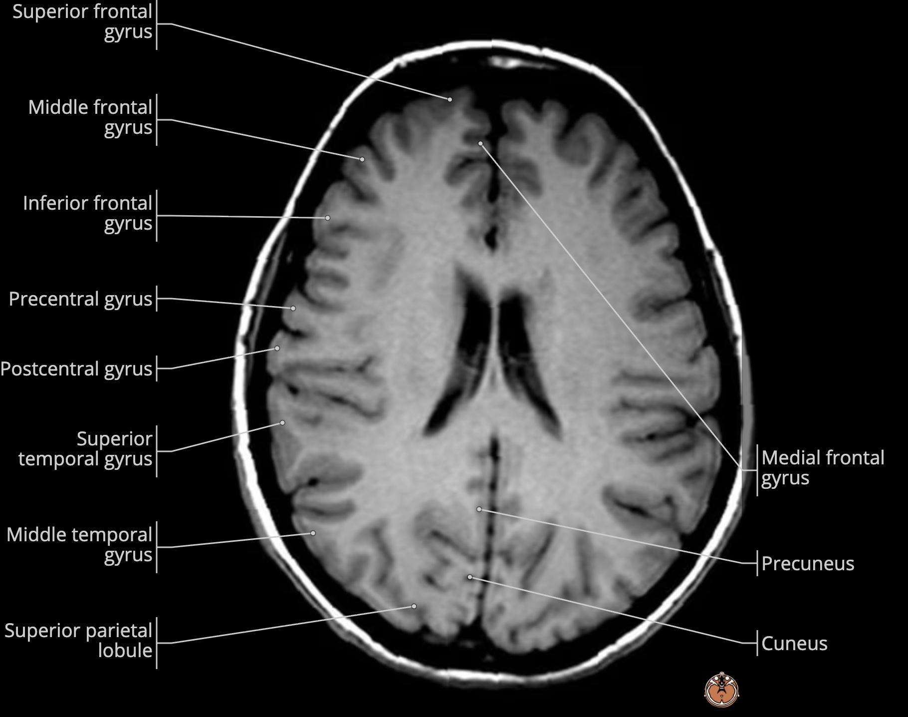
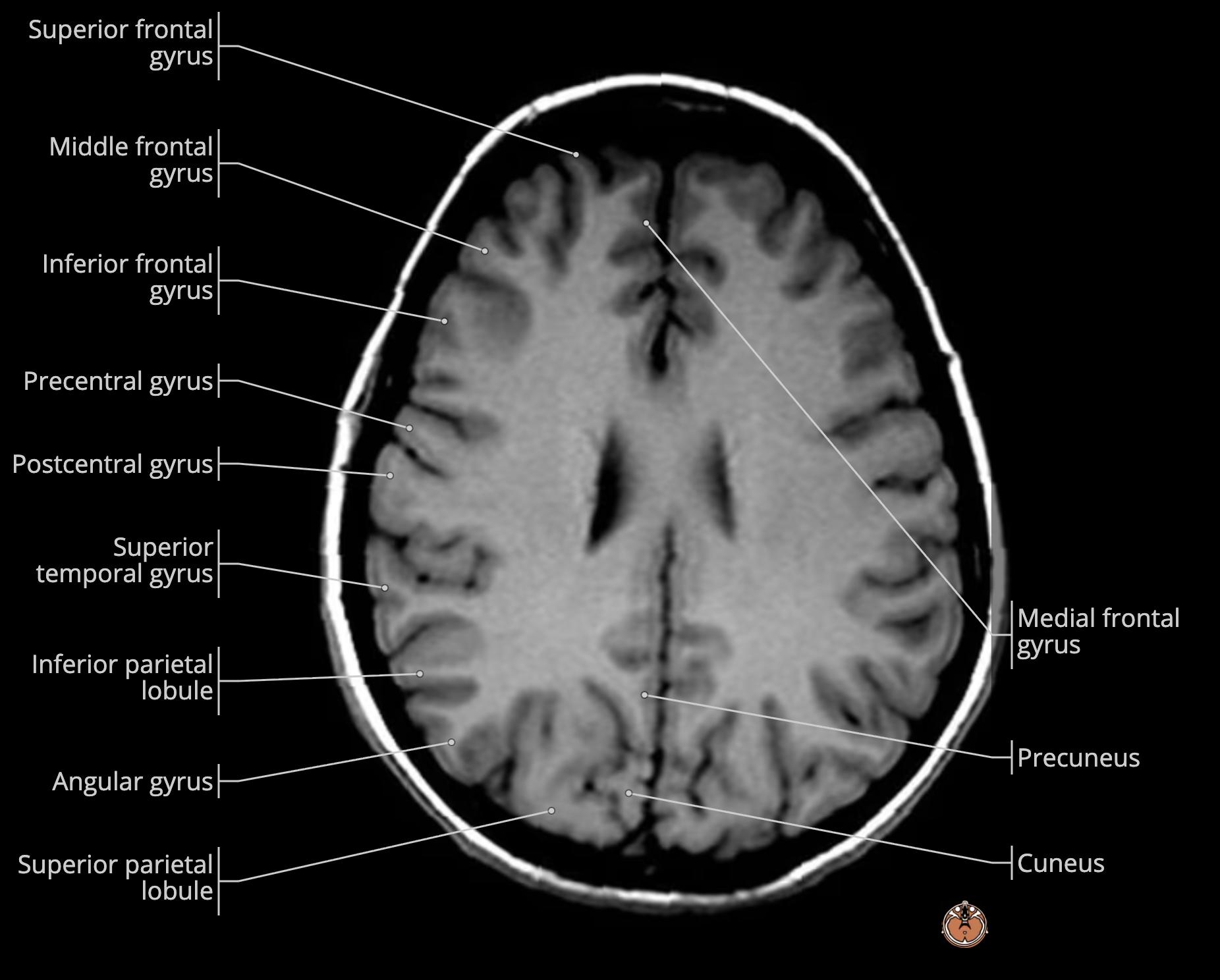
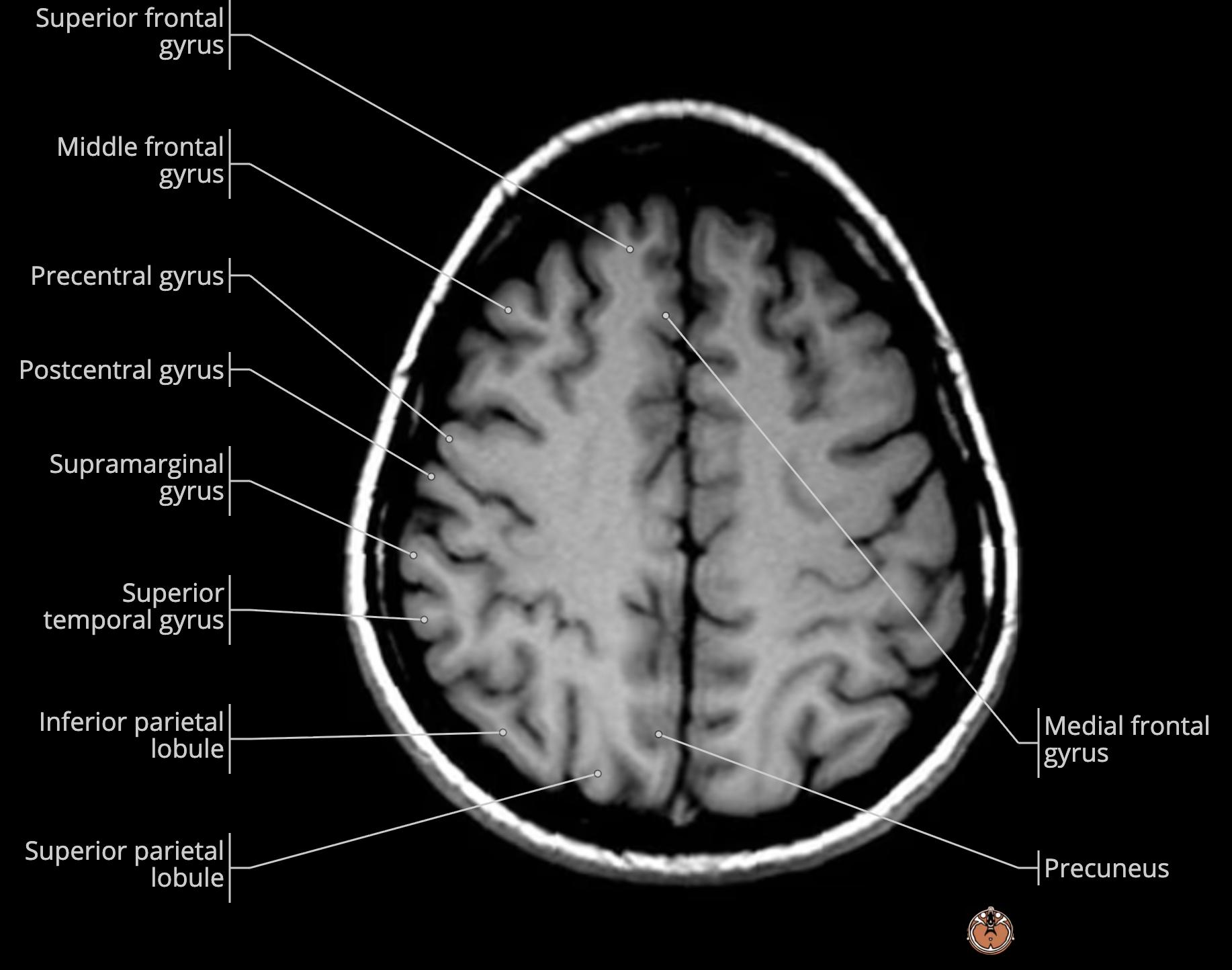
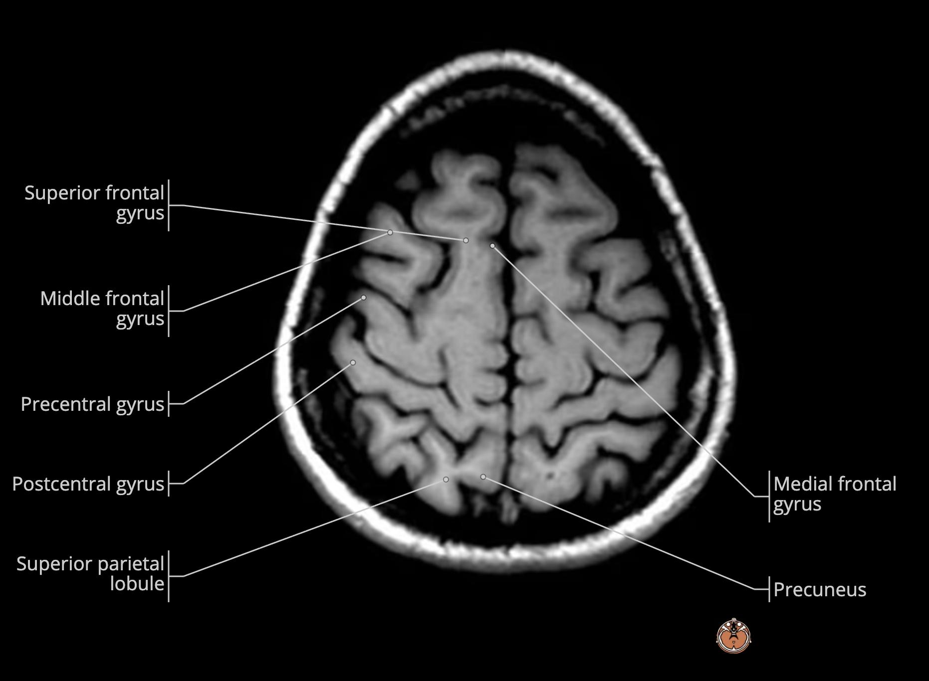
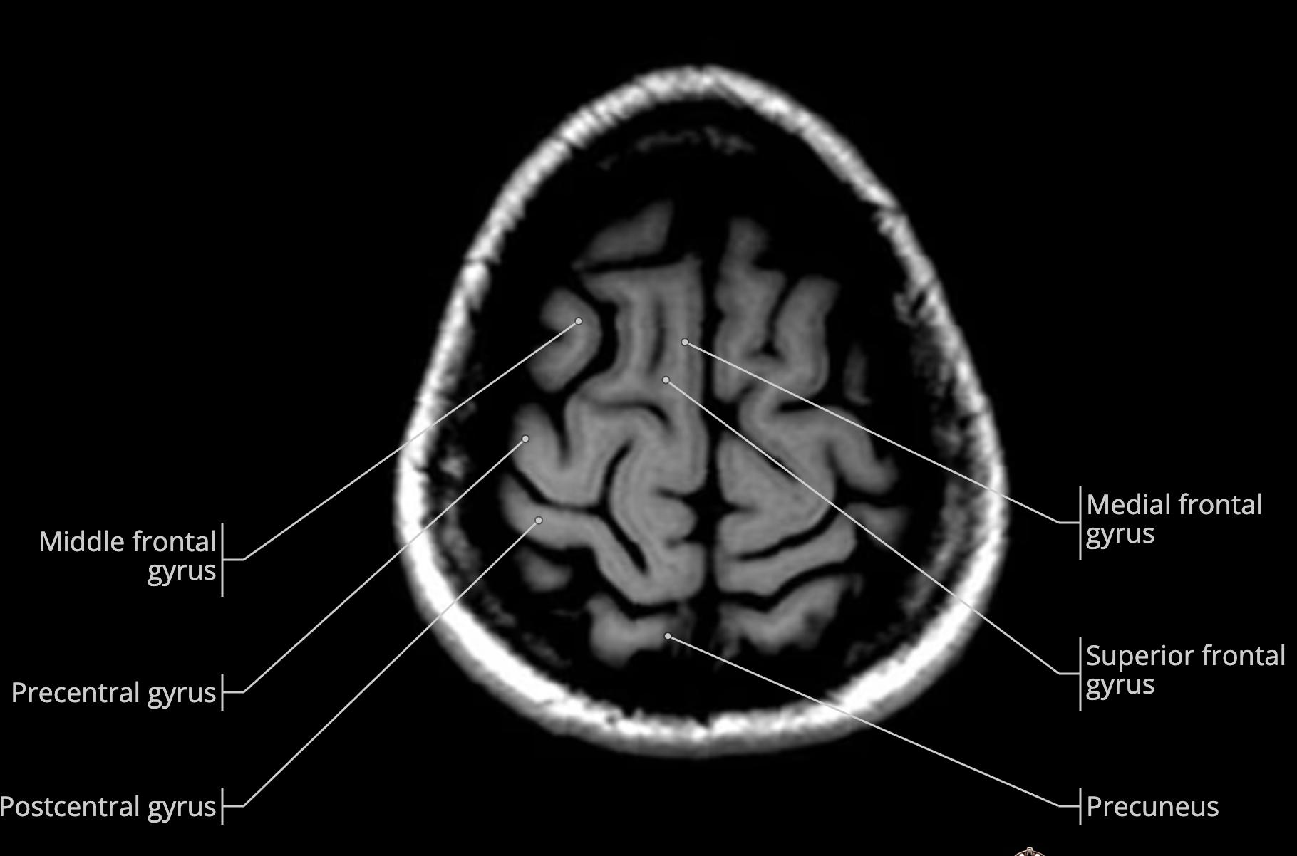
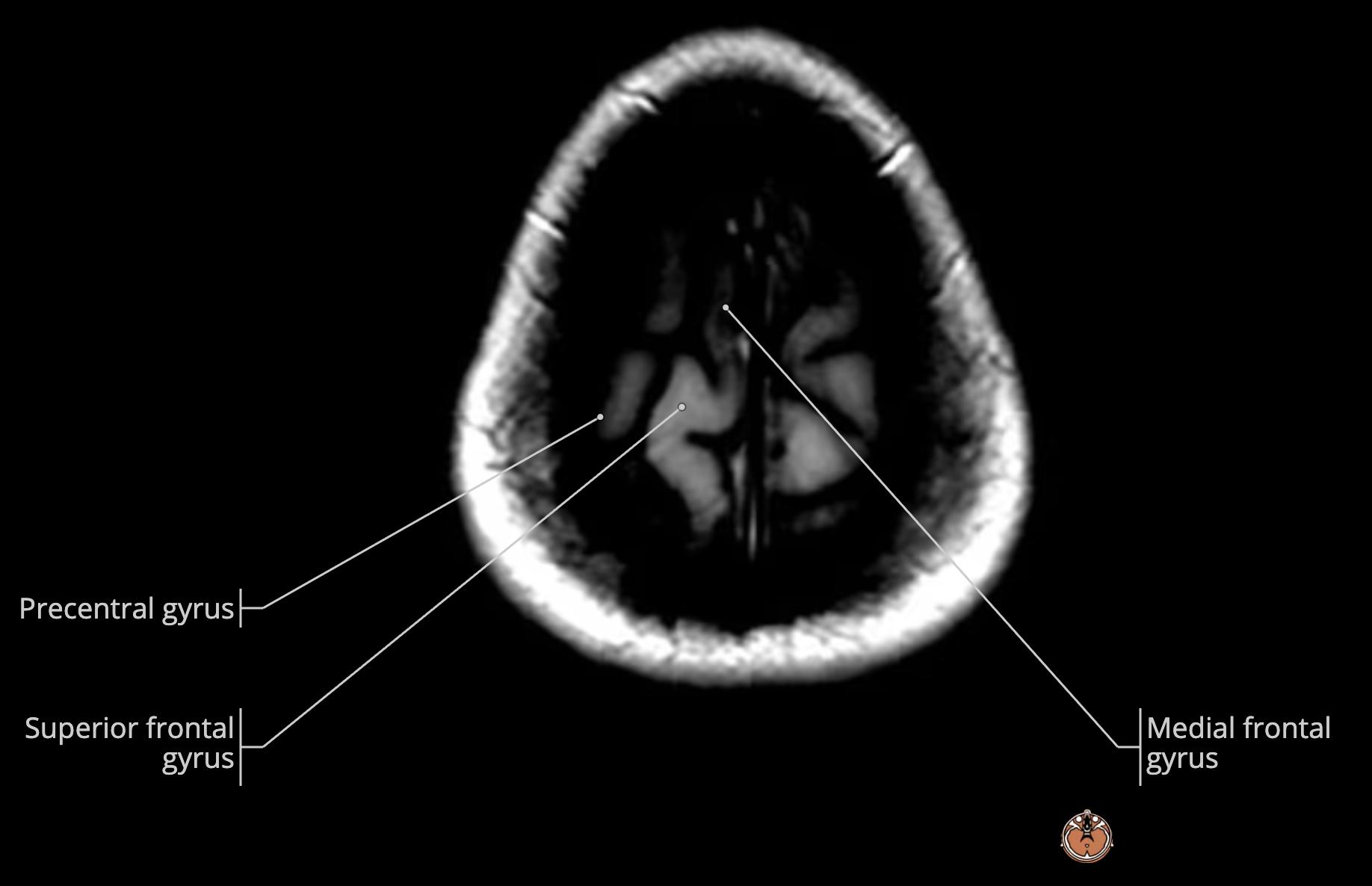
    • orange: precentral gyrus[…] of frontal[…] lobe
    • yellow: postcentral gyrus[…] of parietal[…] lobe

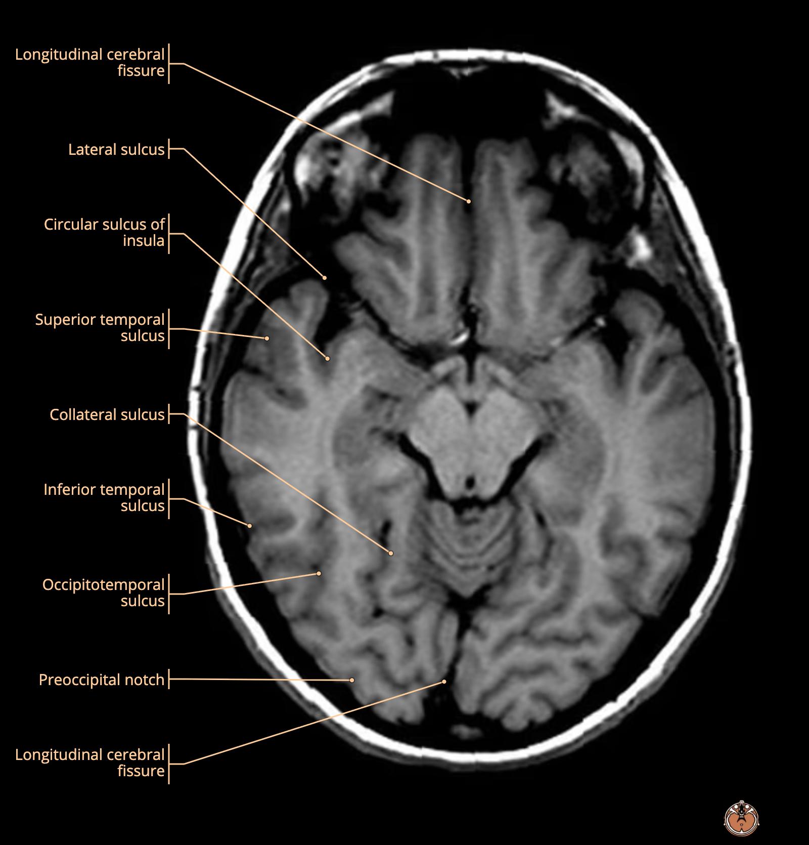
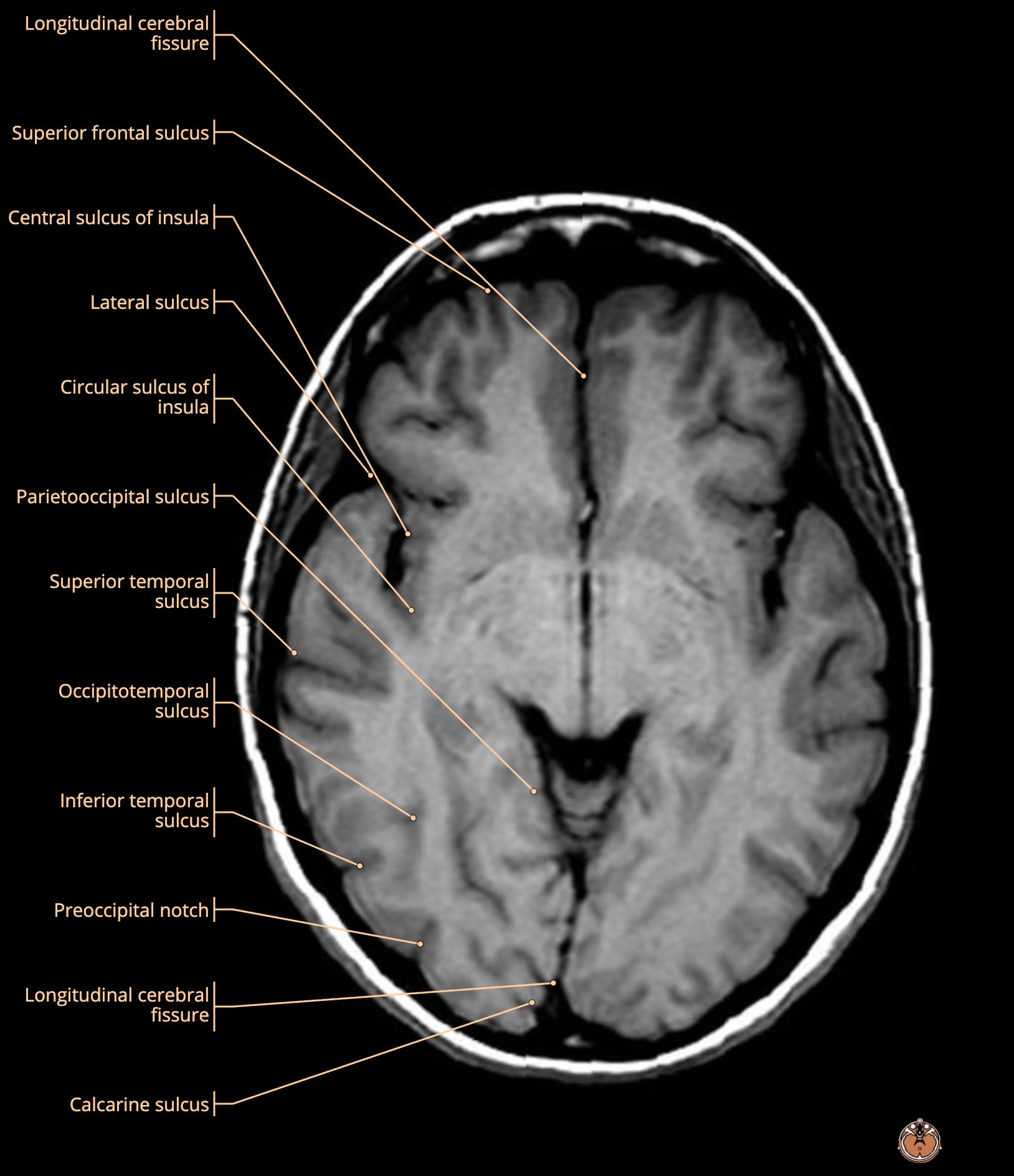
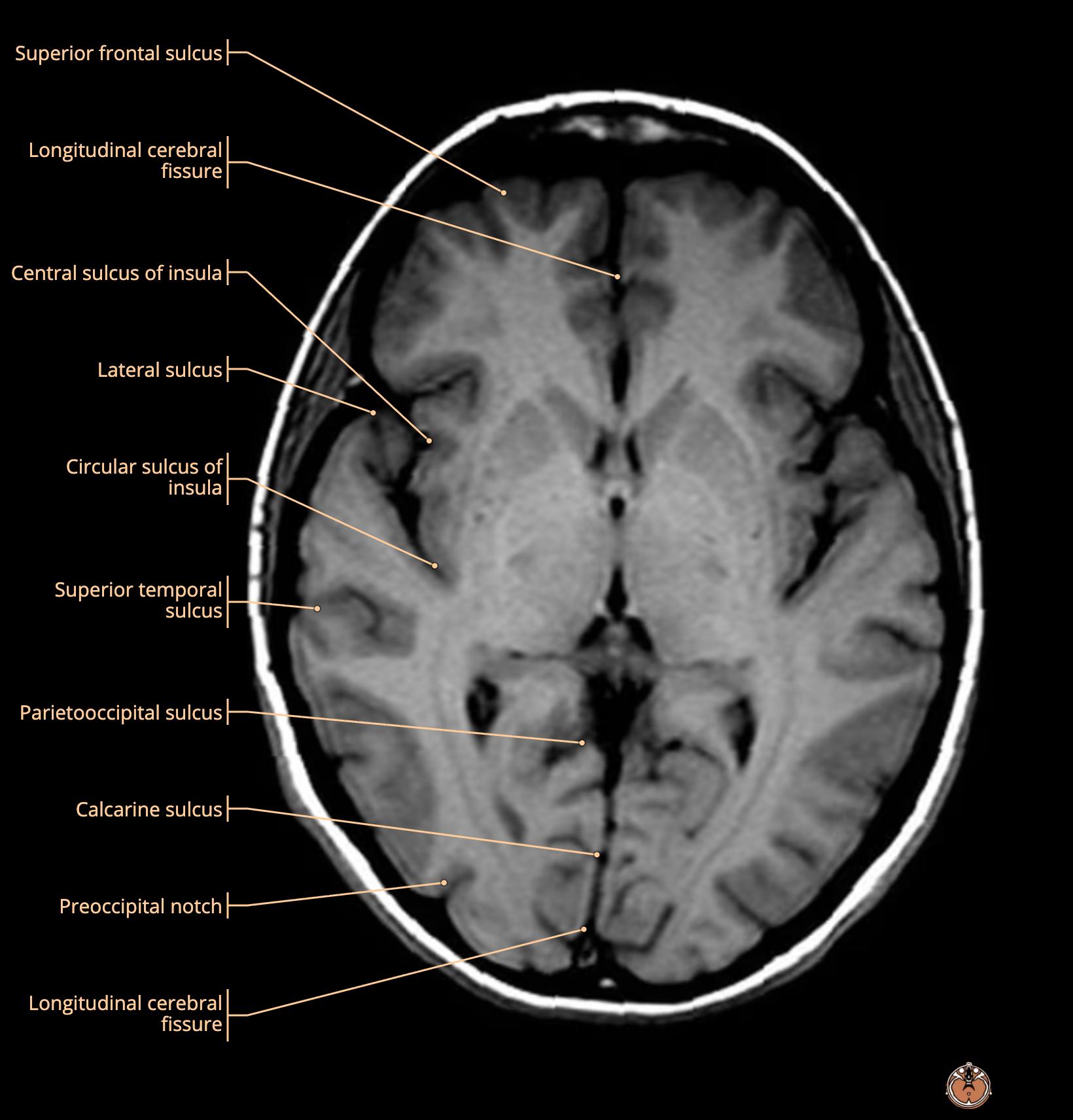
    MRI Brain Anatomy
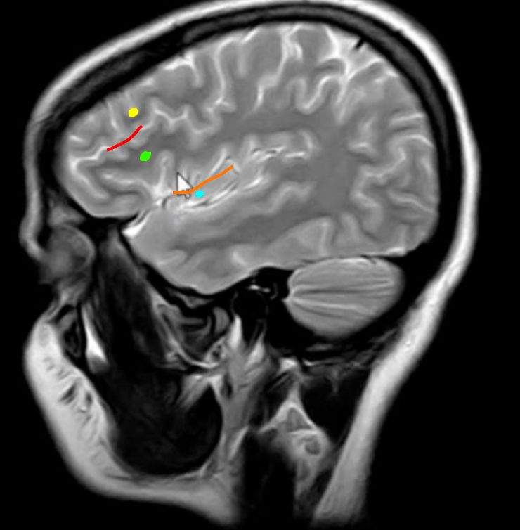
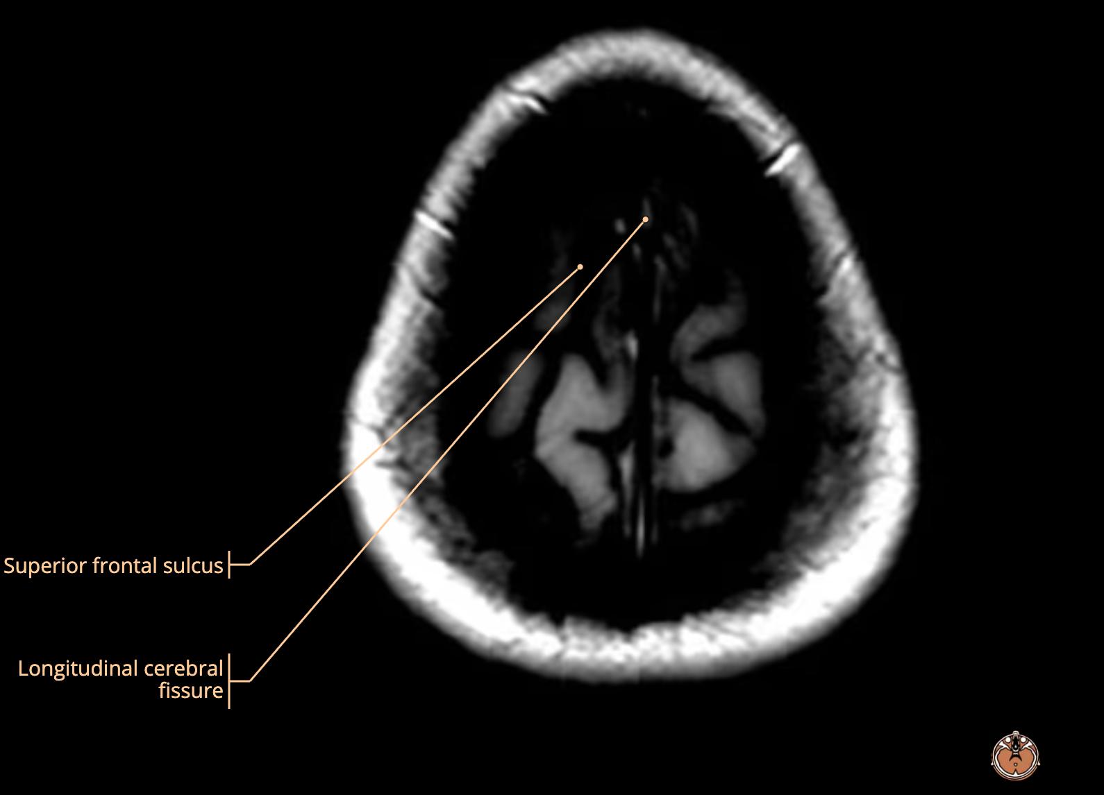
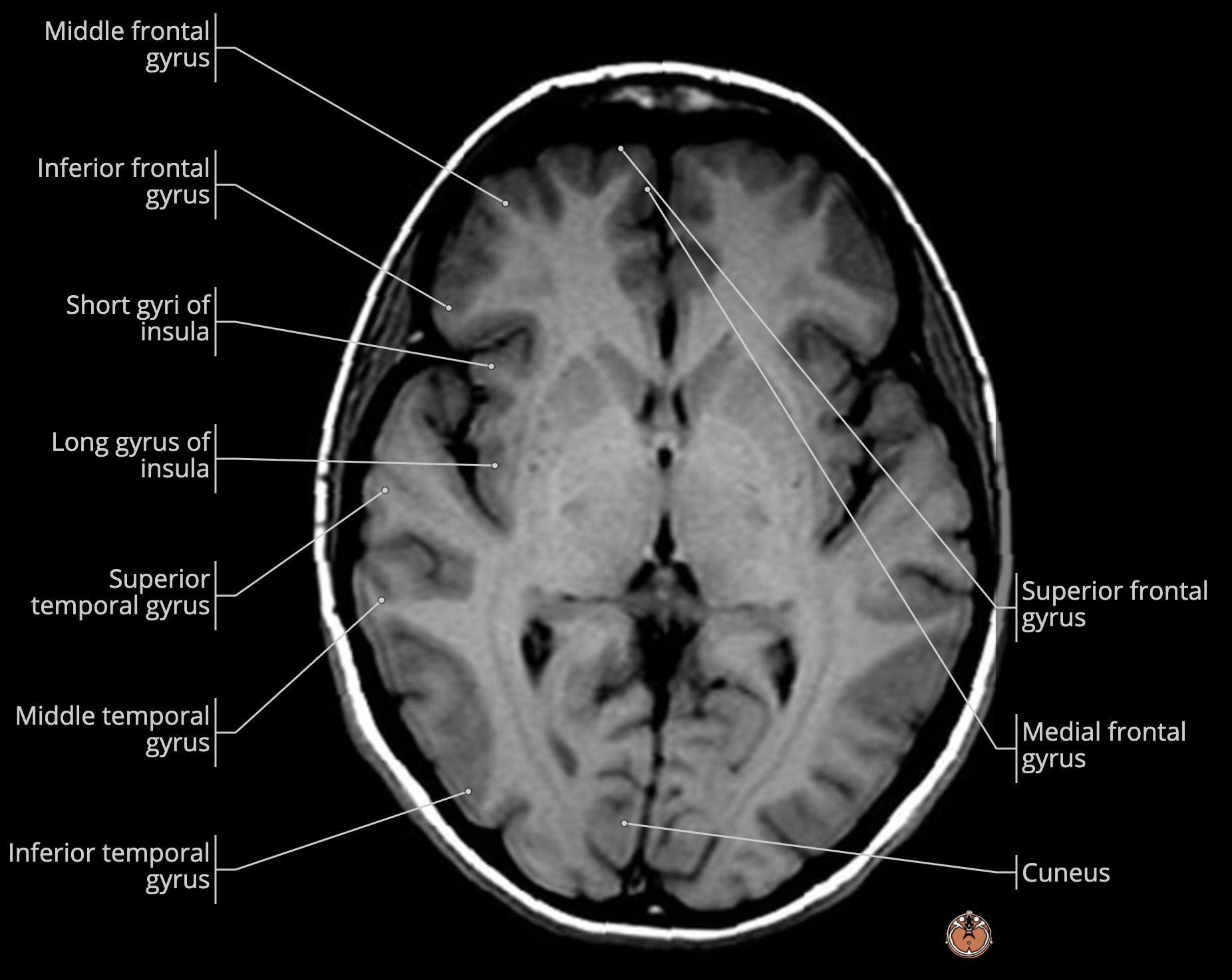
    • yellow: superior frontal gyrus[…]
    • red: superior frontal sulcus[…]
    • green: middle frontal gyrus[…]
    • orange: inferior frontal sulcus[…]
    • light blue: inferior frontal gyrus[…]

    MRI Brain Anatomy
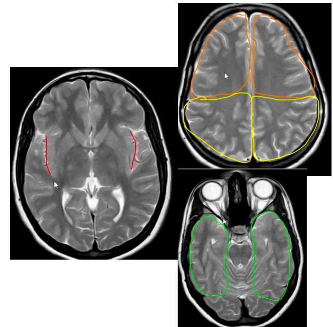
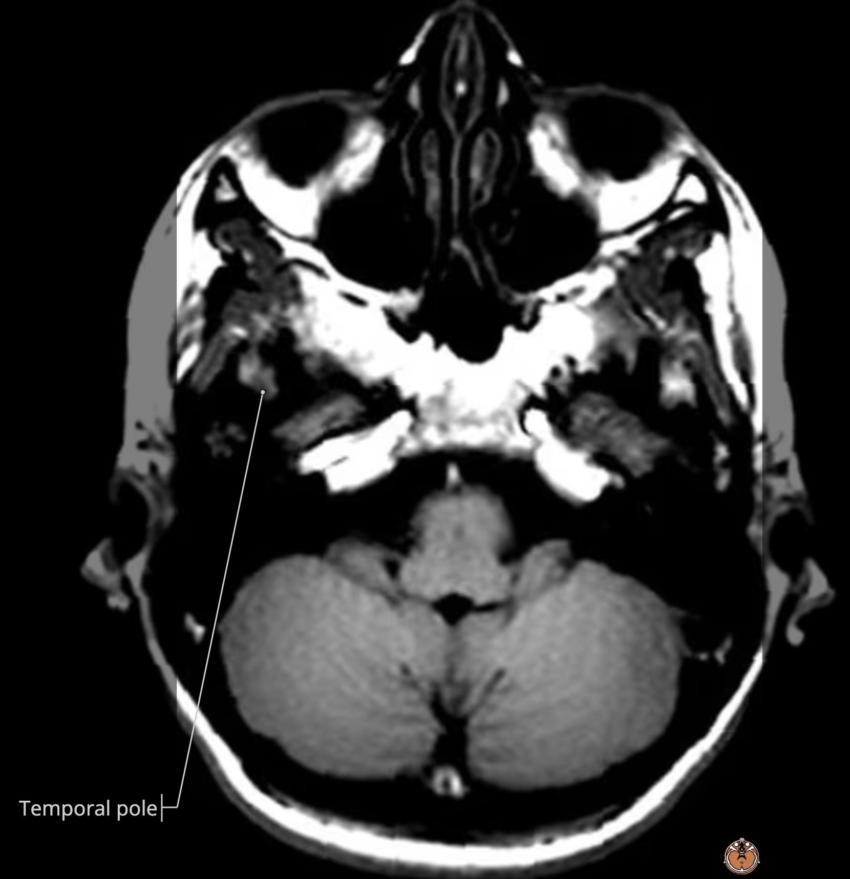
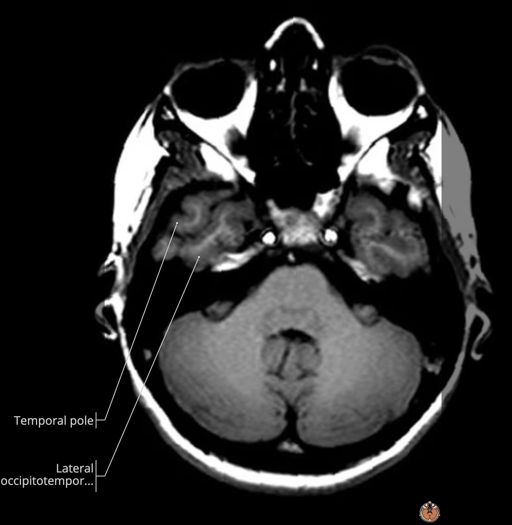
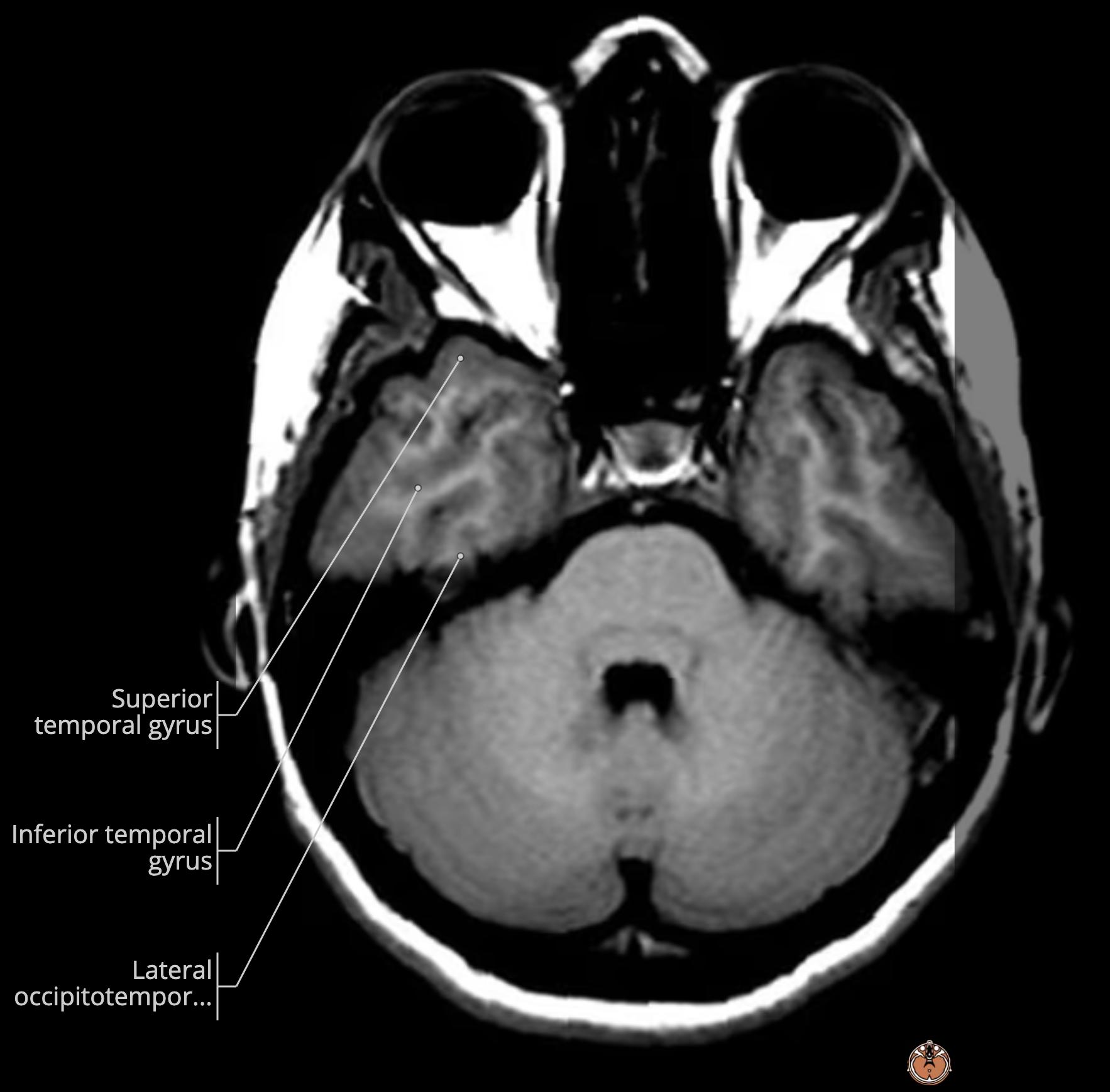
    • red: sylvian fissure[…]
      • everything above it: frontal[…] (orange) or parietal[…] (yellow) lobe
      • everything below it: temporal[…] lobe

    MRI Brain Anatomy
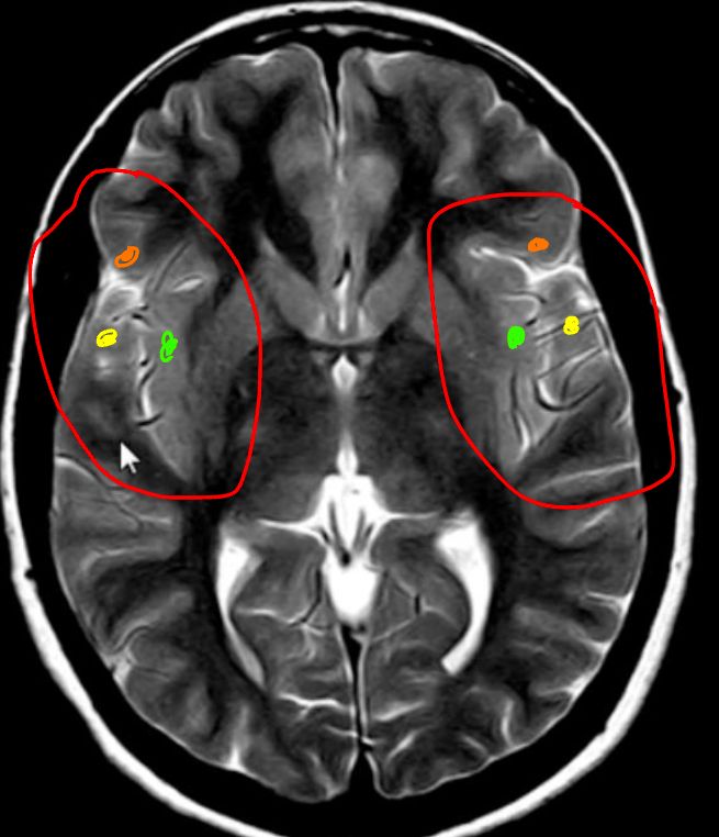
    • red: insula[…] covered by
      • orange: frontal operculum[…]
      • green: temporal operculum[…]
      • yellow: parietal operculum[…]

    MRI Brain Anatomy
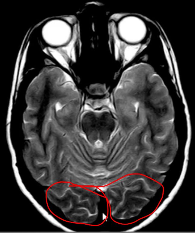
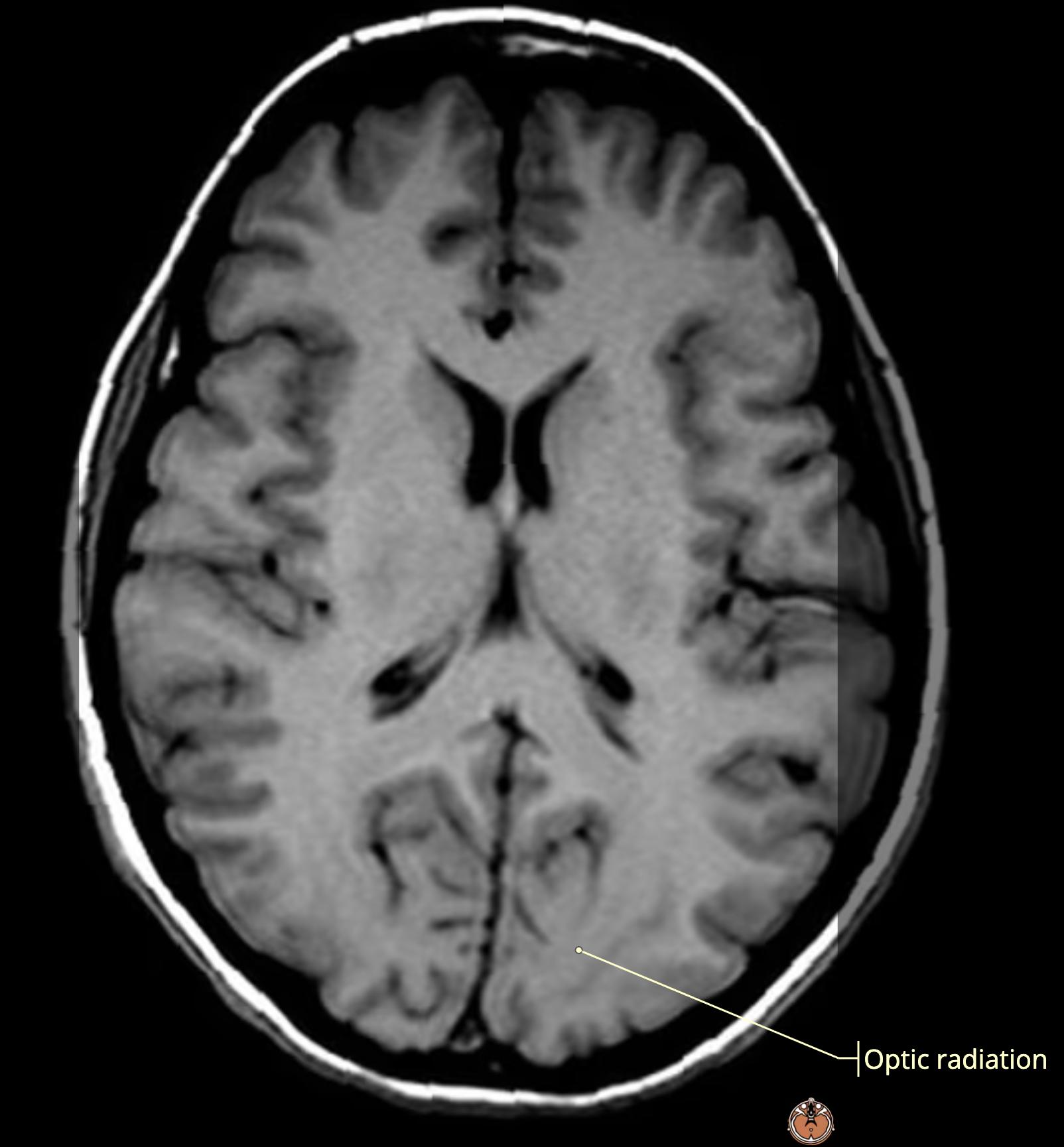
    • occipital[…] lobes

    MRI Brain Anatomy
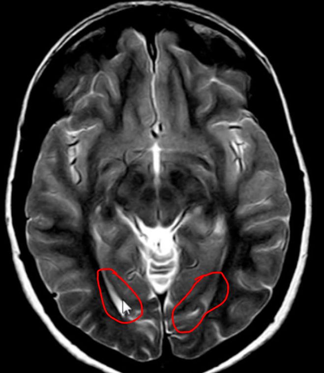
    • ID this: occipital horns, lateral ventricle[…]

    MRI Brain Anatomy
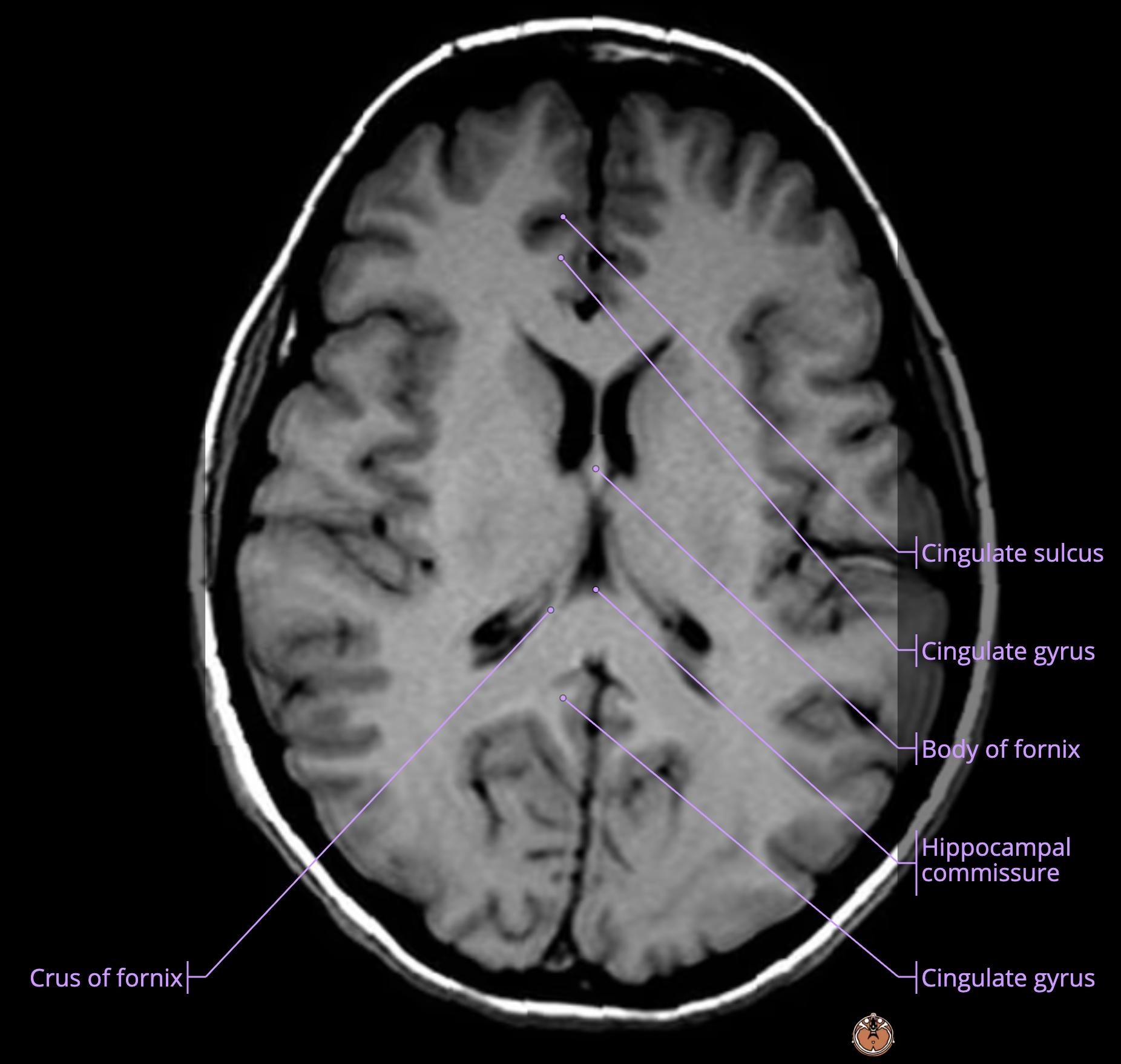
    • yellow: corpus callosum[…]
    • red: pericallosal sulcus[…]
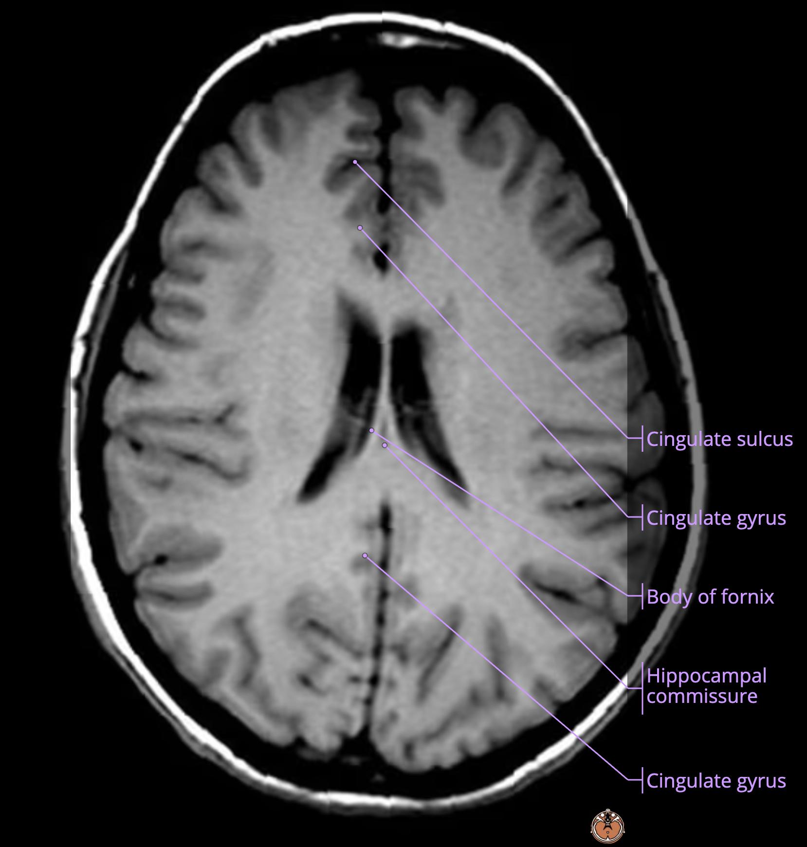
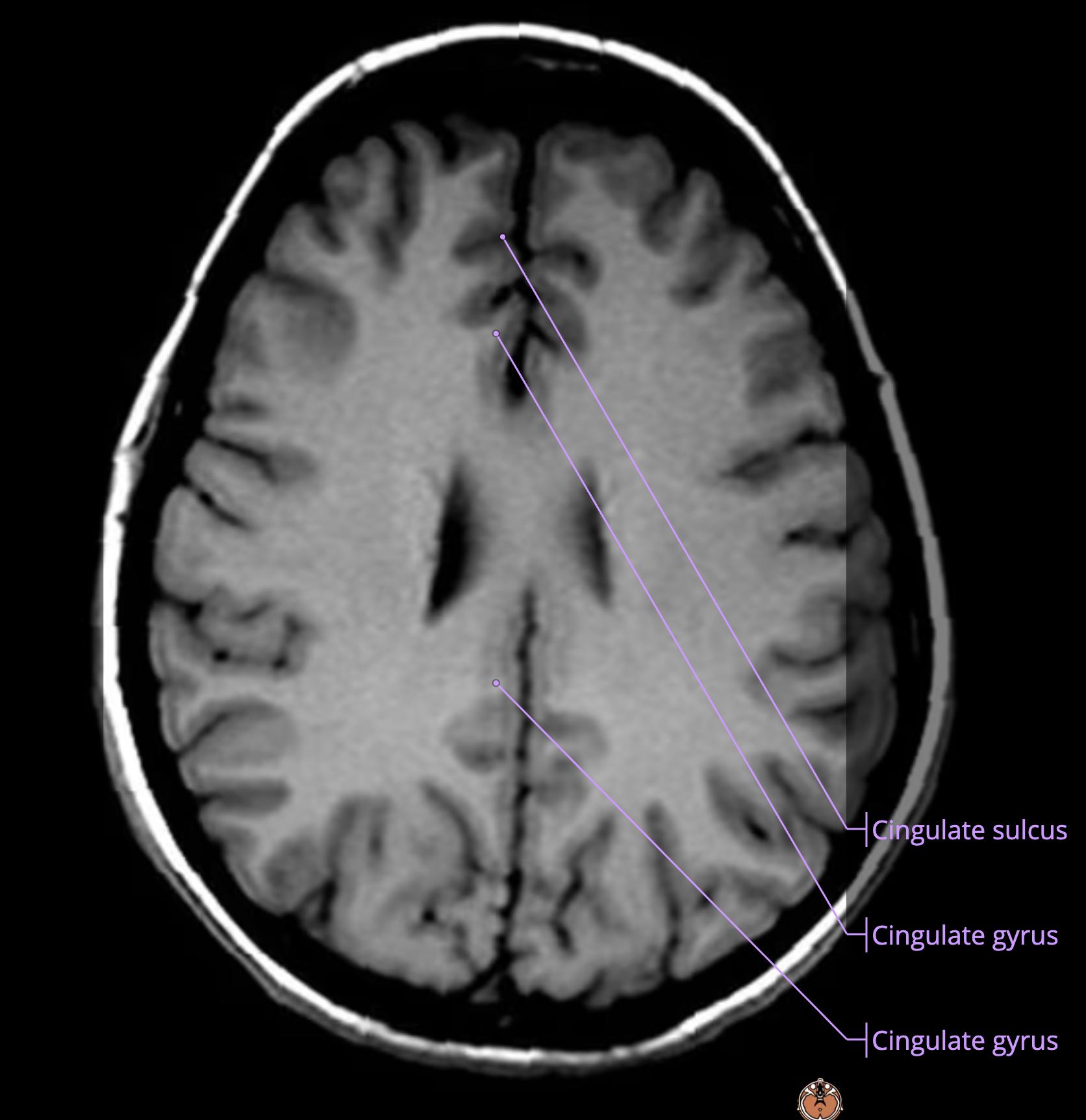
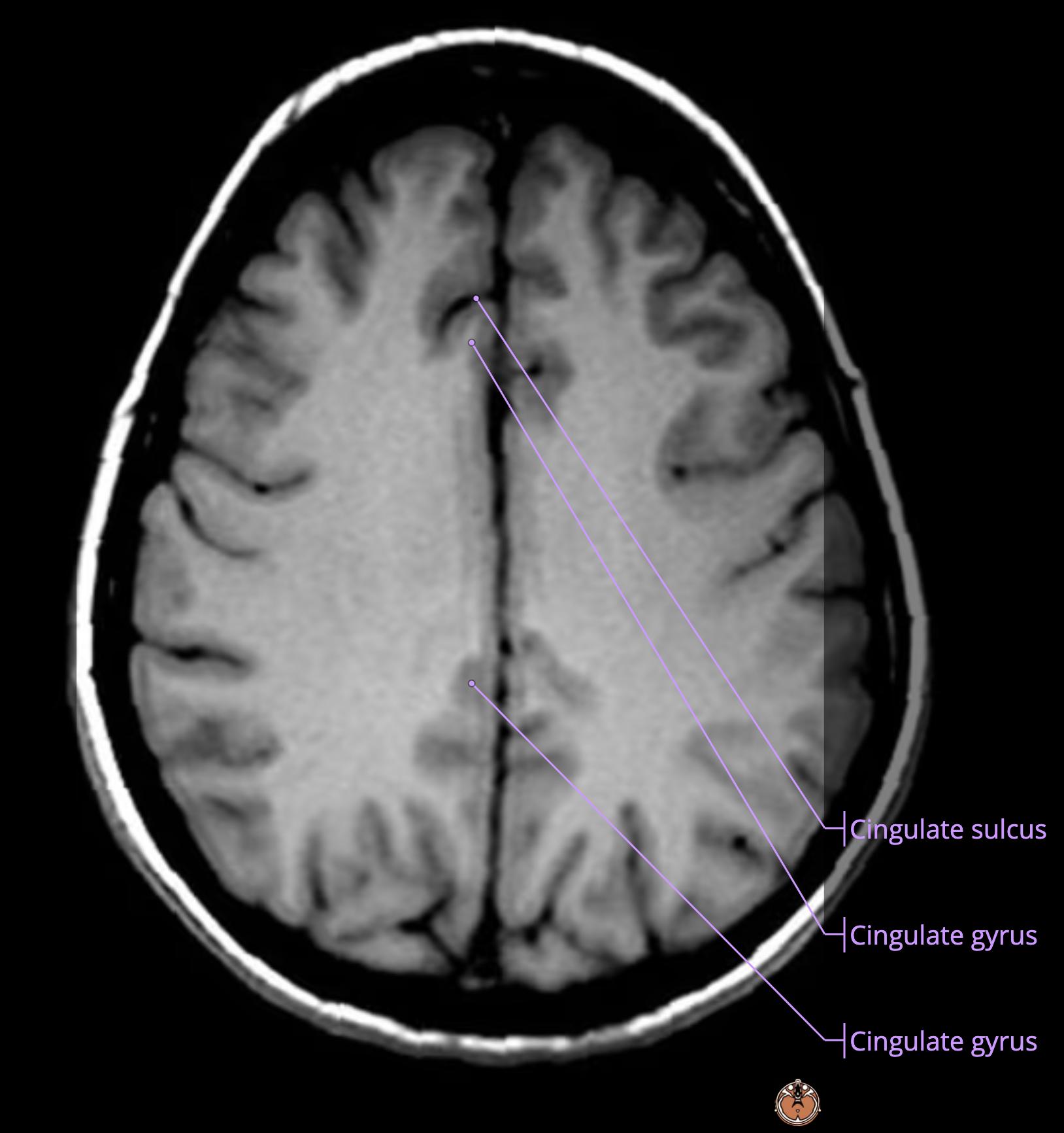
    • green: cingulate gyrus[…]
    • orange: cingulate sulcus[…]
    • light blue: parieto-occipital sulcus[…] separating the parietal[…] lobe from the occipital[…] lobe

    MRI Brain Anatomy
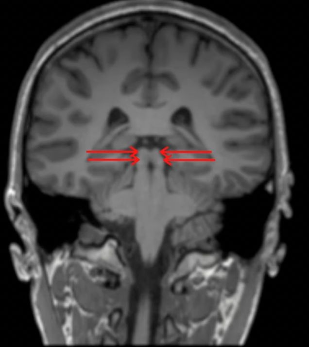
    • middle[…] cerebellar peduncles connecting cerebellum[…] to pons[…]

    MRI Brain Anatomy
    • superior[…] cerebellar peduncles connecting cerebellum[…] to midbrain[…]

    MRI Brain Anatomy
    • inferior[…] cerebellar peduncles (not very visible) connecting cerebellum[…] to medulla[…]

    MRI Brain Anatomy - name this structure
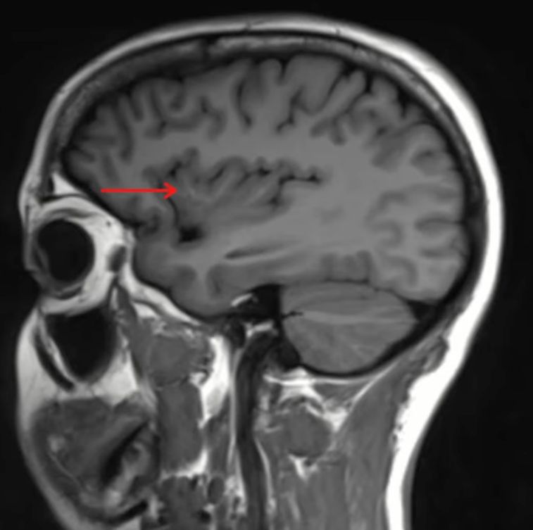
    • insular[…] cortex
    • we are slightly lateral to midline; we know this because we are cutting the eye[…] through the middle

    MRI Brain Anatomy - name this structure
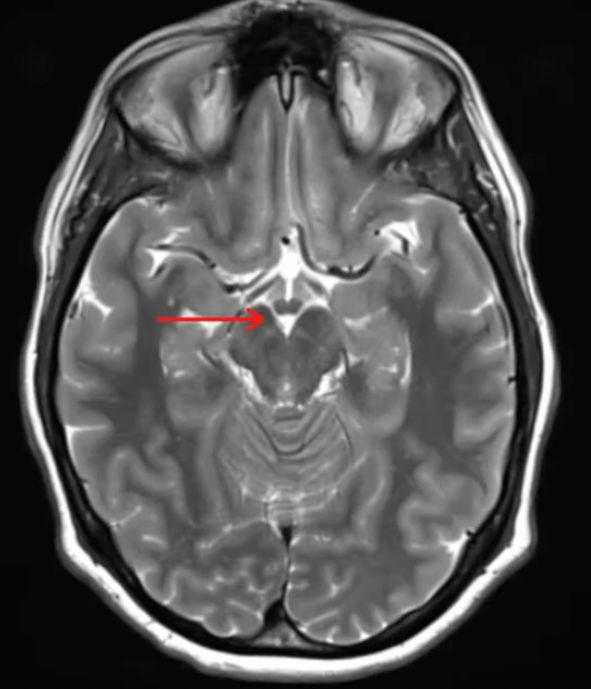
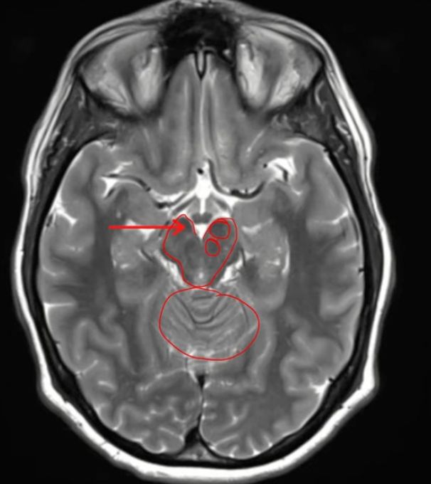
    • right substantia nigra[midbrain anterior structure] which is split further into the pars compacta/reticularis[structures within that you cannot visualize on axial cut MRI]
    • the structure slightly posterior to it is the red nucleus[…]
    • we are at the level midbrain (mickey mouse)[…] and top of cerebellum[…]

    MRI Brain Anatomy - name this structure
    • head of caudate[…] running lateral to the lateral ventricles[…] which encompasses part of the corpus striatum[this structure + lentiform nucleus]
    • we know we're lateral to midline because we are not seeing the entirety of the lateral ventricle[…] but rather the structure running laterally to it

    MRI Brain Anatomy - name this structure
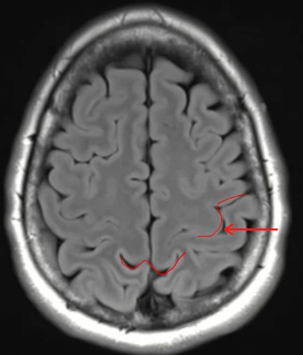
    • left central sulcus[…]
      • as we go further up in the brain, this sulcus gets pushed further posterior[…]ly
    • the midline marginal[…] sulcus creates the "mustache[…]" sign nearby 
      • anterior to this is the postcentral gyrus[…]
      • anterior to this is the central sulcus[…]
      • anterior to this is the precentral gyrus[…]

    MRI Brain Anatomy - name this structure
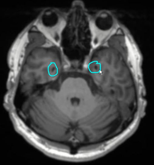
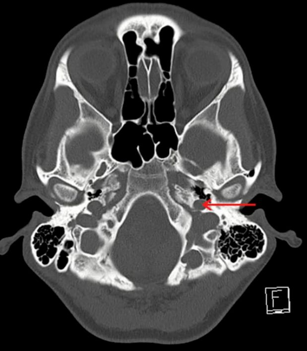
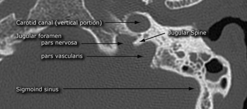
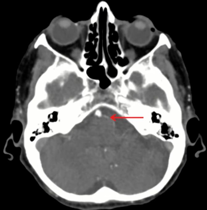
    • this is the left carotid canal[…]
    • posterior to it immediately is the jugular spine[…]

    MRI Brain Anatomy - name this structure
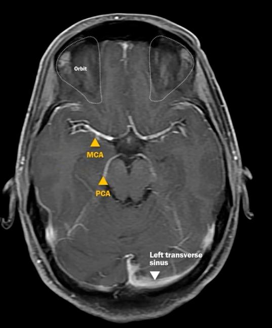
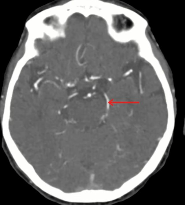
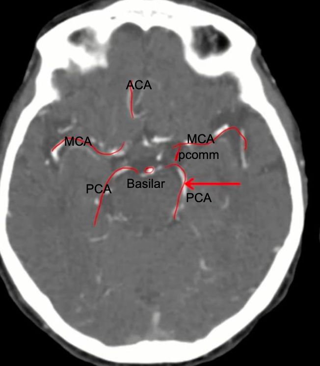
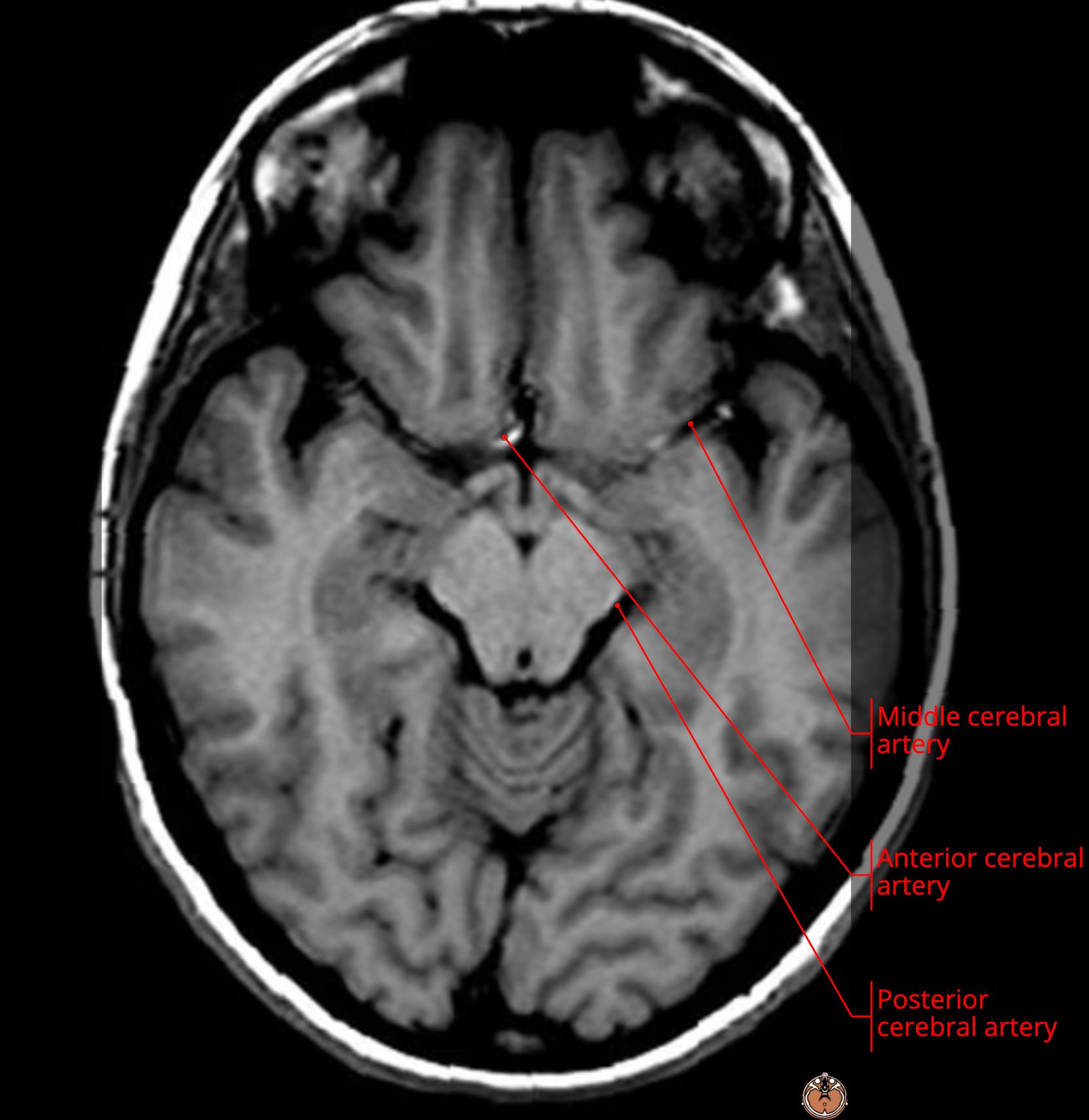
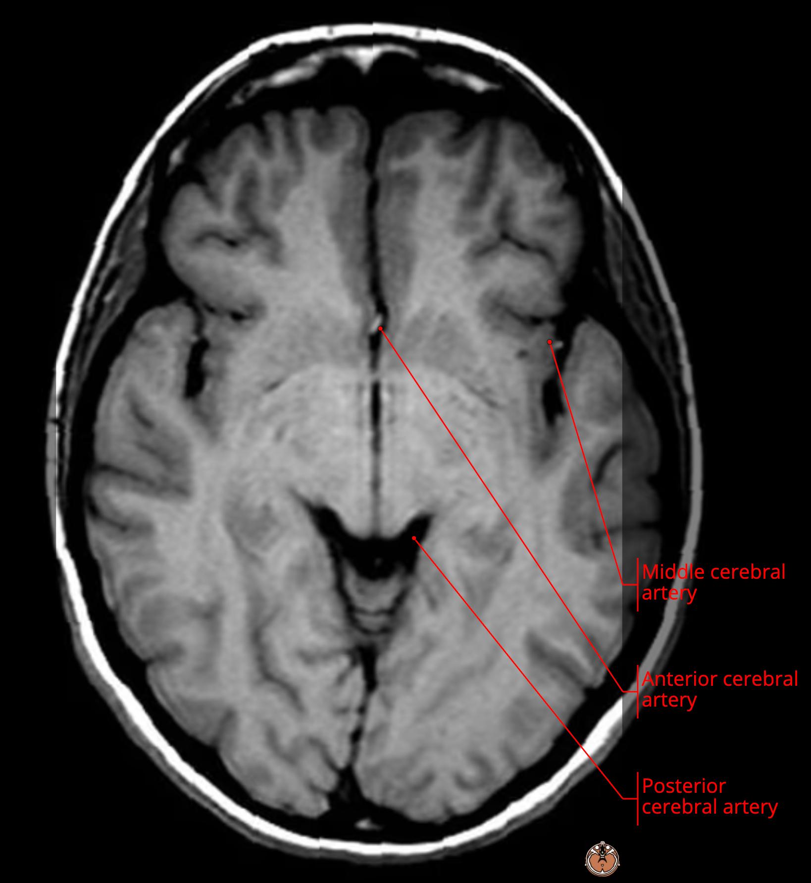
    • this is the left PCA[…]

    MRI Brain Anatomy - name this structure
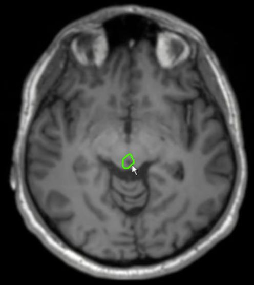
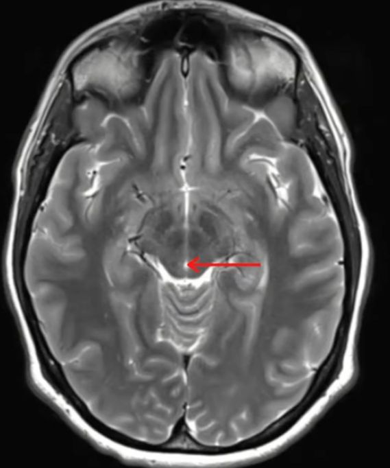
    • this is the cerebral aqueduct of Sylvius[…] 
    • at the level of midbrain[…]
      • can see the SN[…] anteriorly and Red Nucleus[…] posteriorly at this level
    • posteriorly can see the superior portion of the cerebellum[…]

    MRI Brain Anatomy - name this structure
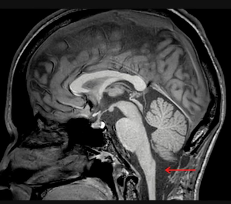
    • this is the cisterna magna[…], the largest cistern 

    MRI Brain Anatomy - name this structure
    • this is the left AICA[…] coming off the Basilar[…]

    MRI Brain Anatomy - name this structure
    • these are the quadrigeminal bodies[…] sitting on the quadrigeminal plate[…] located on the midbrain tectum[…]
      • includes the superior colliculi[…] and inferior colliculi[…]

    MRI Brain Anatomy - name this structure
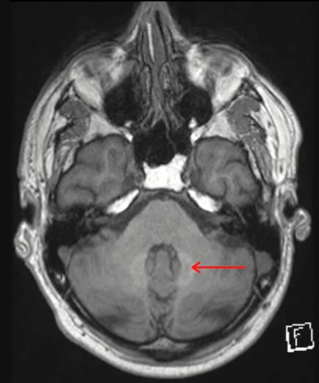
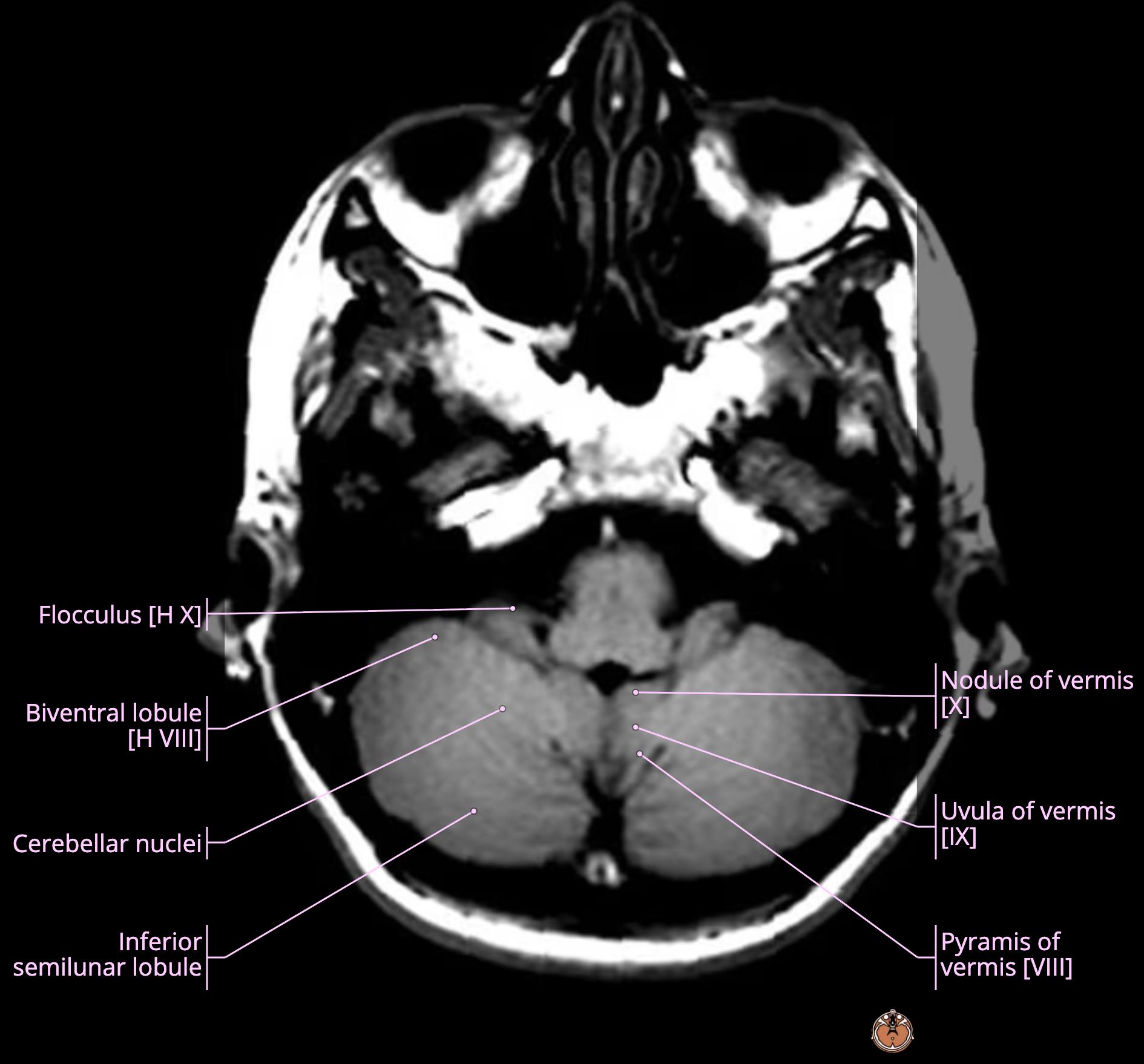
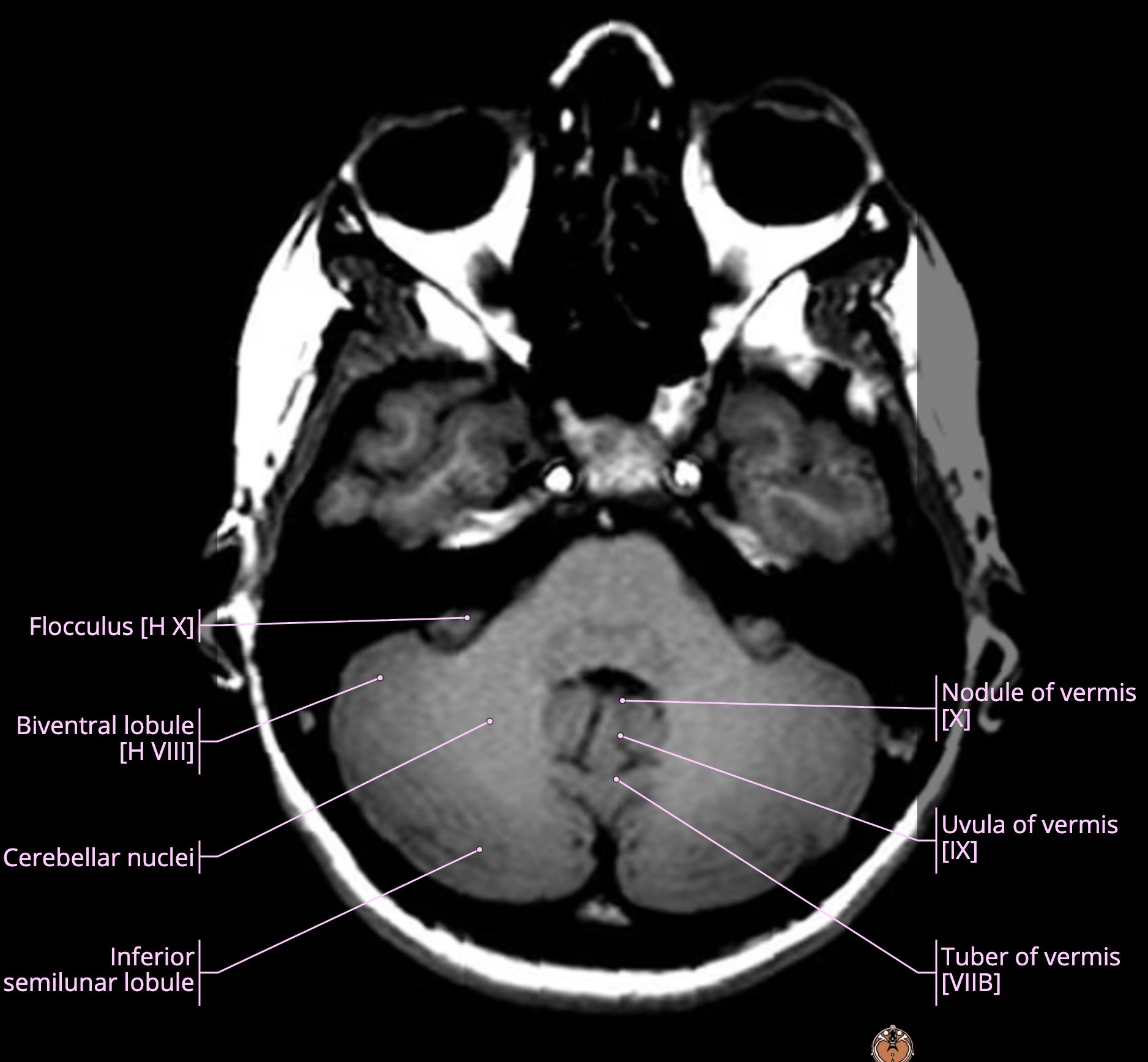
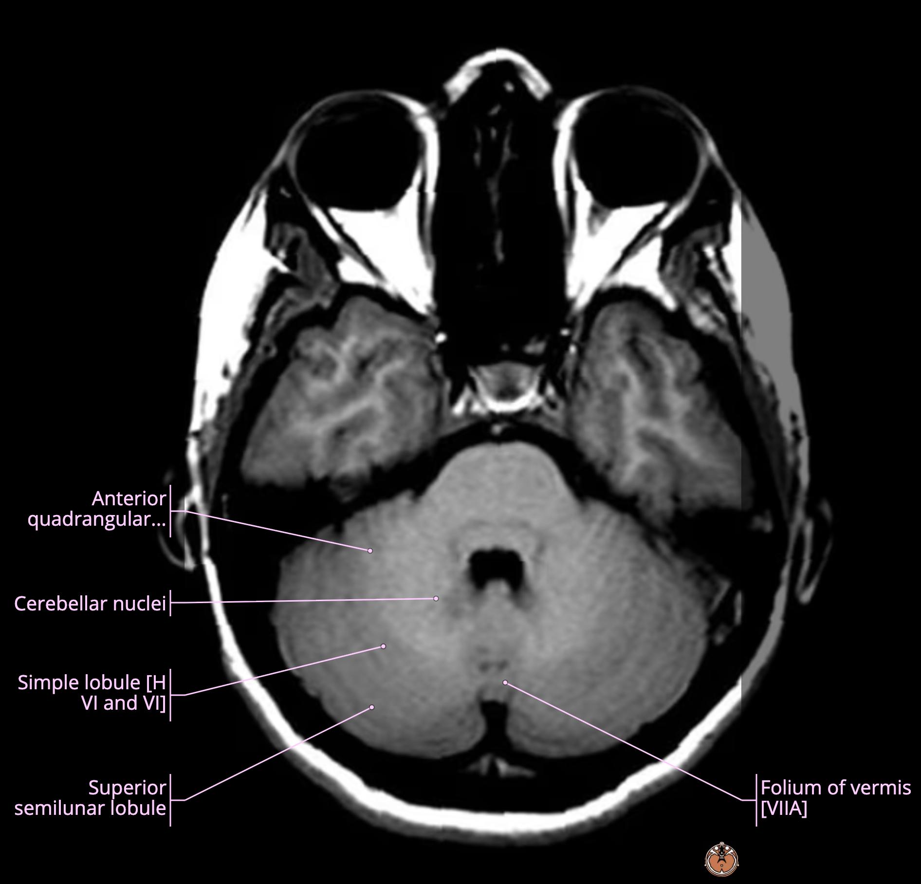
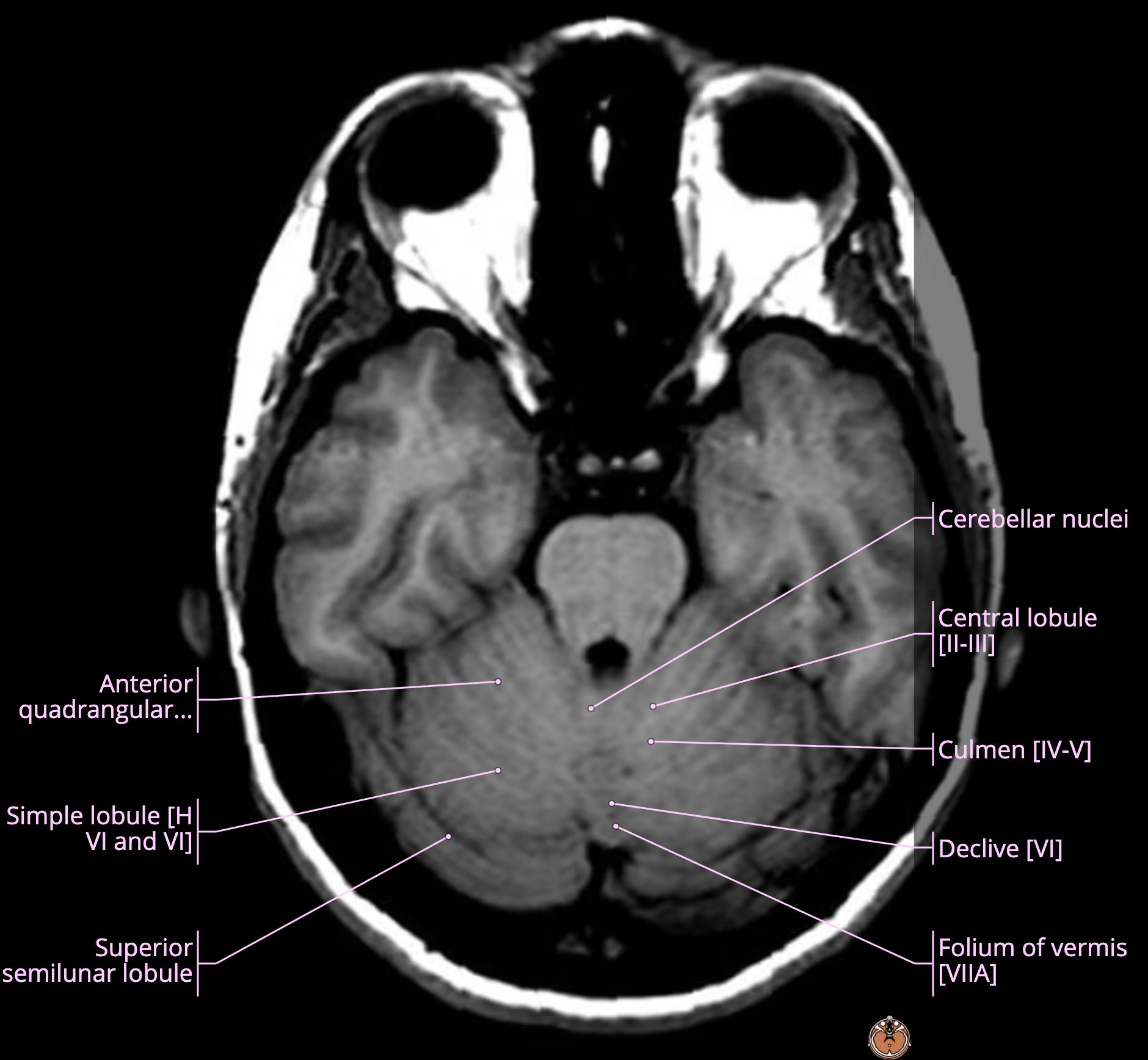
    • this is the dentate nucleus[…] of cerebellum 

    MRI Brain Anatomy - name this structure
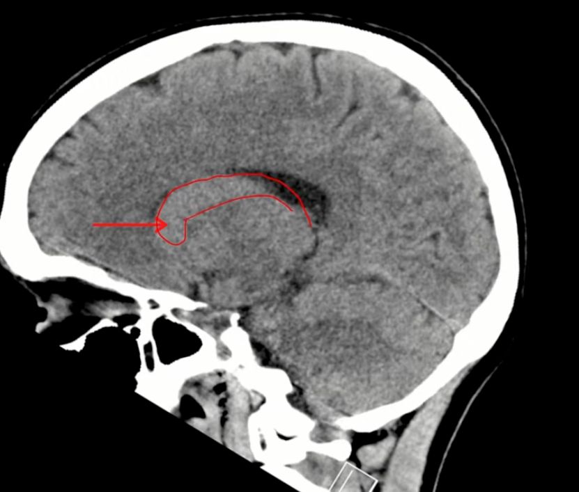
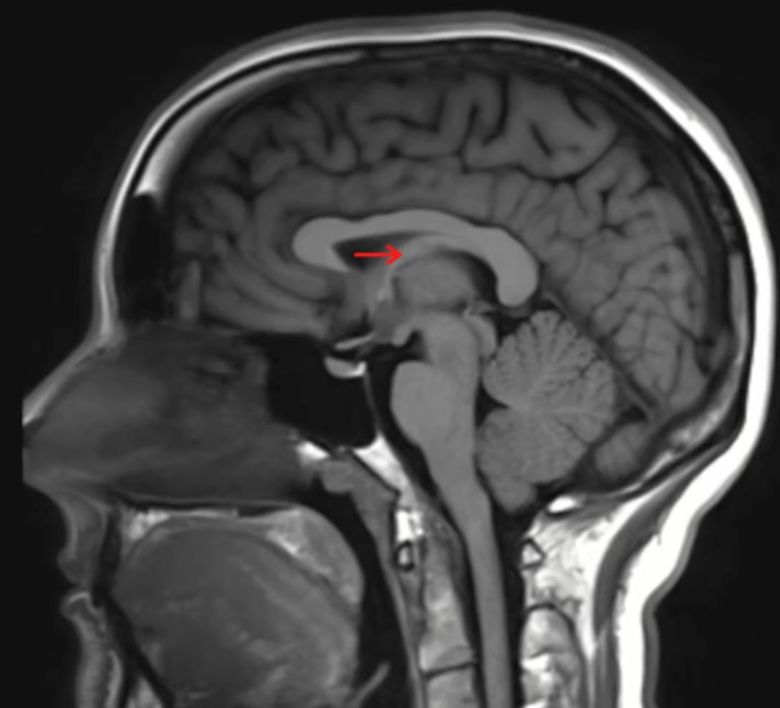
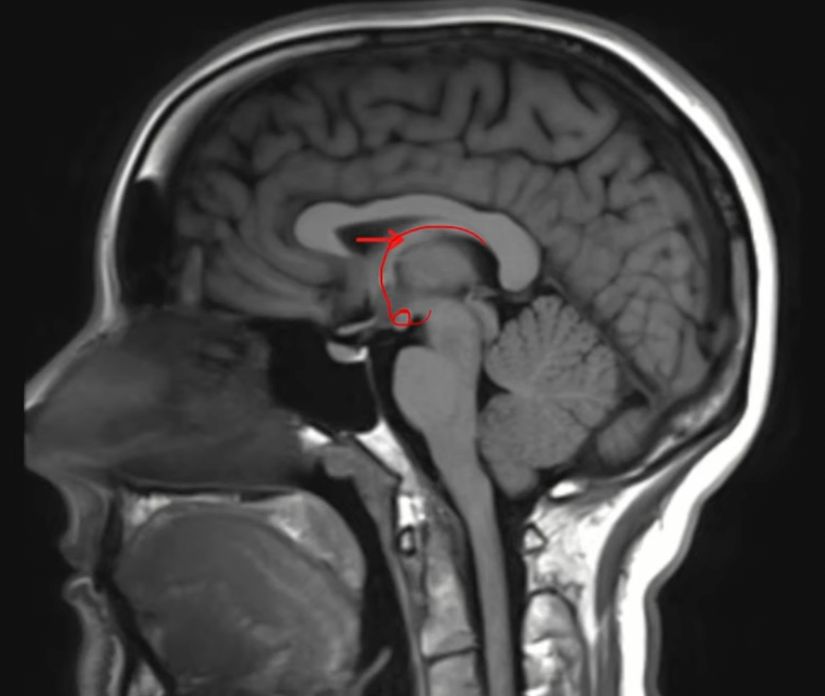
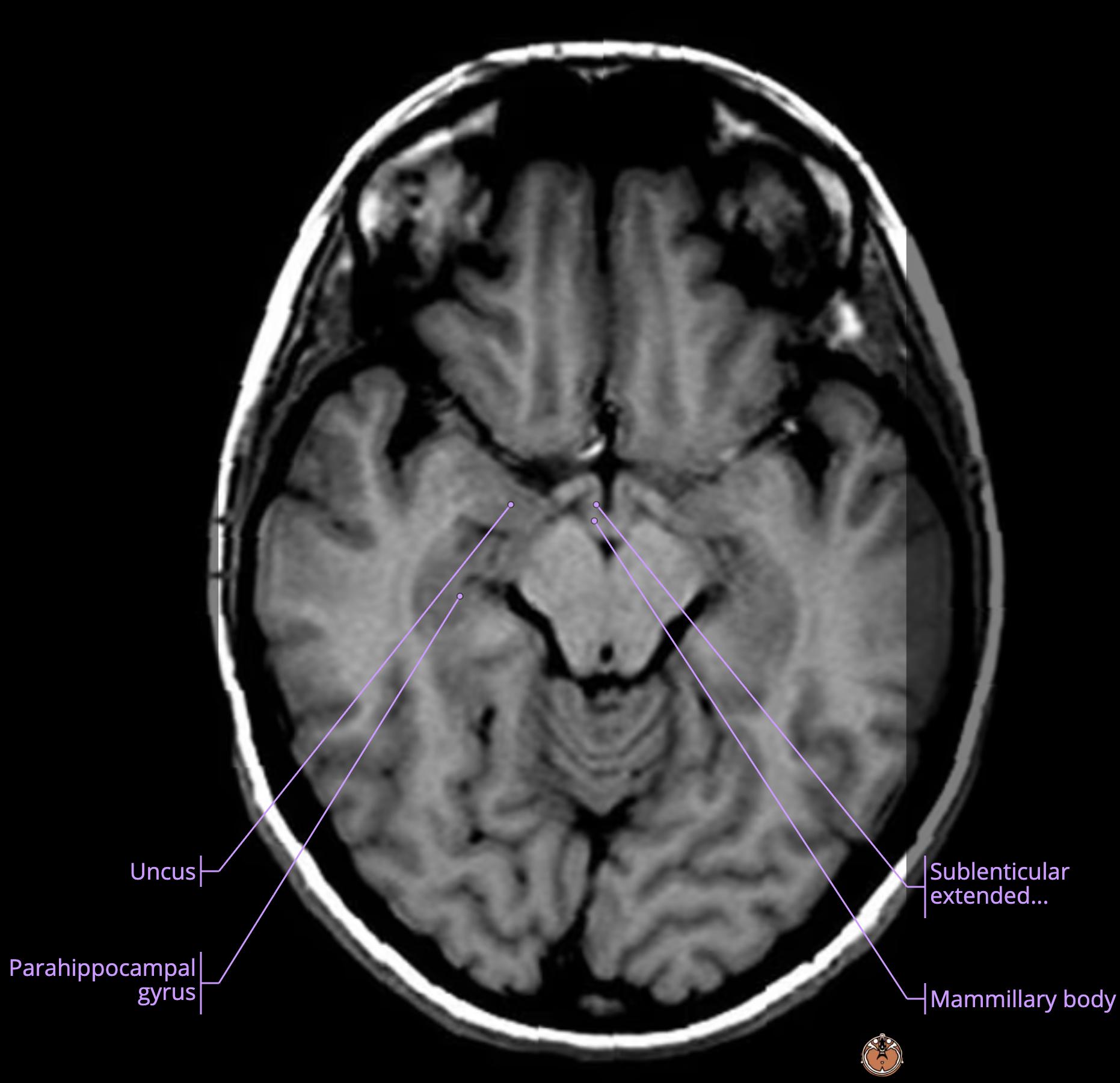
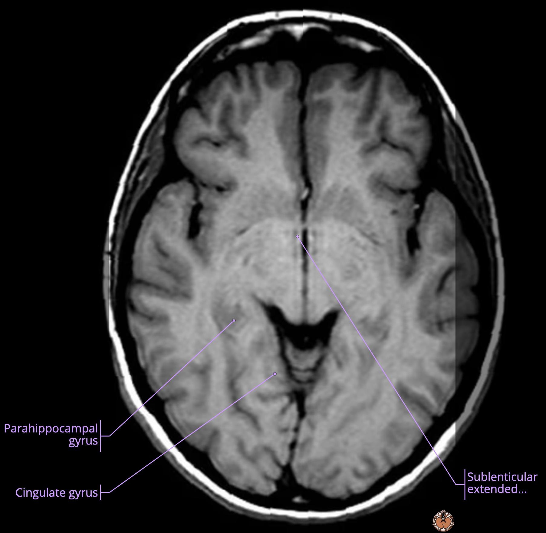
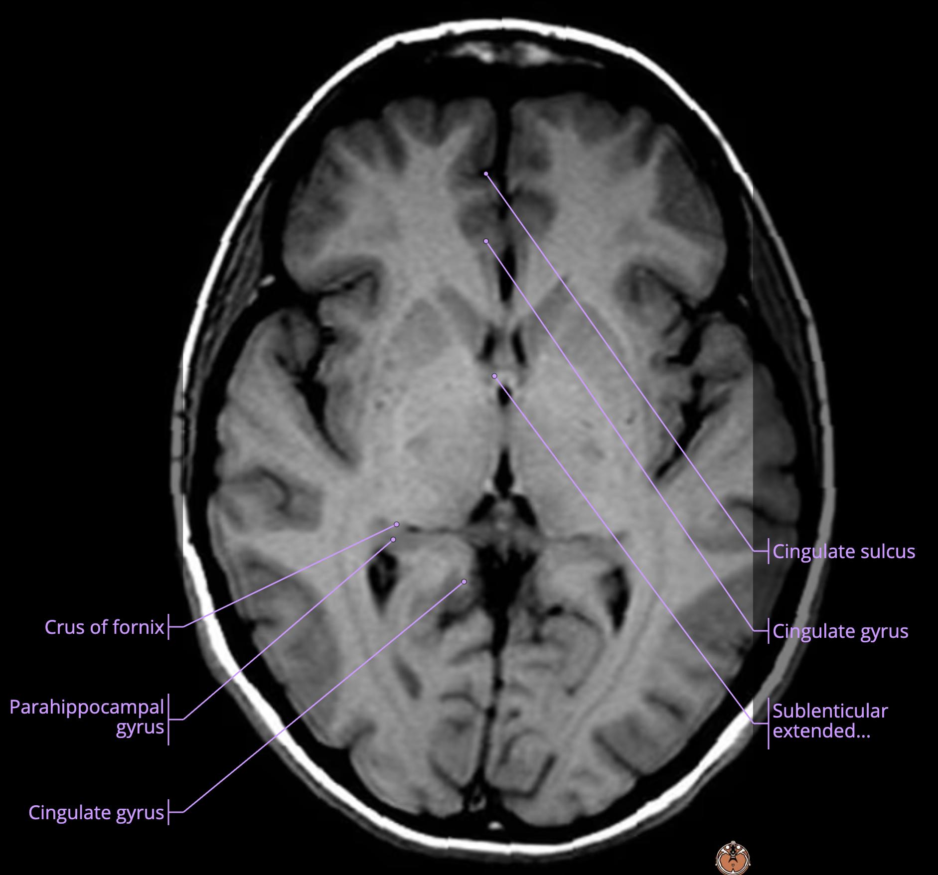
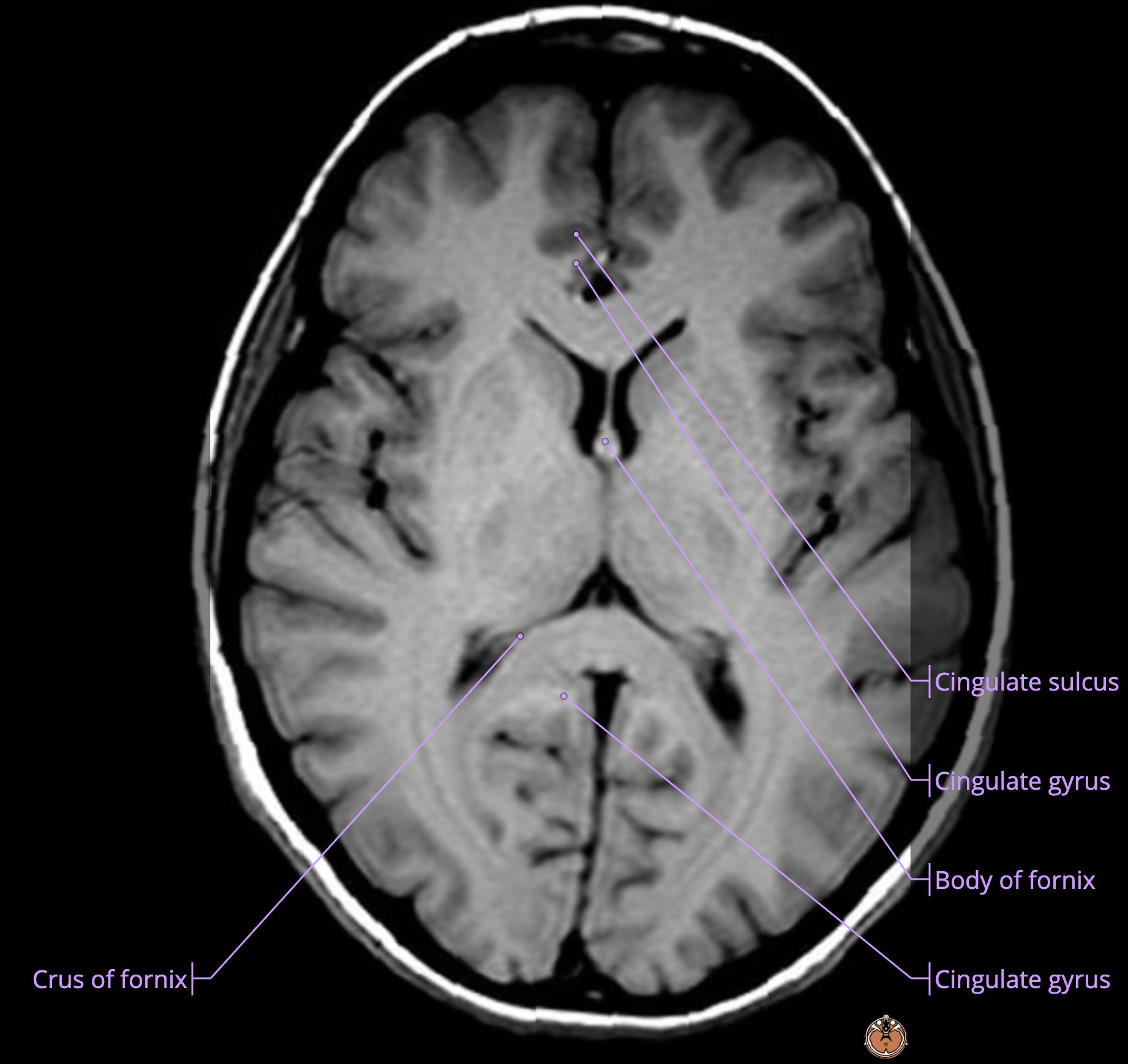
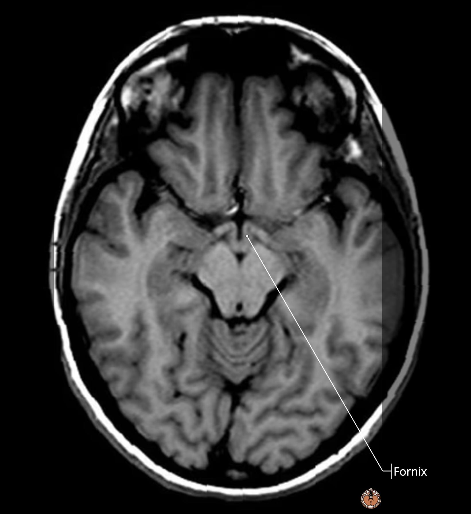
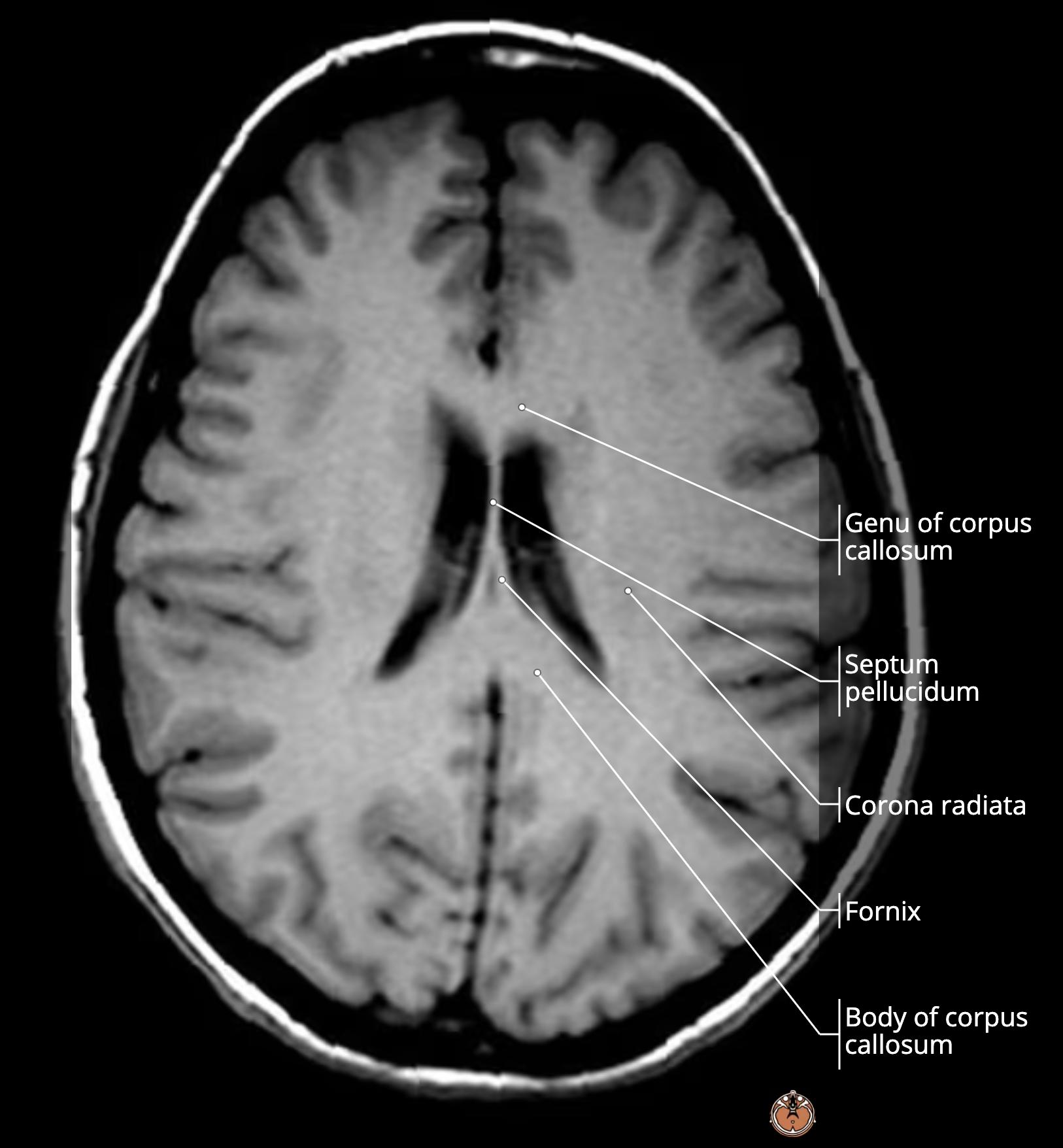
    • this is the body of fornix[…] 
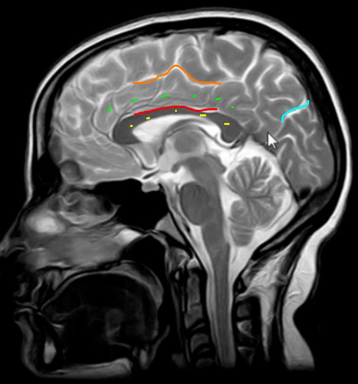
      • this structure runs anteriorly then inferiorly along the column[…] of fornix[…] to the mammillary bodies[…] then up to the ATN[…] of thalamus[…] as part of the papez[…] circuit
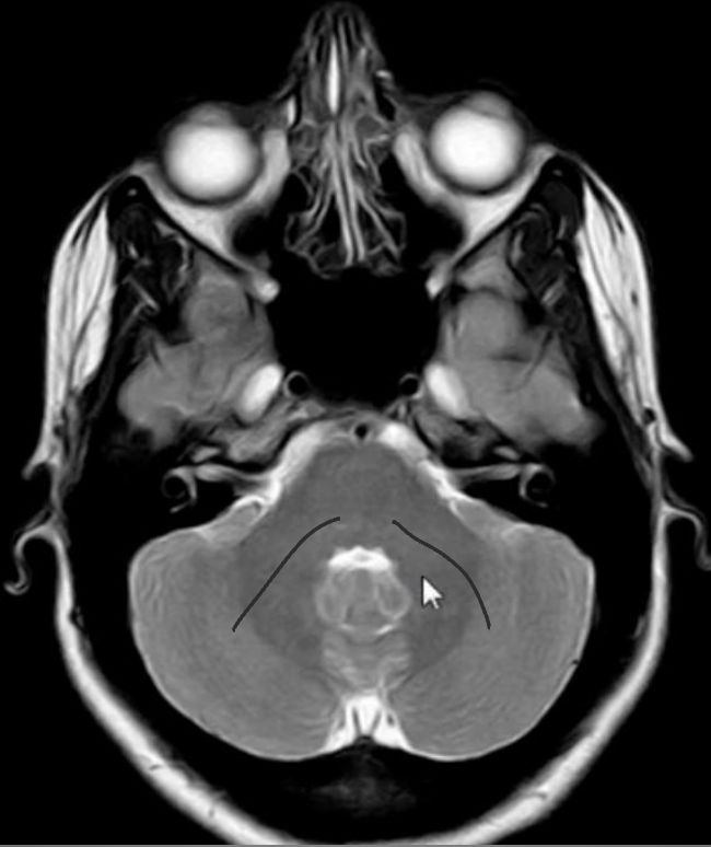
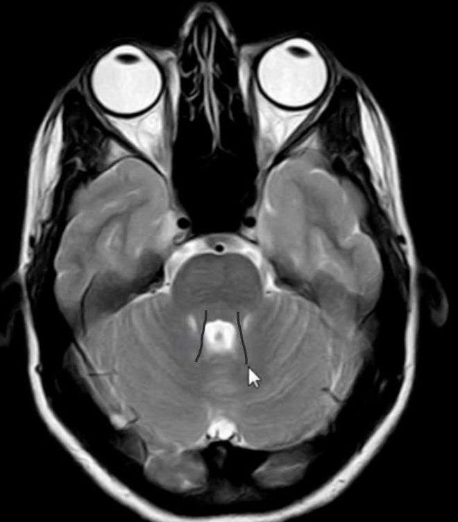
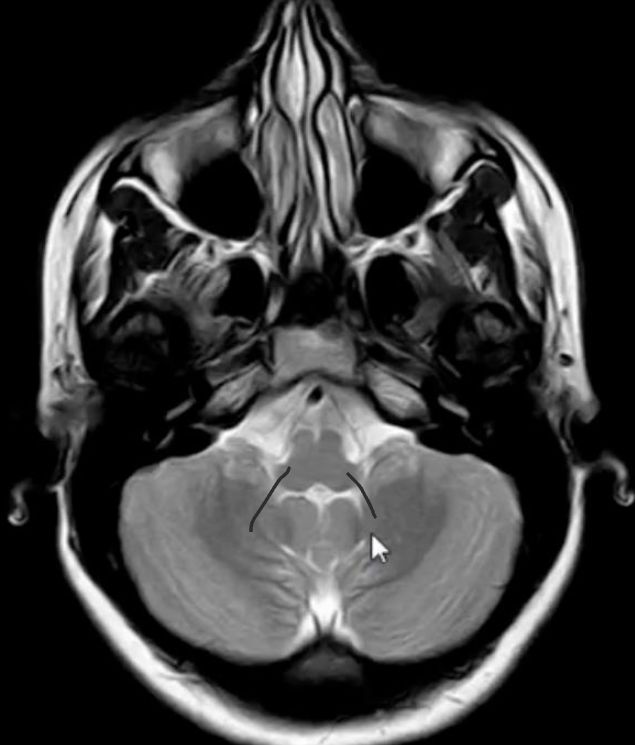
    • we are inferior to corpus callosum[…] 

    MRI Brain Anatomy - name this structure
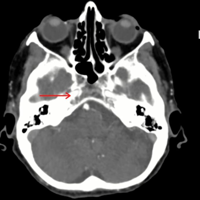
    • this is the R petrous ICA (C2)[vessel, segment]

    MRI Brain Anatomy - name this structure
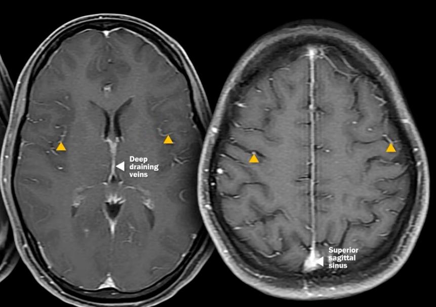
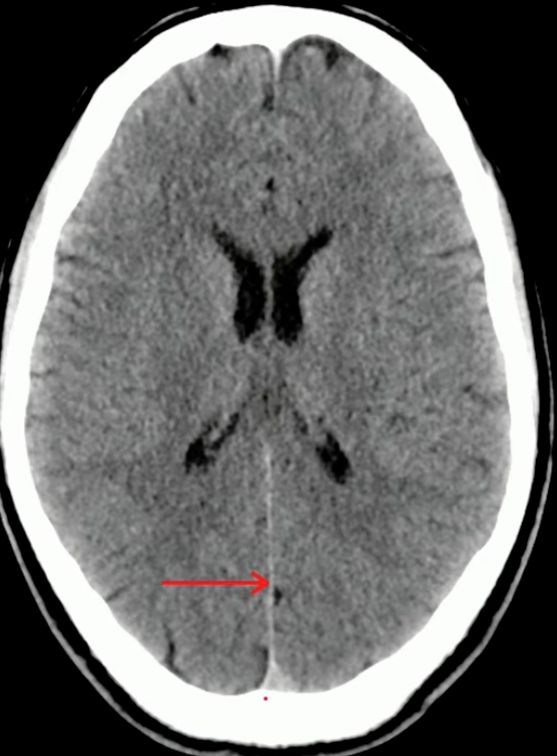
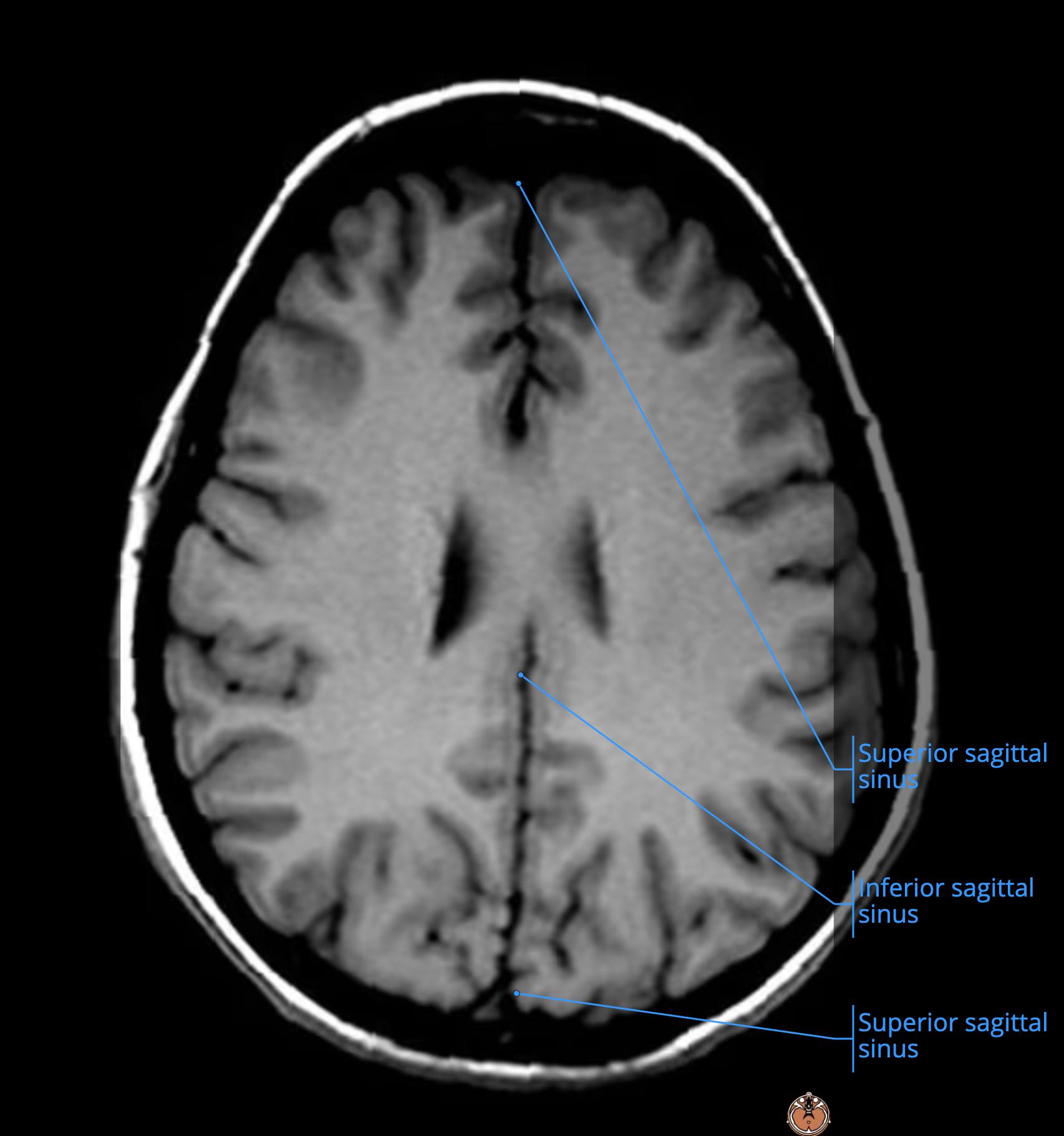
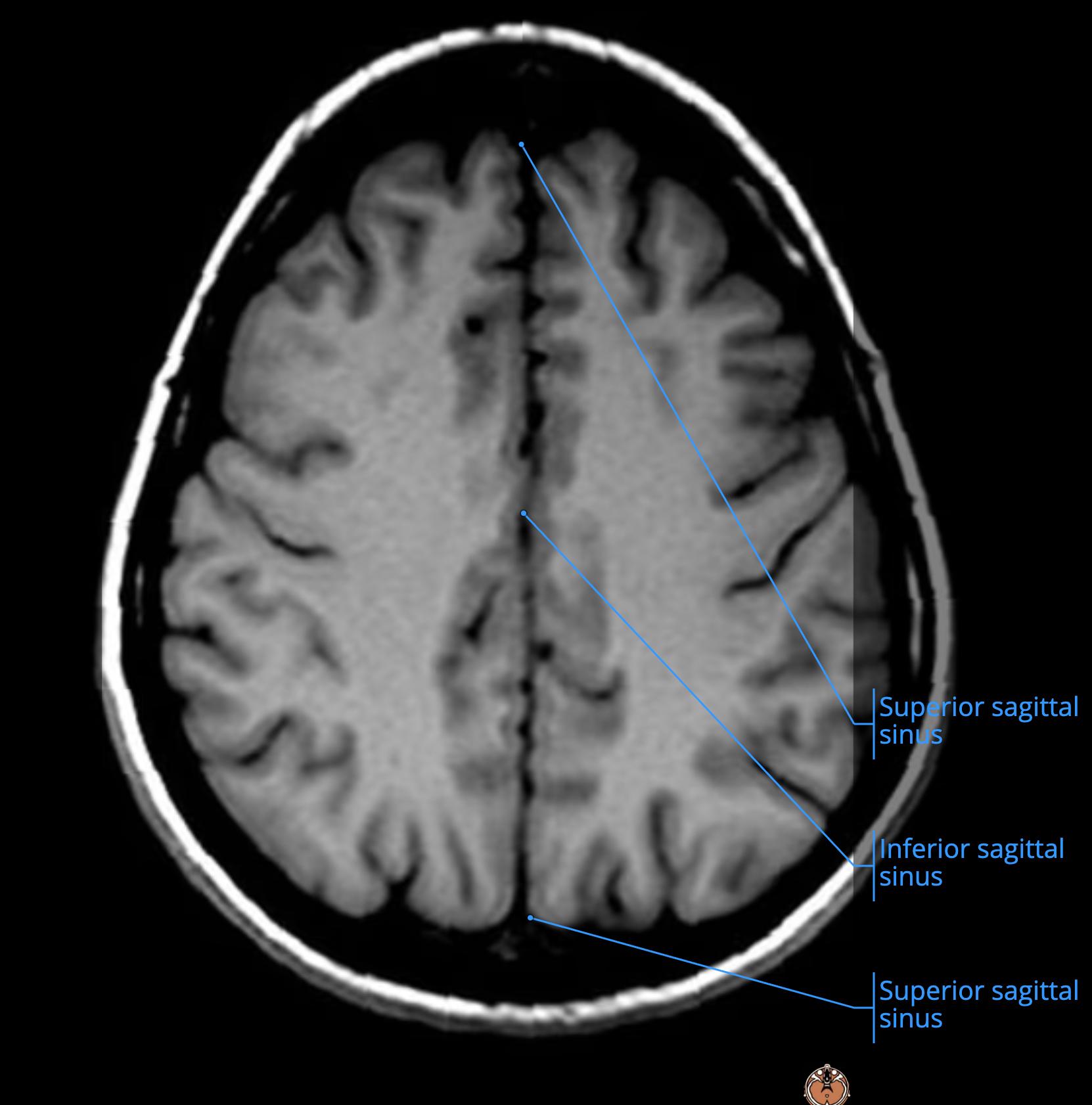
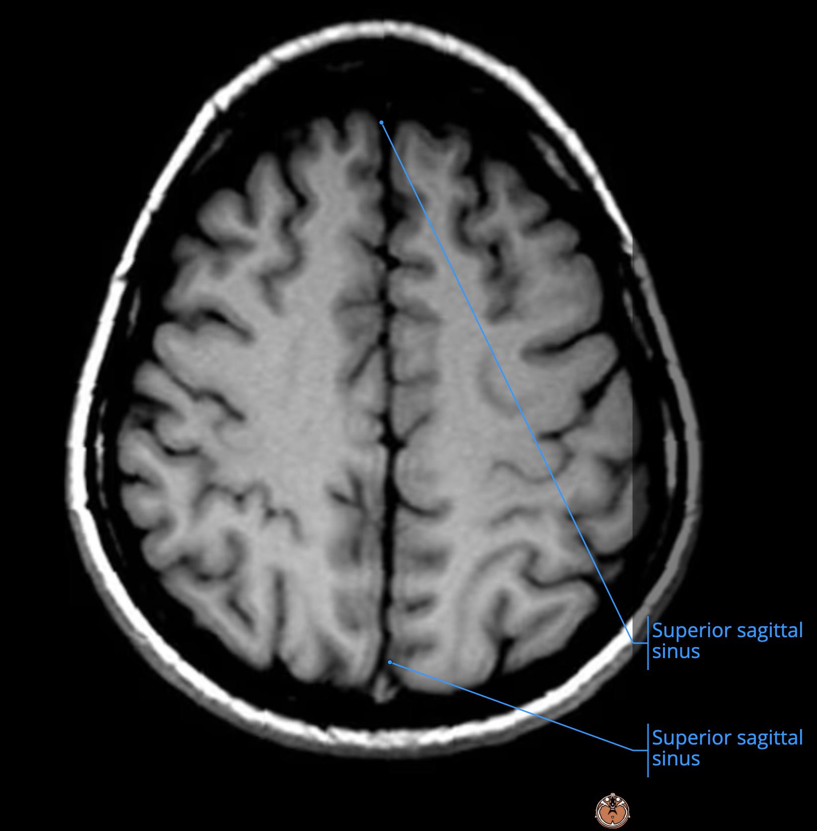
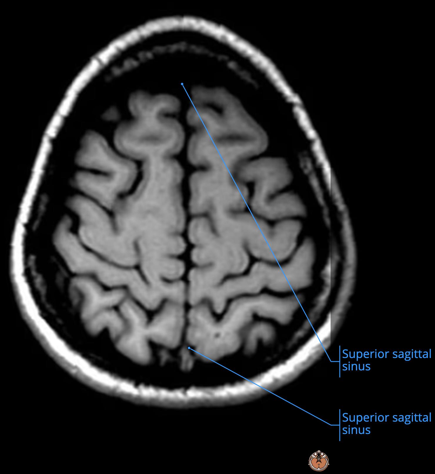
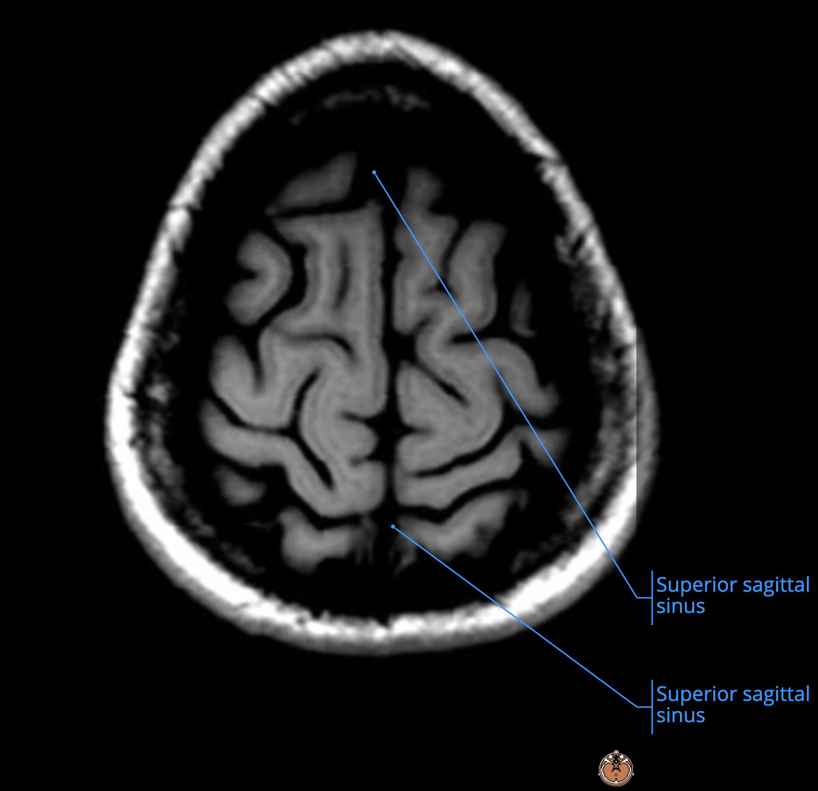
    • this is the posterior falx cerebri[…] where it invaginates and gives rise to the SSS[…]

    MRI Brain Anatomy - name this structure
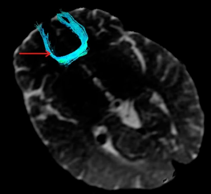
    • this DTI image shows the forceps minor (anterior forceps)[…] which are white matter tracts that connect the frontal lobe[…]genu of CC[…] > contralateral frontal lobe[…] to connect them

    MRI Brain Anatomy - name this structure
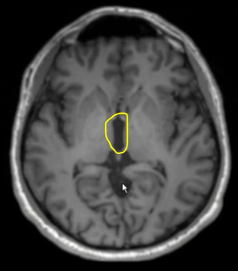
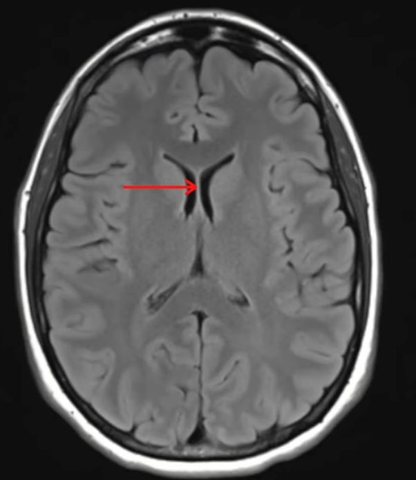
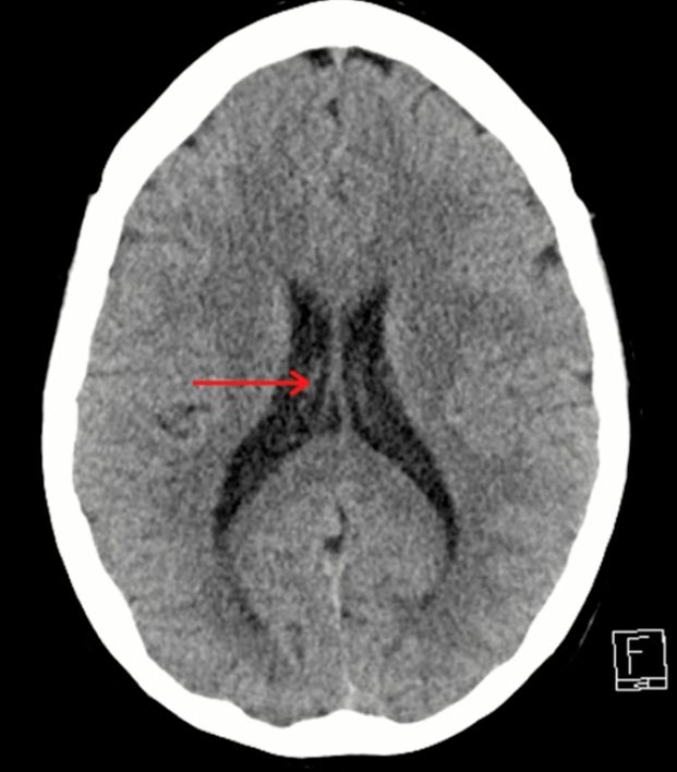
    • this is the septum pellucidum[…]
      • at rest they are 2[…] separate tissue planes with a potential[…] space in between them
      • sometimes if these planes of tissue are separated physiologically this is called cavum septum pellucidum[…]

    MRI Brain Anatomy - name this structure
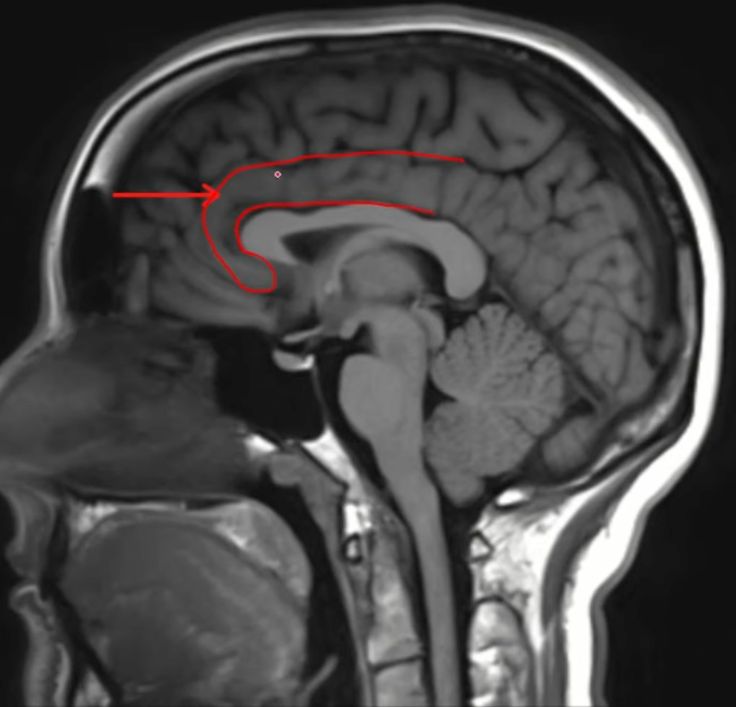
    • this is the cingulate gyrus[…] which helps us regulate emotions

    MRI Brain Anatomy - name this structure
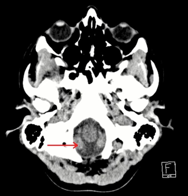
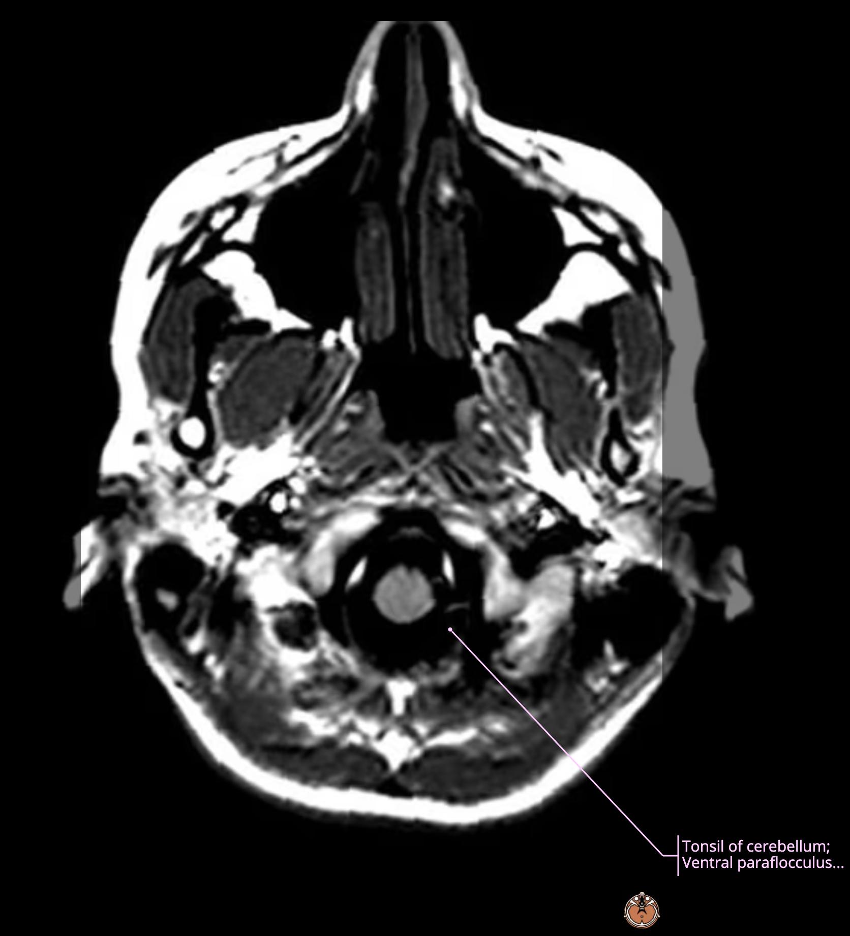
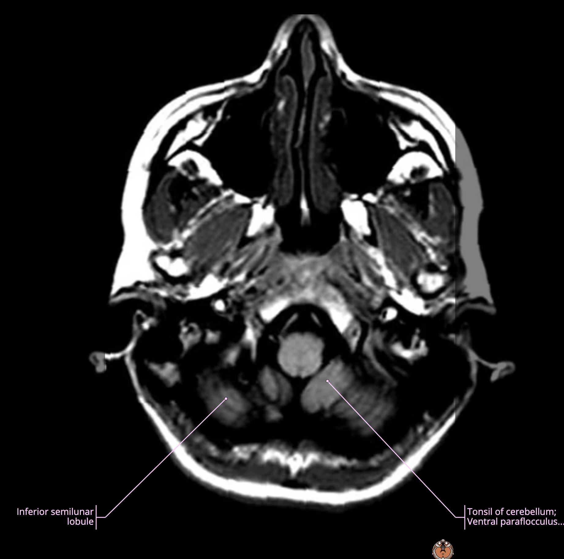
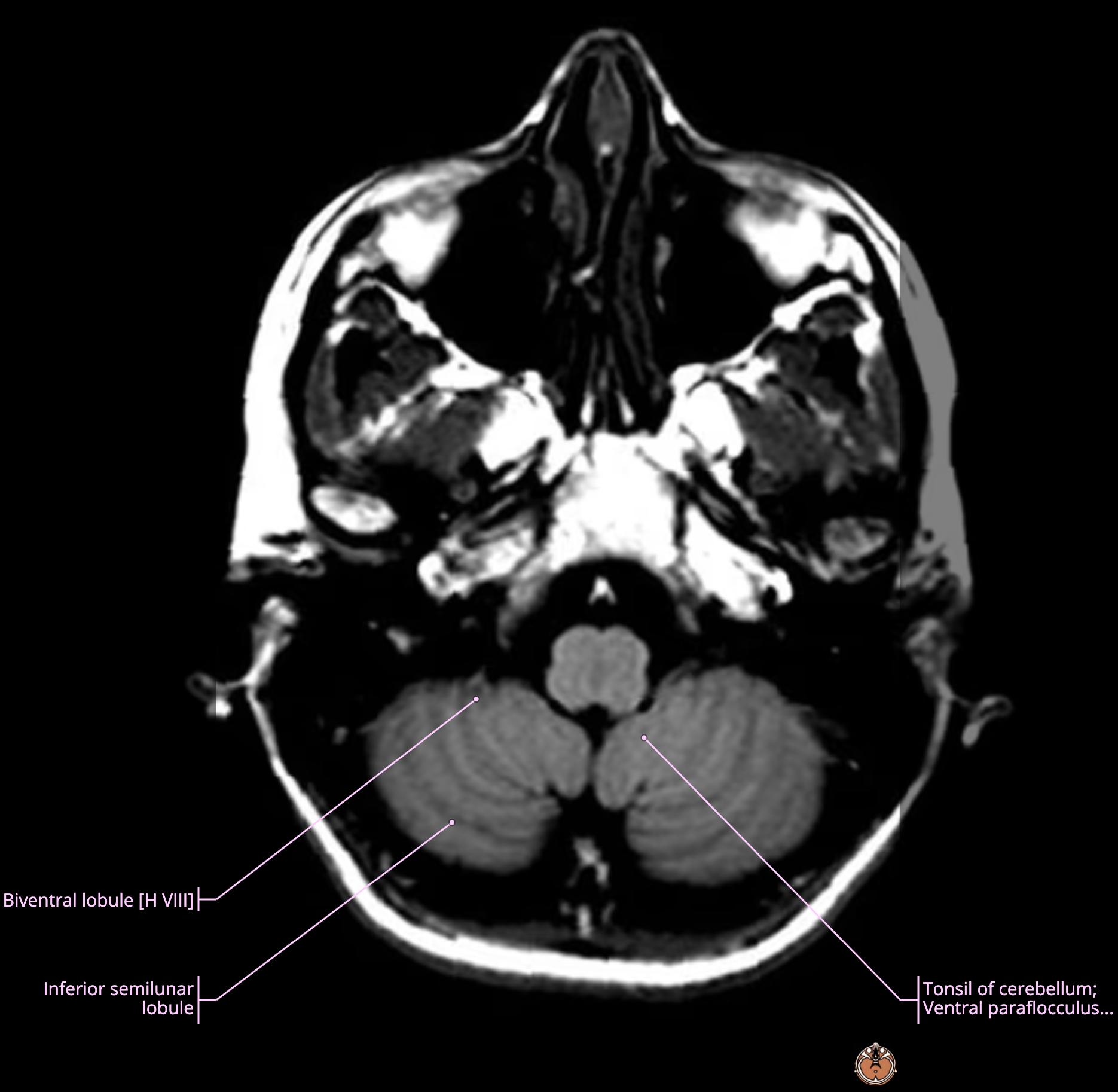
    • this is the right cerebellar tonsil[…] located at the level of foramen magnum[…]
      • in cases of high ICP[…], see how far it pushes down via foramen magnum

    MRI Brain Anatomy - name this structure
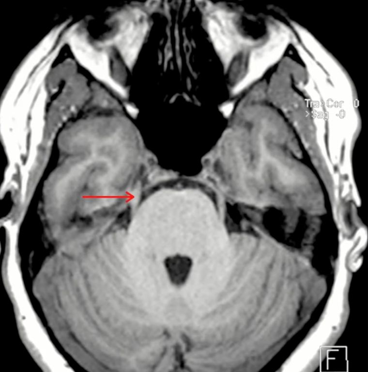
    • this is the right trigeminal nerve[…] that extends into Meckel's Cave[…]

    MRI Brain Anatomy - name this structure
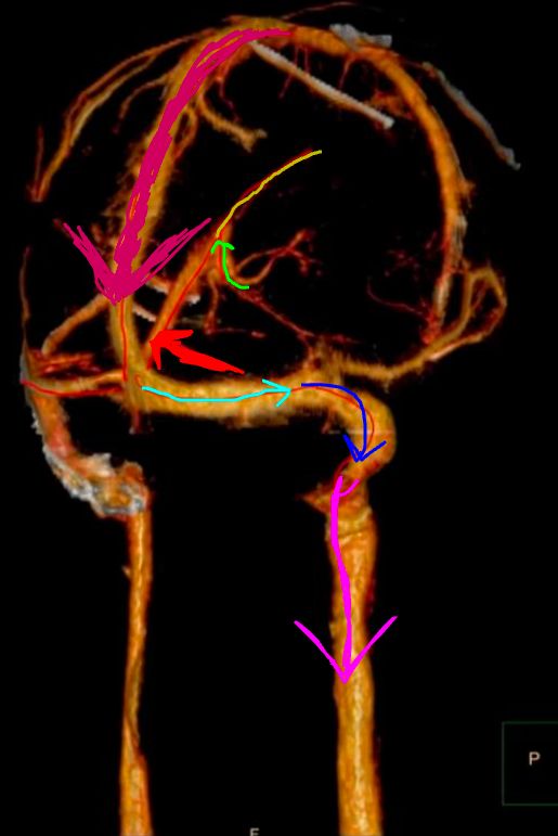
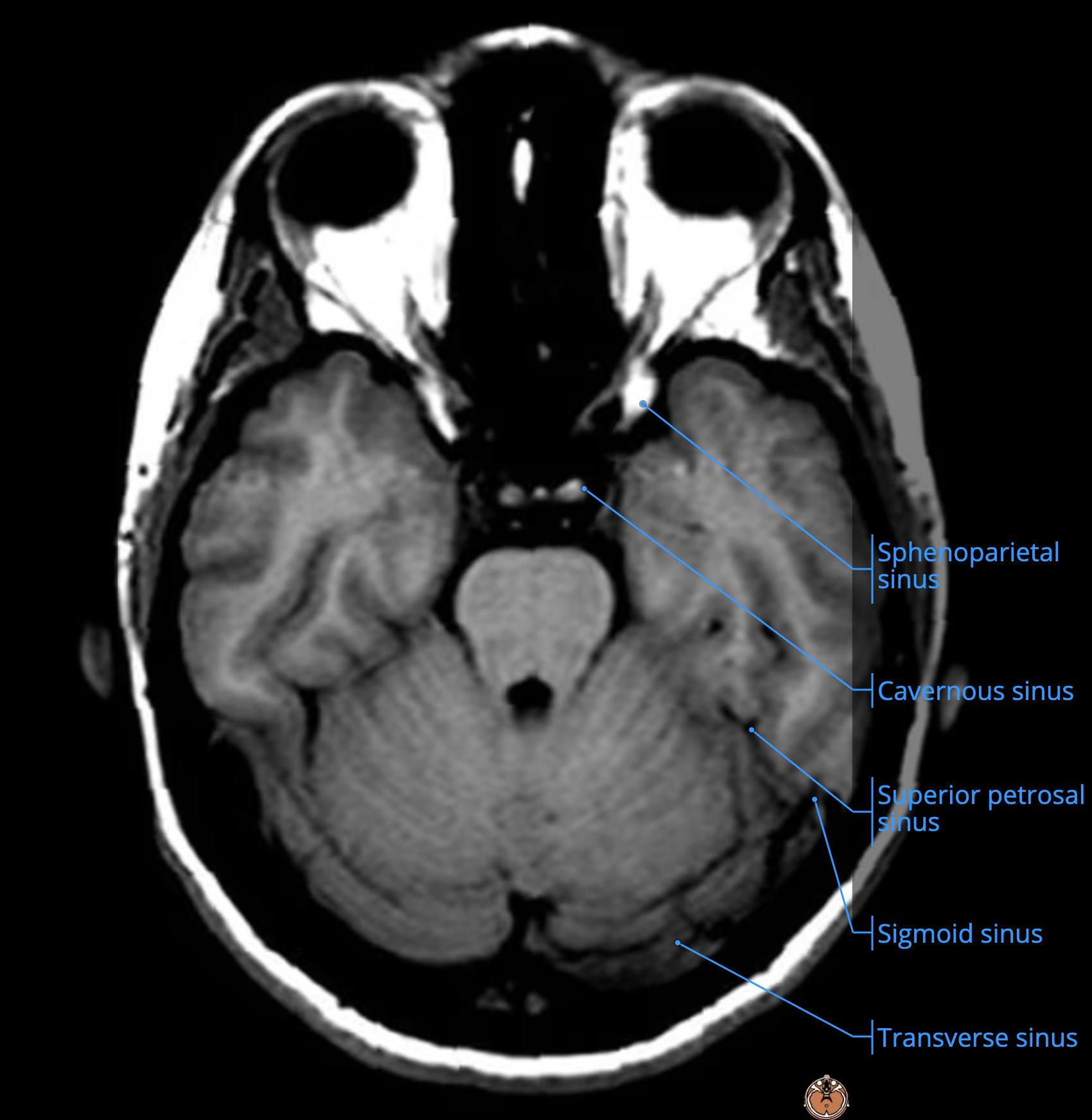
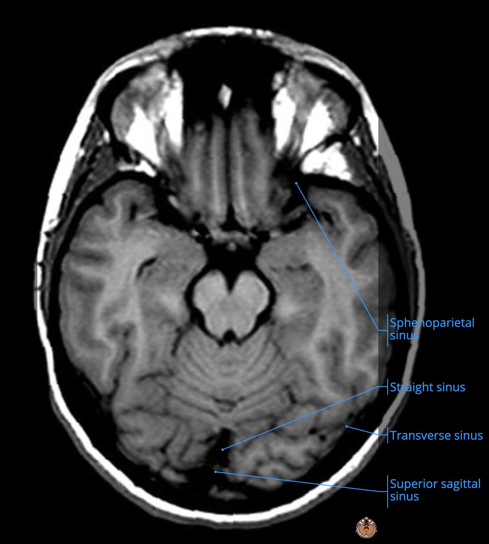
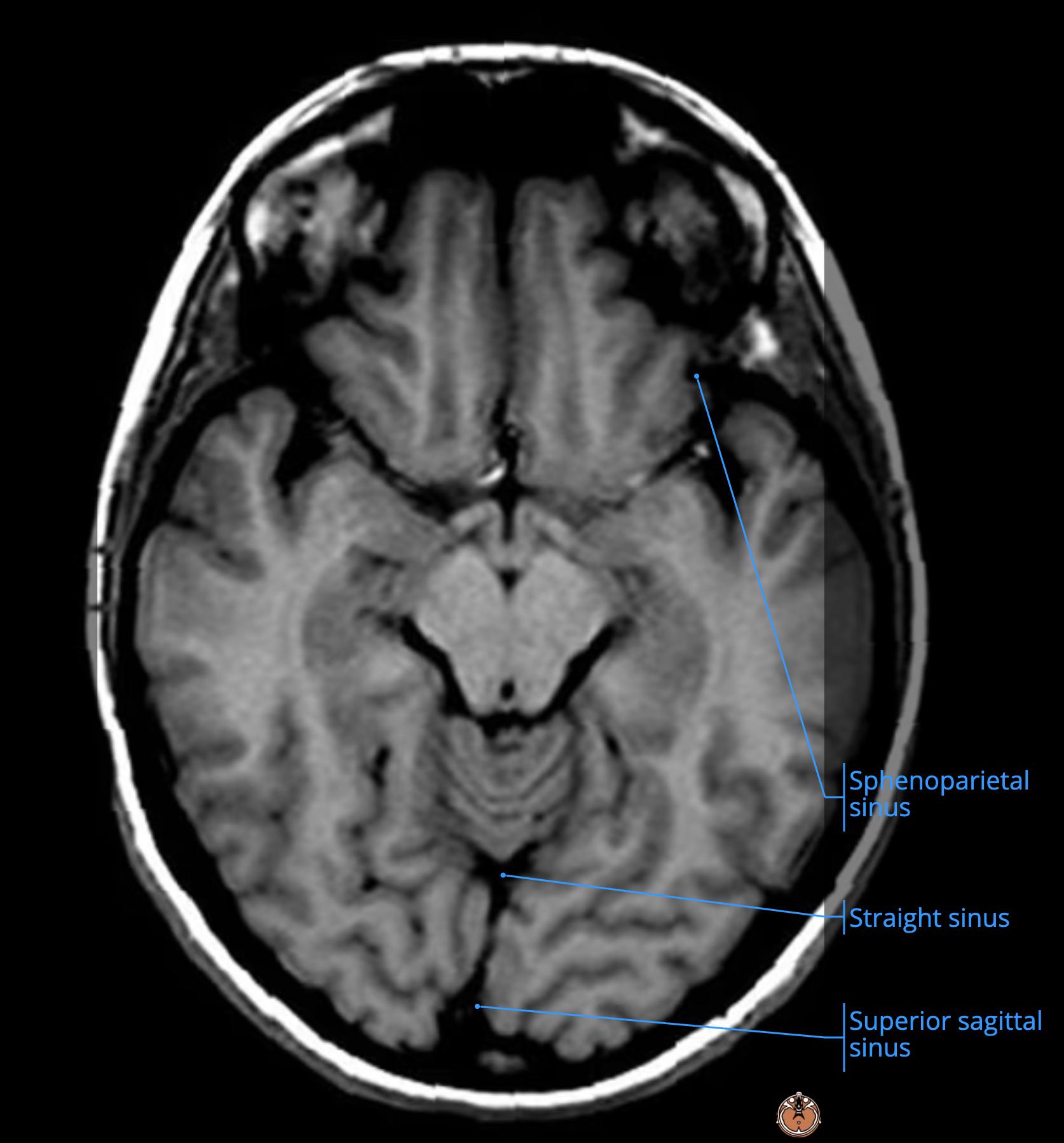
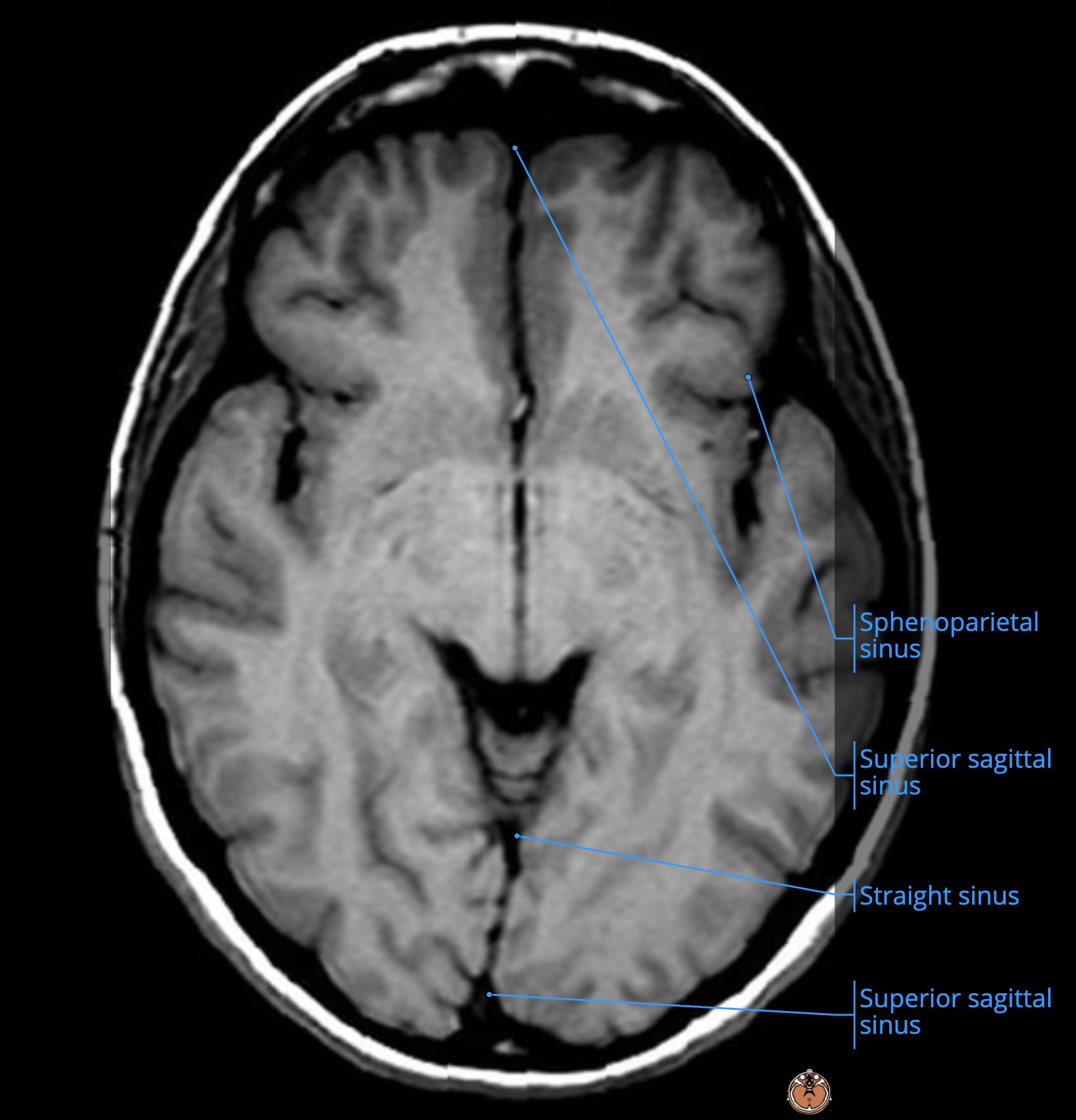
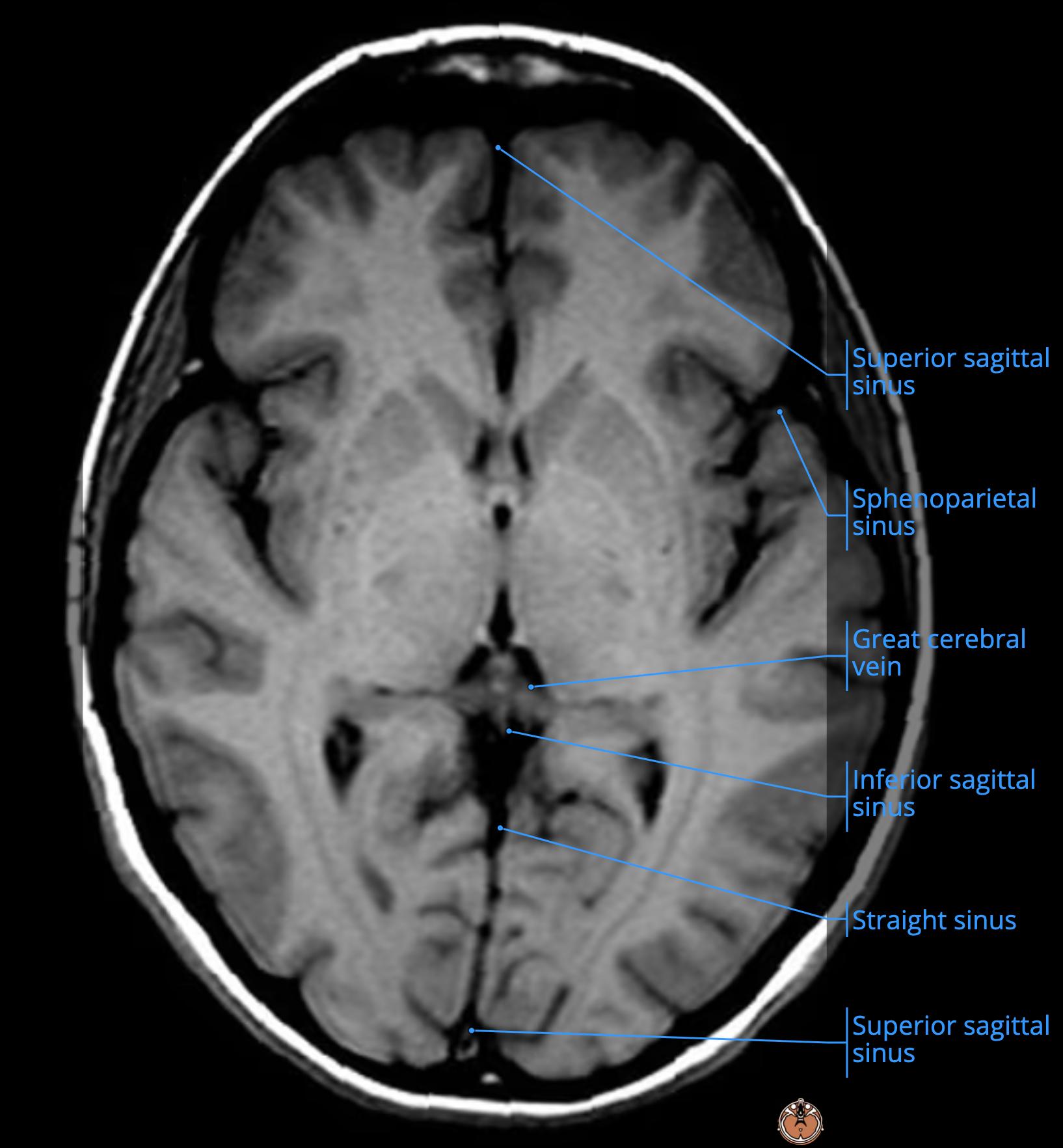
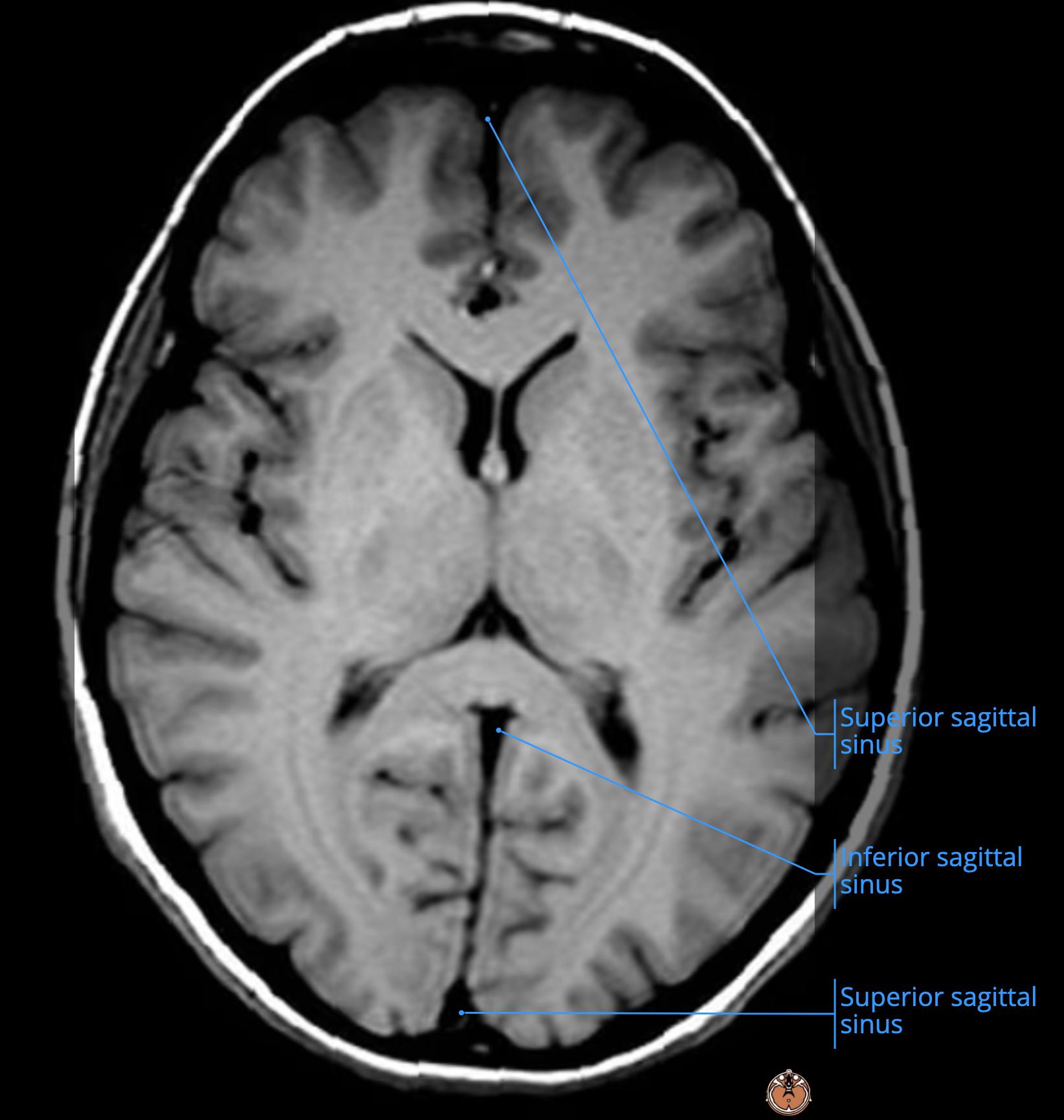
    • this is the straight sinus[…] (red) where the inferior sagittal sinus[…] (yellow) connects with the Great Cerebral Vein of Galen[…] (light green)
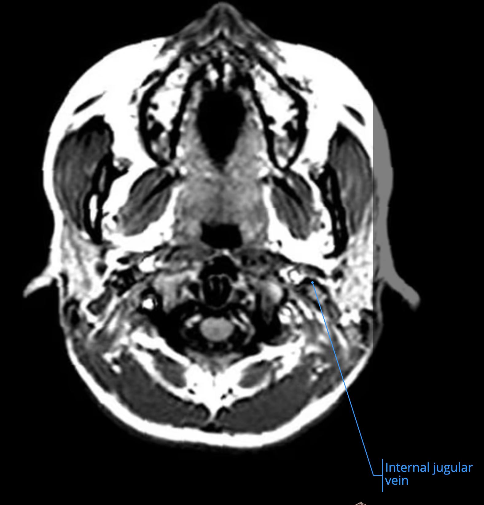
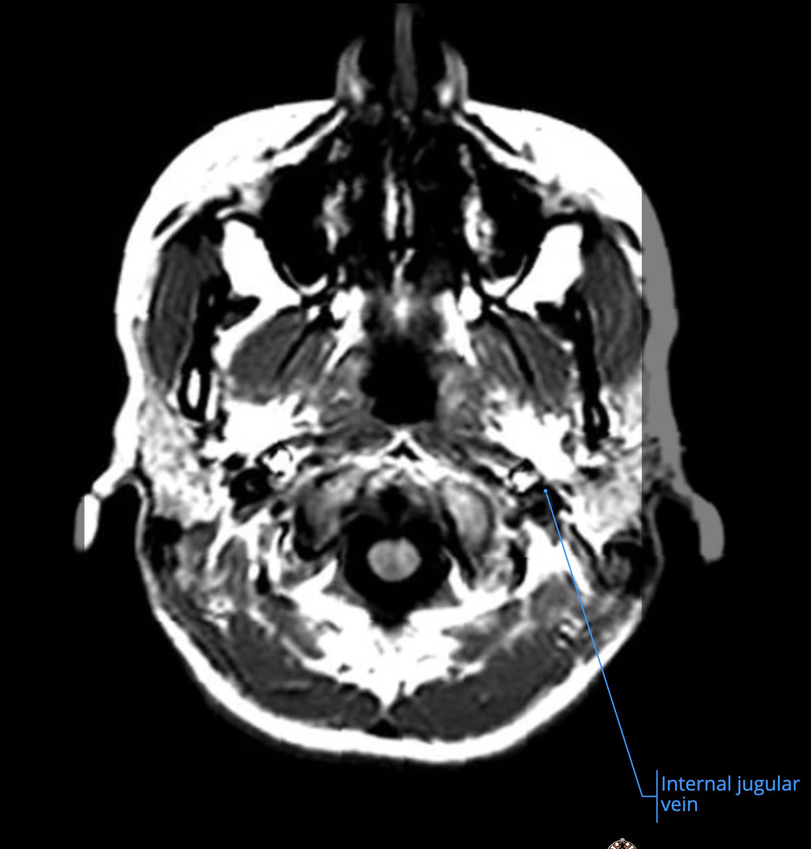
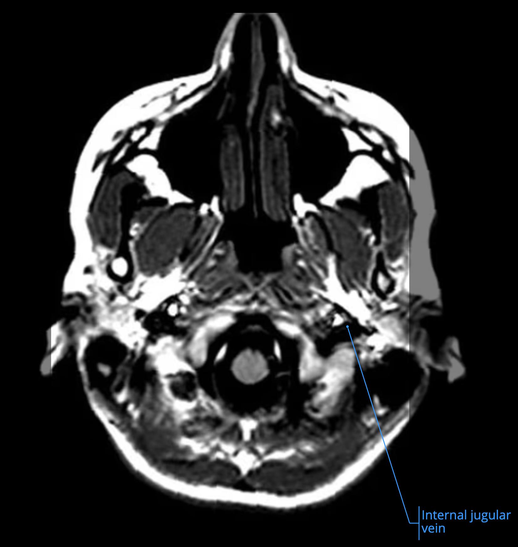
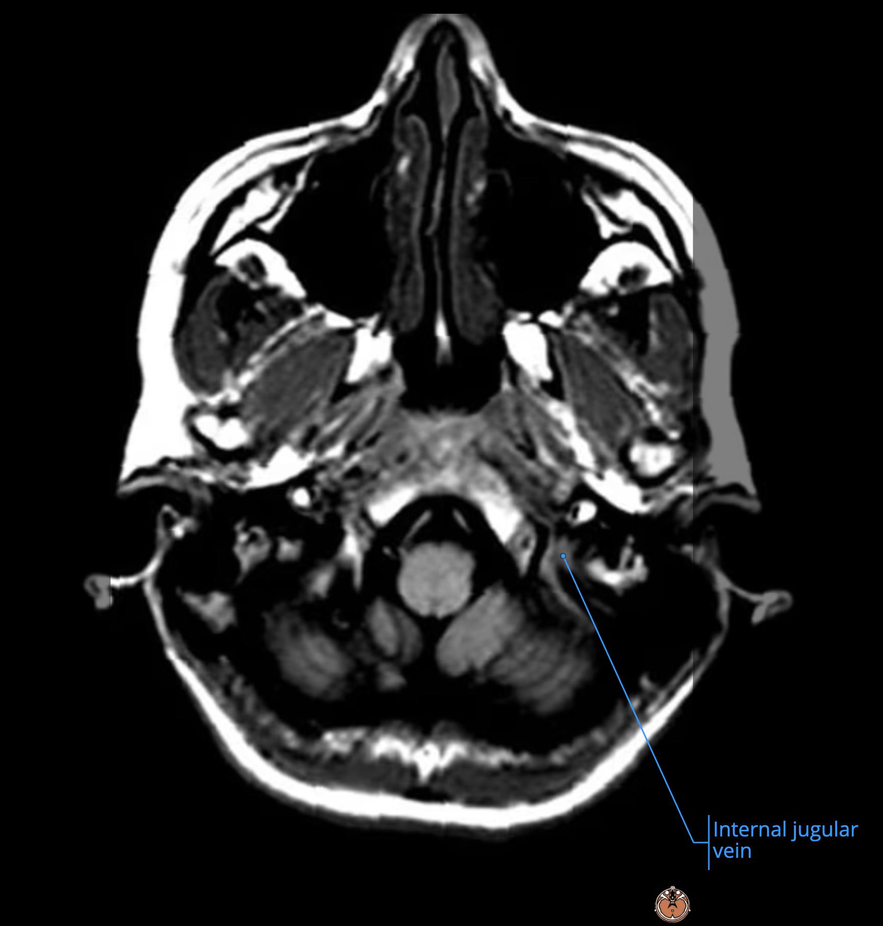
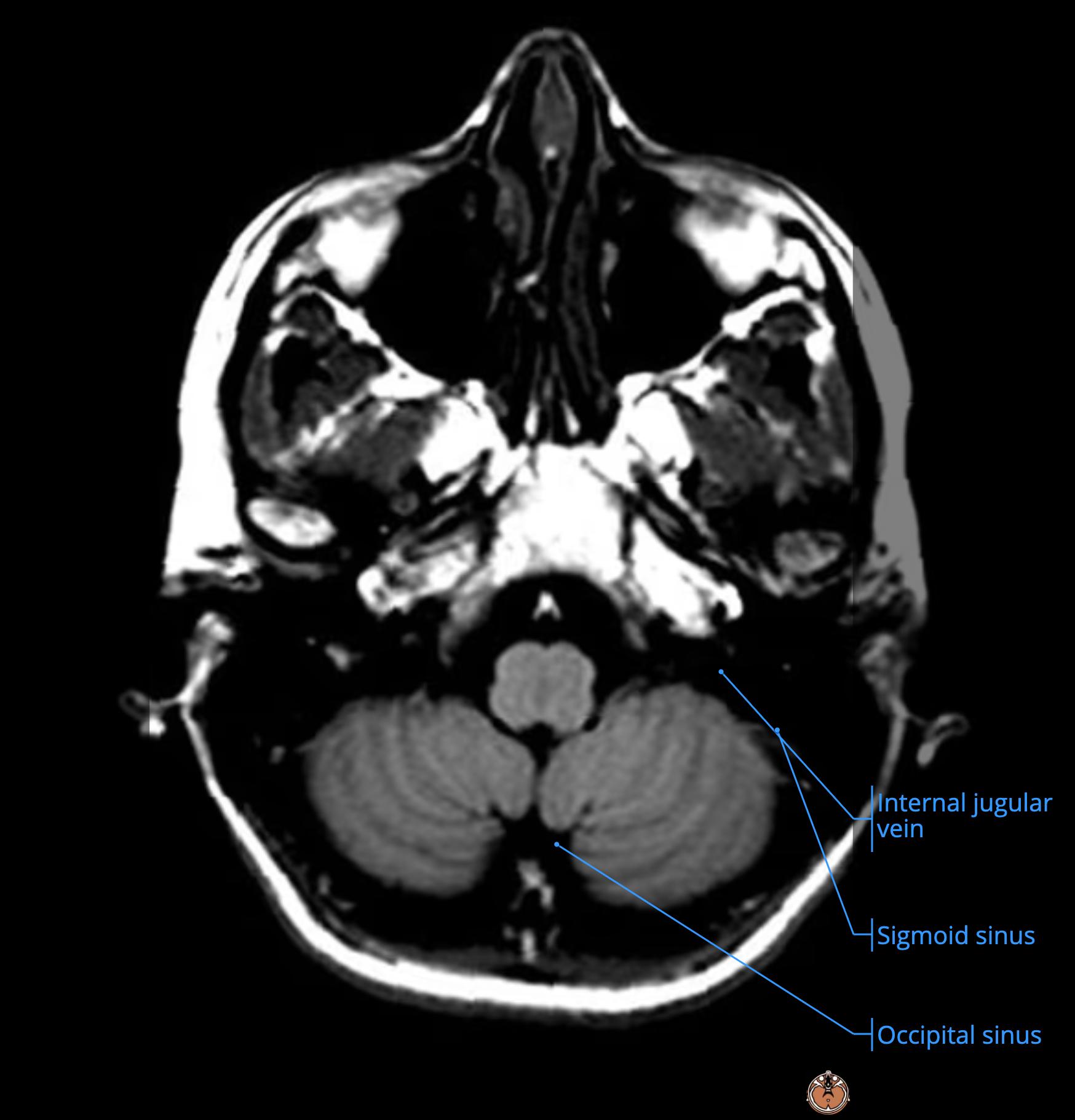
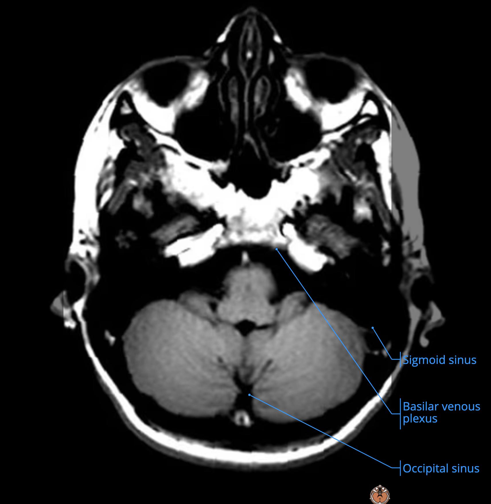
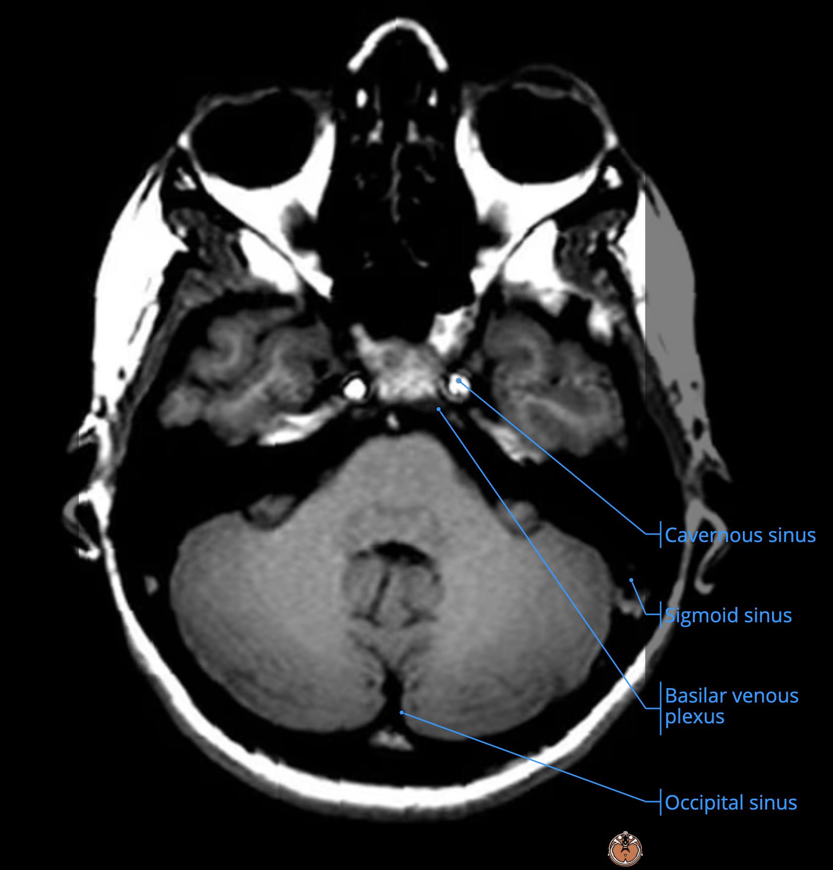
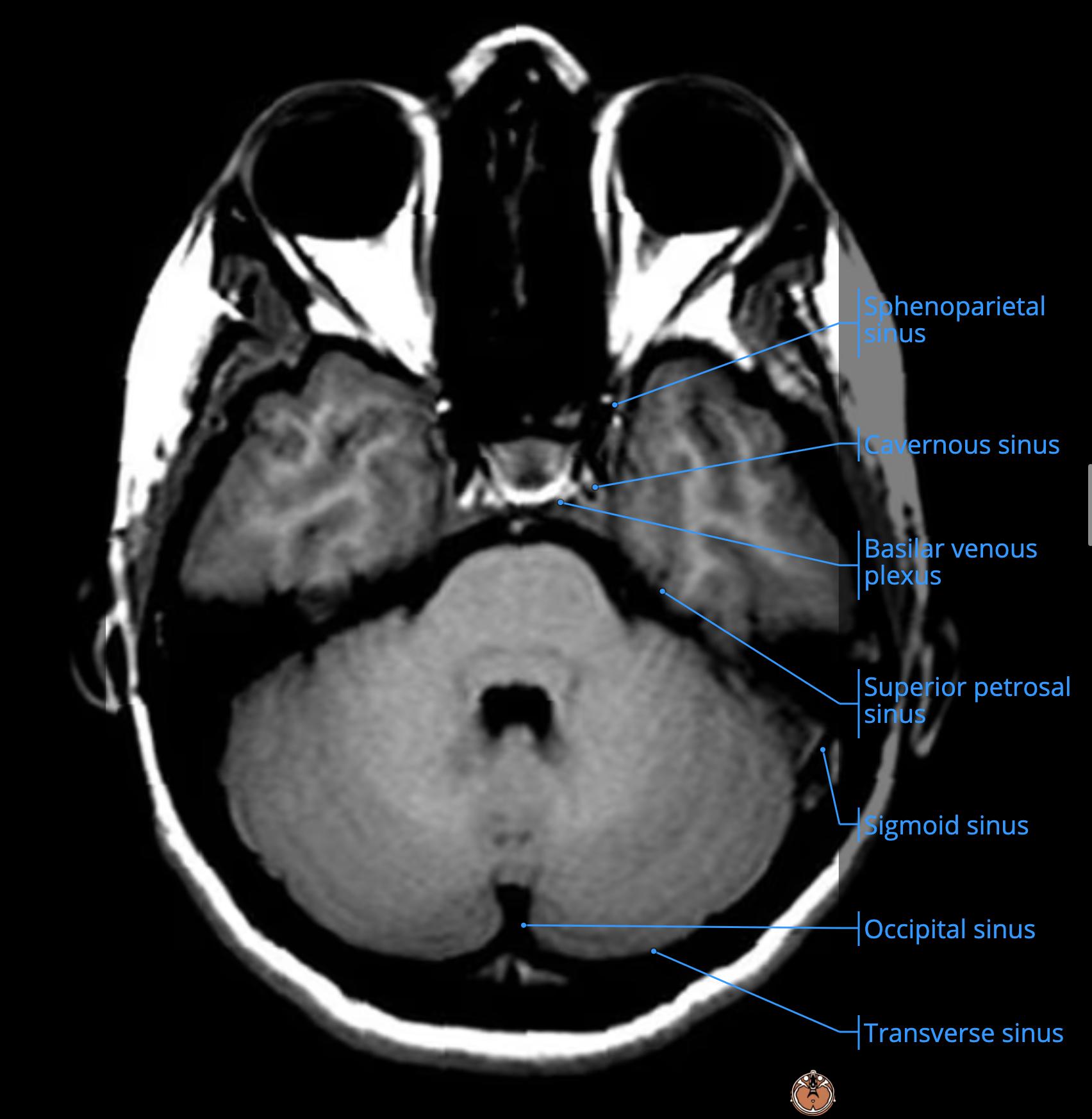
      • this then joins with the SSS[…] (dark pink) to form the Confluence of Sinuses[…] which is then drained away by the Transverse Sinuses[…] (light blue) into the Sigmoid Sinuses[…] (dark blue) into the IJV[…] (medium pink)

    MRI Brain Anatomy - name this structure
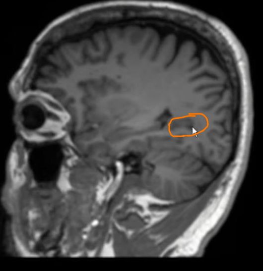
    • this is the R choroid plexus[…]

    MRI Brain Anatomy - name this structure
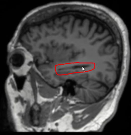
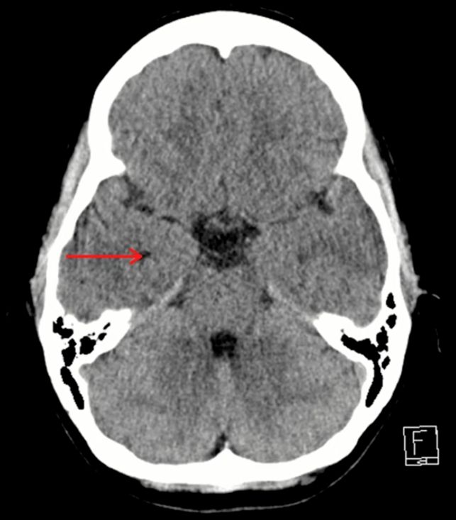
    • this is the R temporal horn, lateral ventricle[…]

    remember the 3D shape of the ventricle
    MRI Brain Anatomy - name this structure
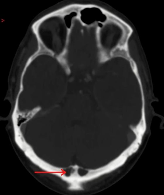
    • this is an intraosseous arachnoid granulation[…]
      • these are physiologic variants

    MRI Brain Anatomy - name this structure
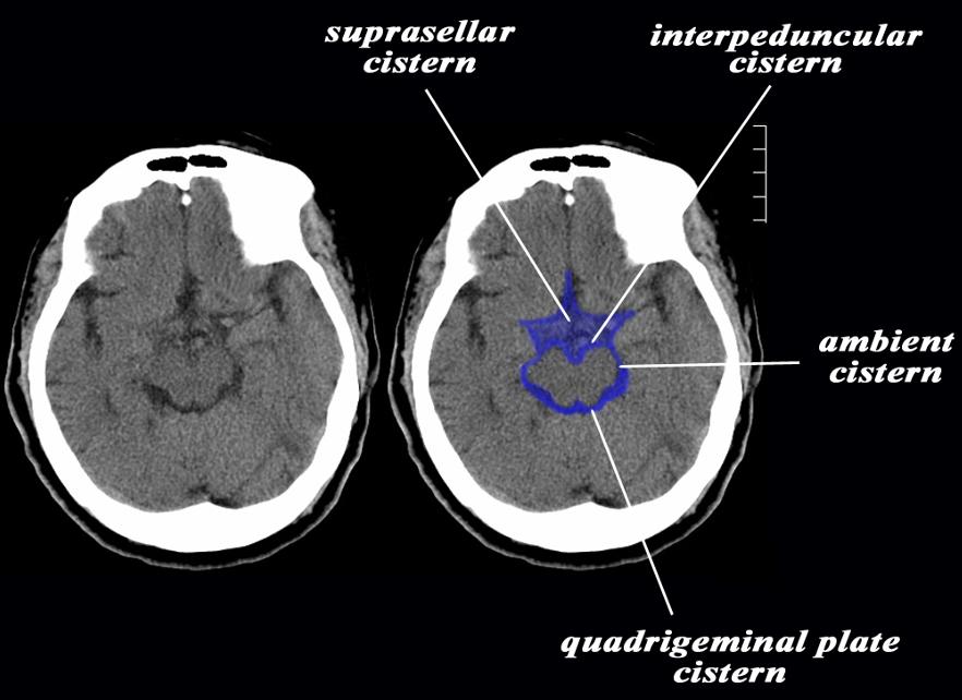
    • the 3 cisterns pictured here are
      • interpeduncular[…] anteriorly
      • ambient[…] laterally wrapping around the midbrain[…]
      • quadrigeminal[…] posteriorly

    MRI Brain Anatomy - name this structure
    • this is the right cerebral peduncle[…]
      • the cistern anteriorly is the interpeduncular[…] cistern

    MRI Brain Anatomy
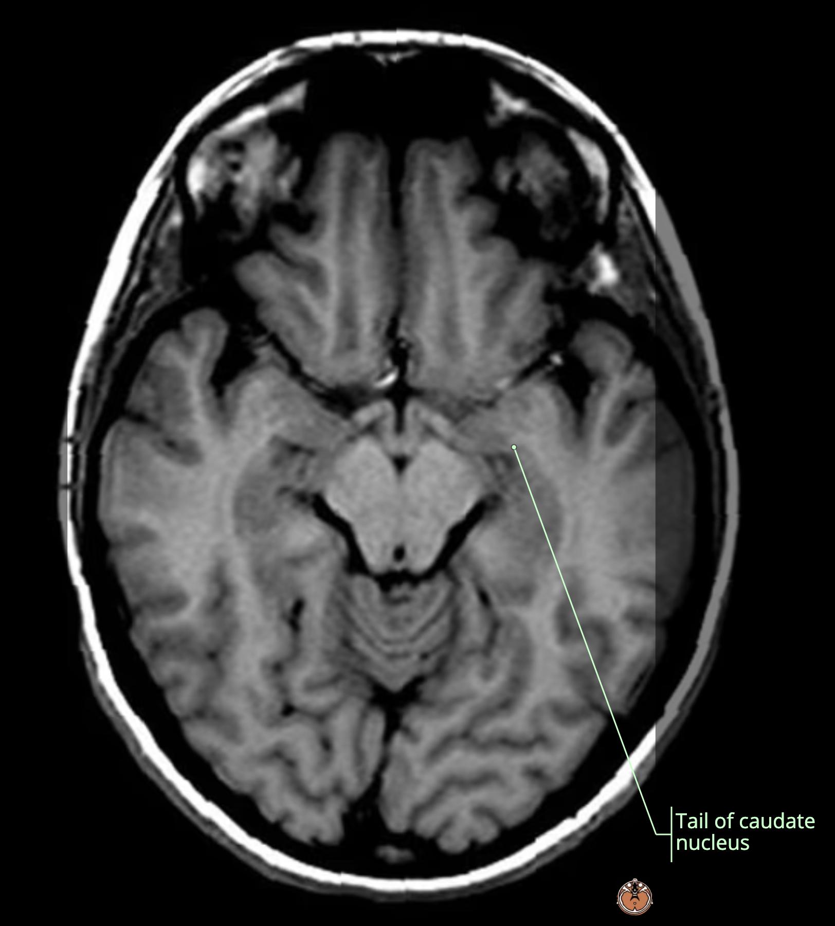
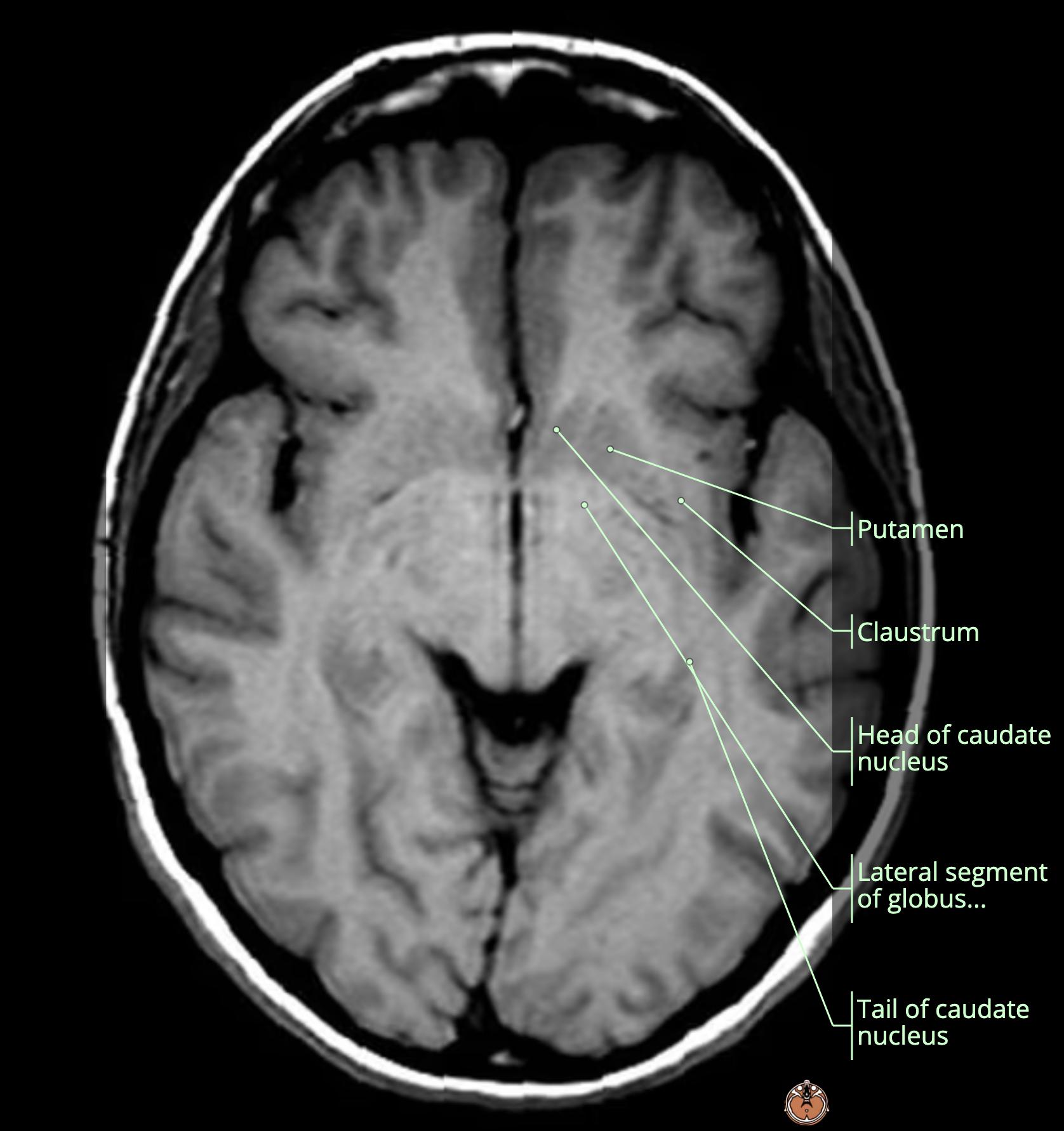
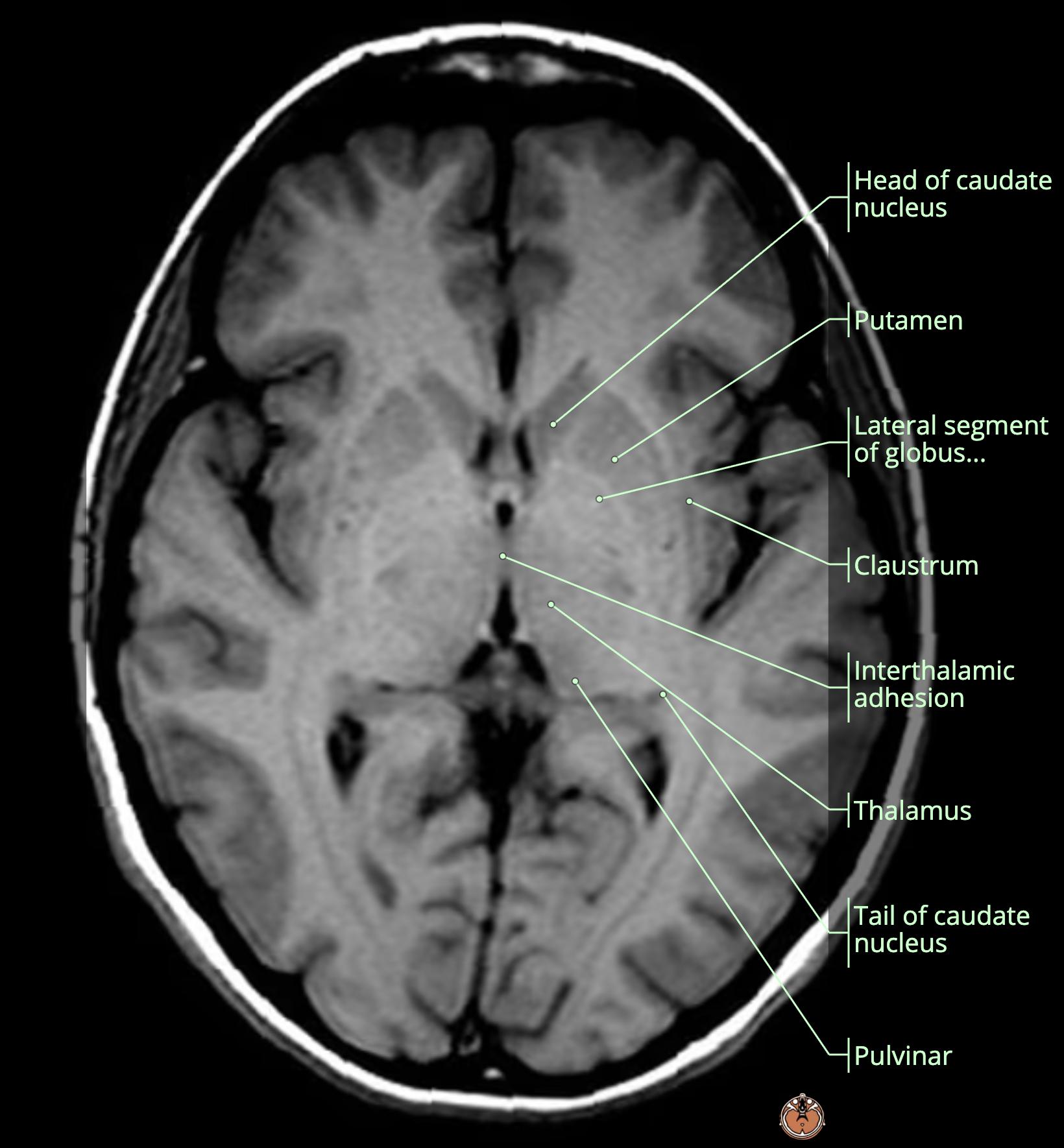
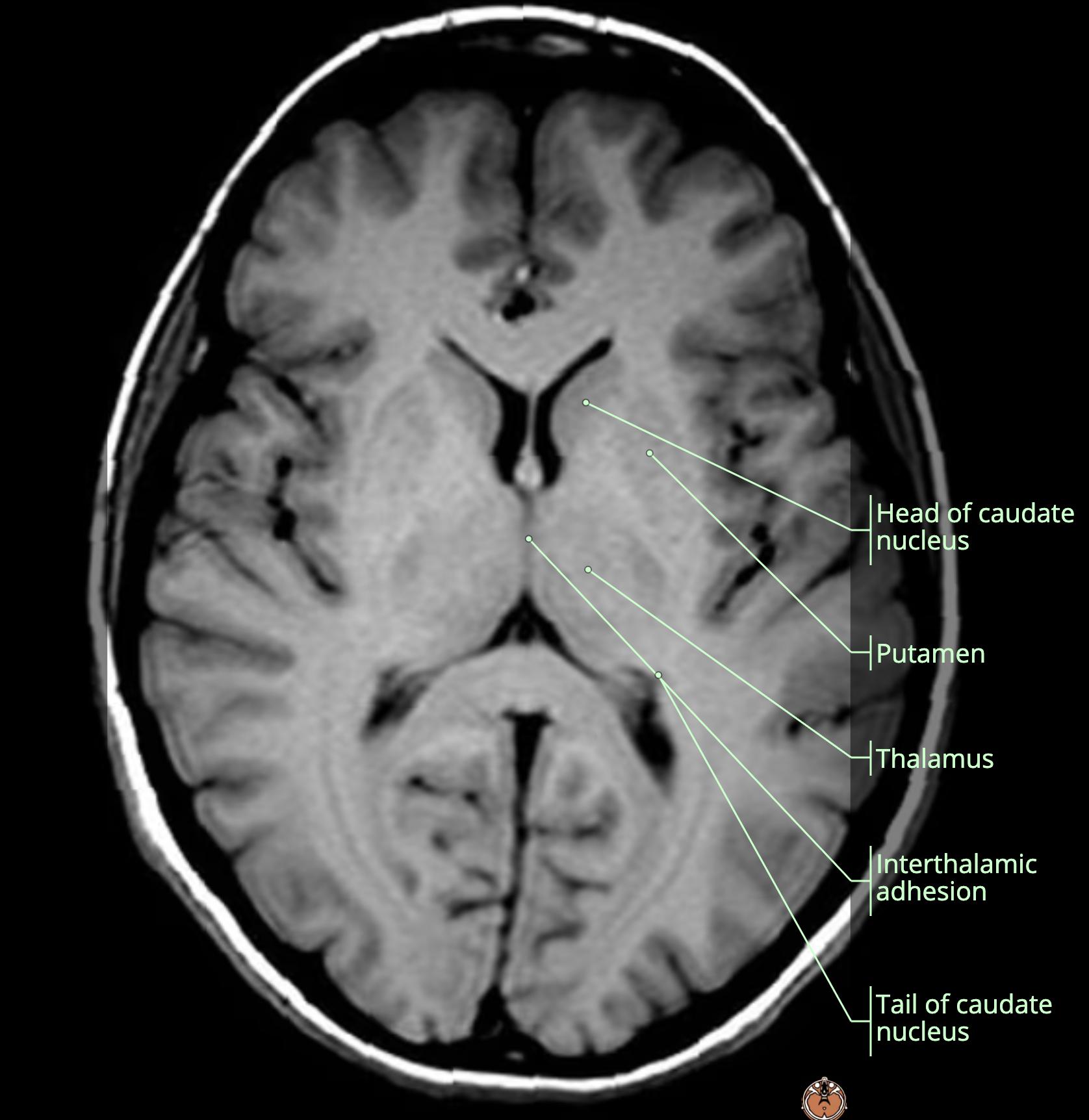
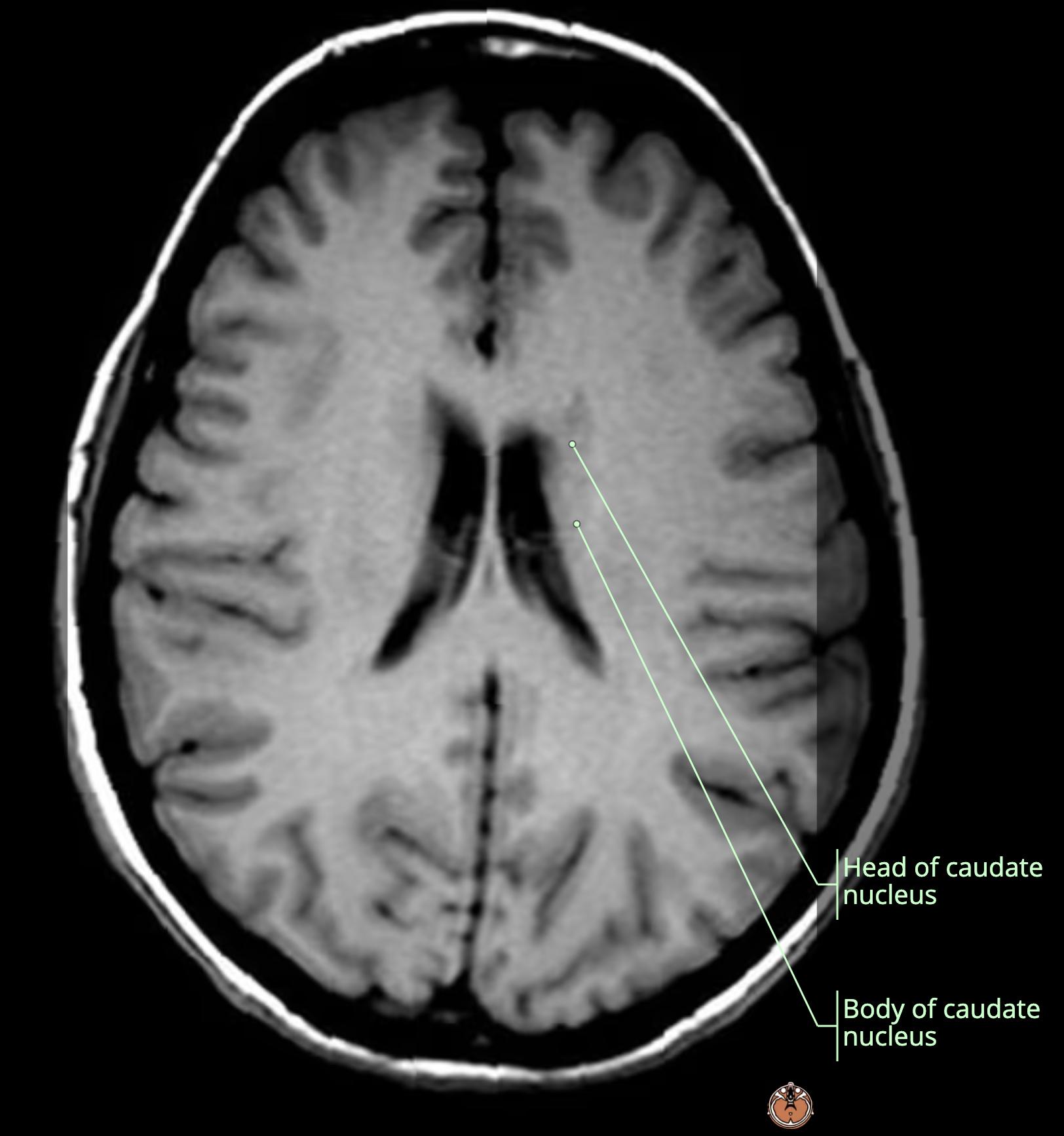
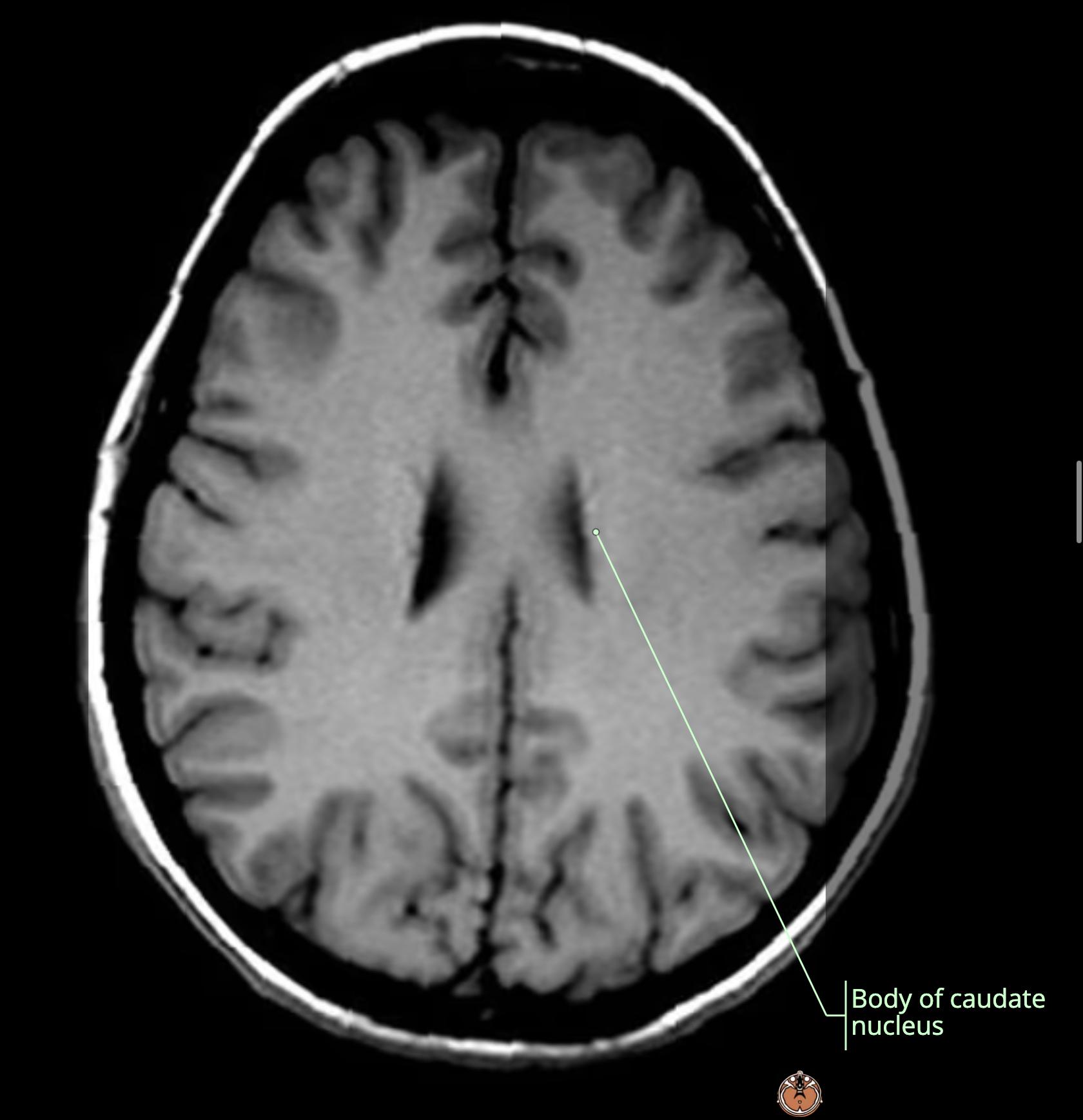
    • red: body of caudate[…] running posteriorly and laterally to the lateral ventricles[…] > down to the occipital horns where it becomes the tail of caudate nucleus[…]

    MRI Brain Anatomy
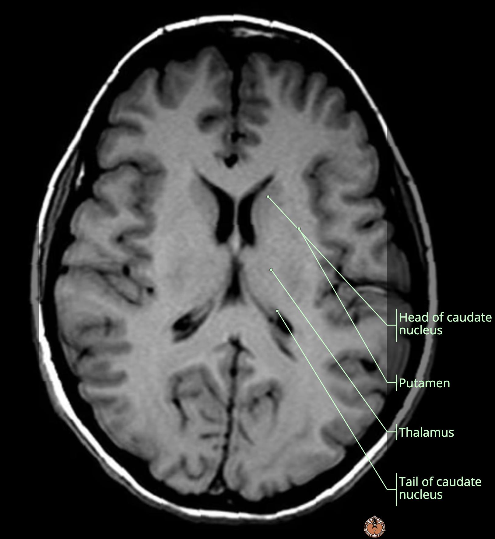
    • inferiorly we see that the putamen[…] and the head of caudate[…] start off almost touching
    • as we rise superiorly, the putamen[…] ends up more separated posteriorly with the IC[…] now splitting the 2 structures

    MRI Brain Anatomy
    • red: putamen[…]
    • orange: Gpe/i[…]
    • yellow: anterior limb/genu/posterior limb IC[…]
    • green: thalamus[…]

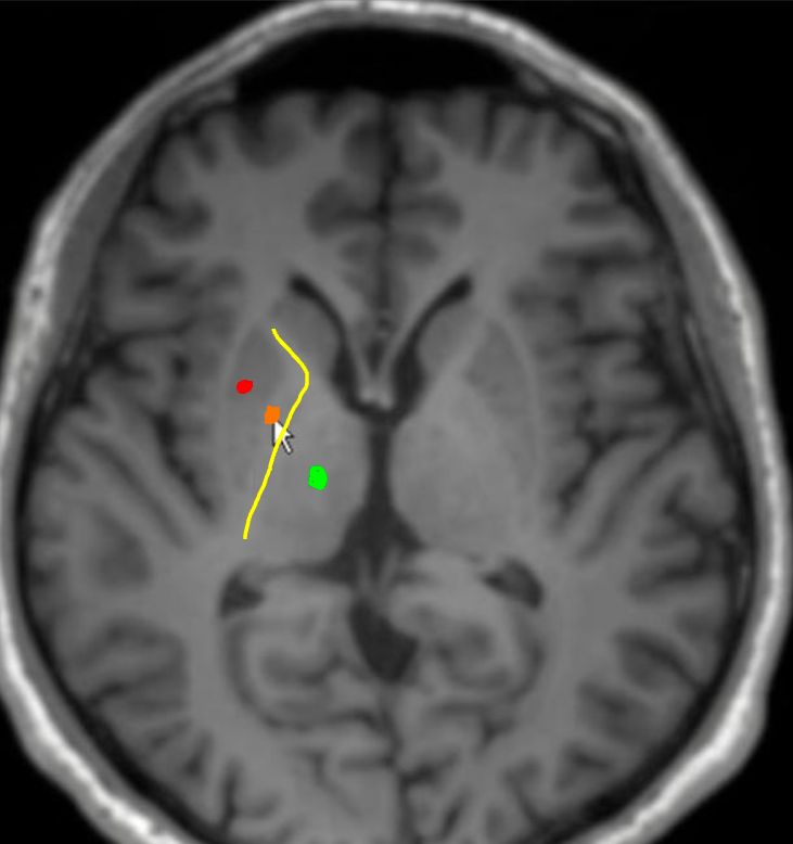
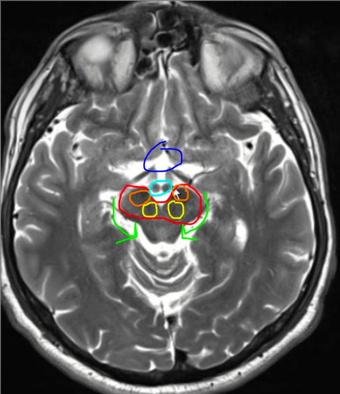
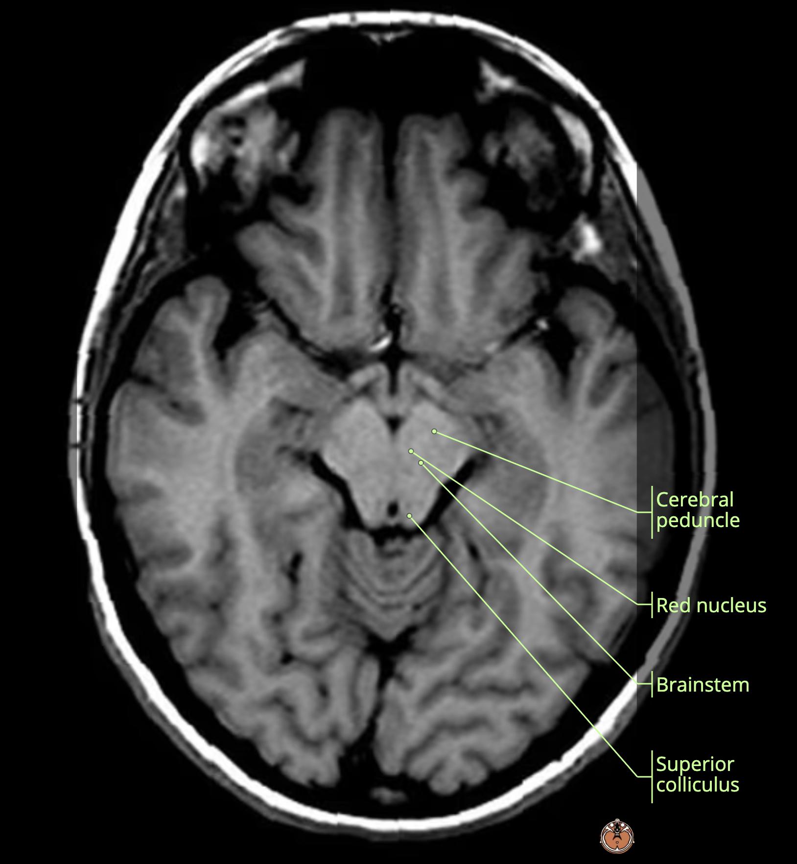
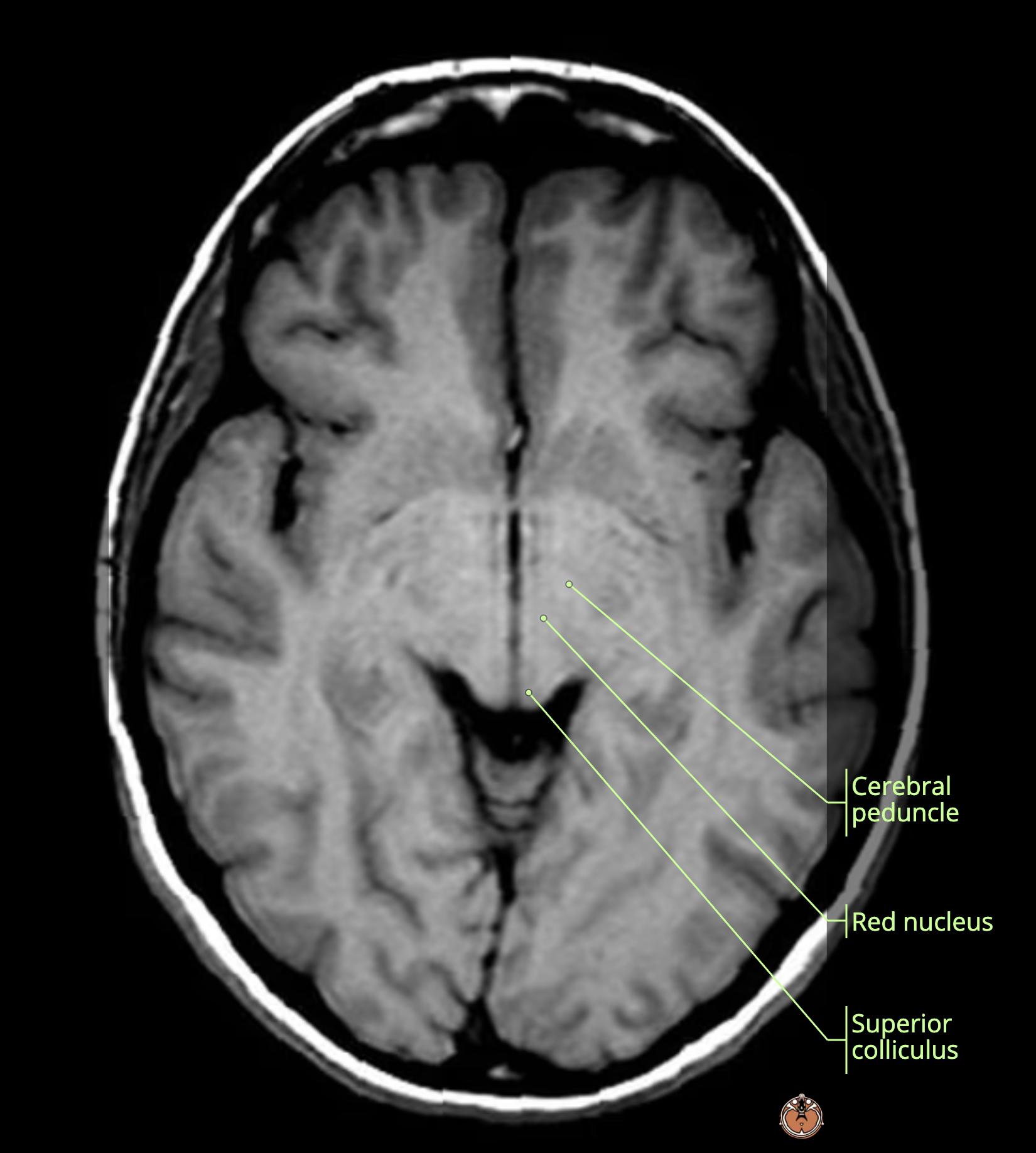
    MRI Brain Anatomy
    • red: cerebral peduncles[…] containing the 
      • orange: SN[…]
      • yellow: red nuclei[…]
    • light green: ambient cisterns[…]
    • light blue: mammillary bodies[…] which form the inferior[…] border of the hypothalamus[…]
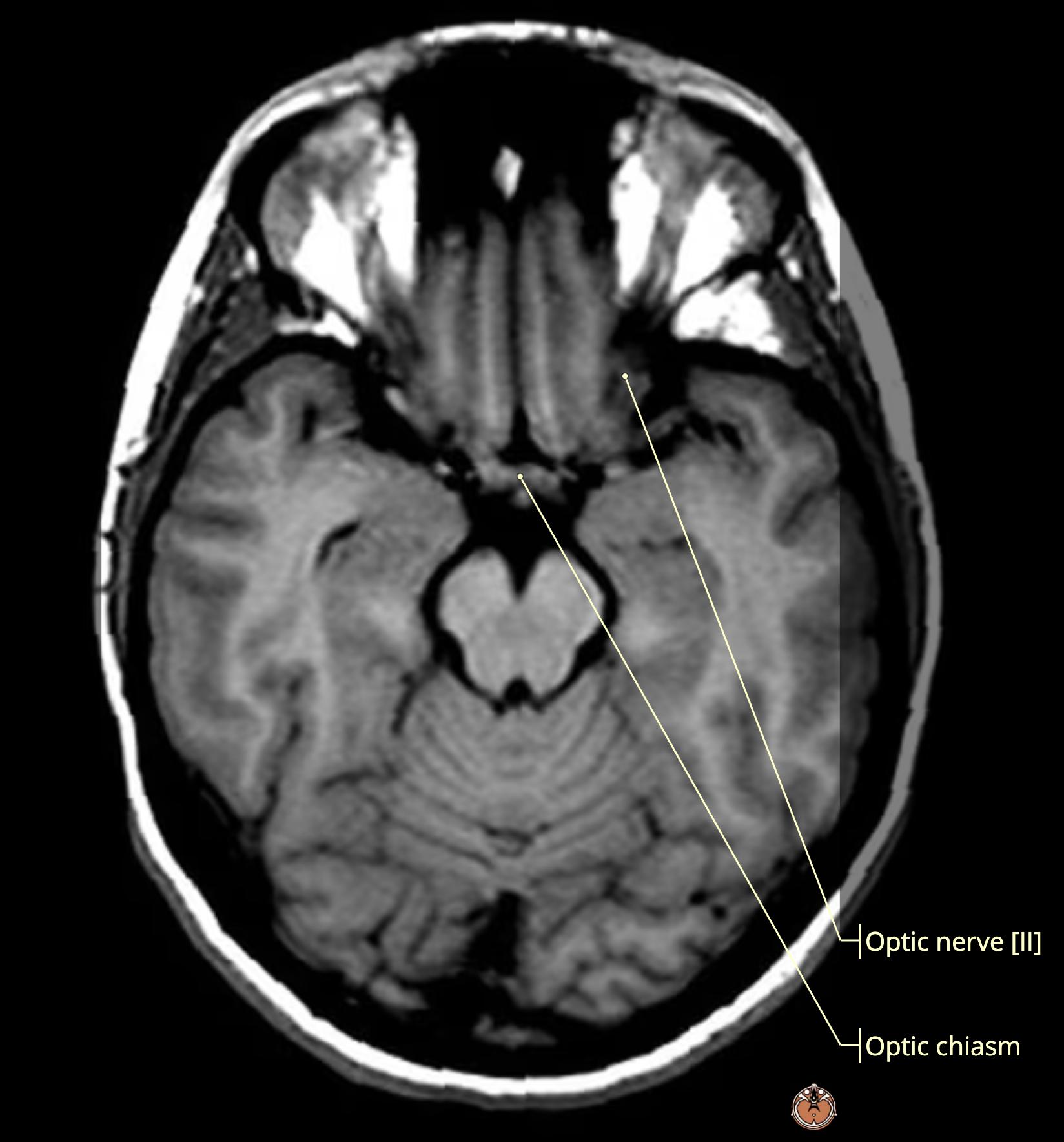
    • dark blue: anterior to the mamillary bodies[…] is the optic chiasm[…]

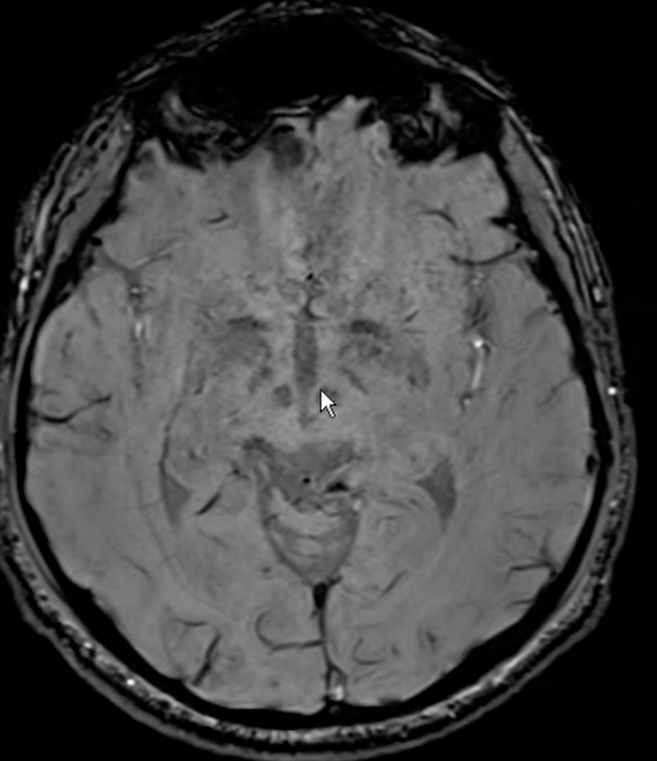
    MRI Brain Anatomy
    • SWI[…] sequence MRI is best for visualizing the substantia nigra which is DA rich

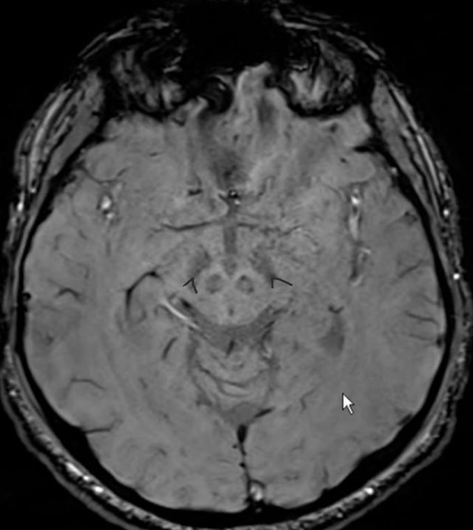
    MRI Brain Anatomy
    • normally in the SN there should be a small "split" at the posterior limb which is called the "swallow tail[…]" sign
      • this is lost in diseases such as PD[…]

    MRIb Anatomy
    • red: superior colliculi[…]
    • orange: inferior colliculi[…]
    • yellow: thalamus[…]

    MRI Brain wo Anatomy
    • this is the subthalamic nucleus[…] sitting right under the thalamus[…]

    Rapid Review 
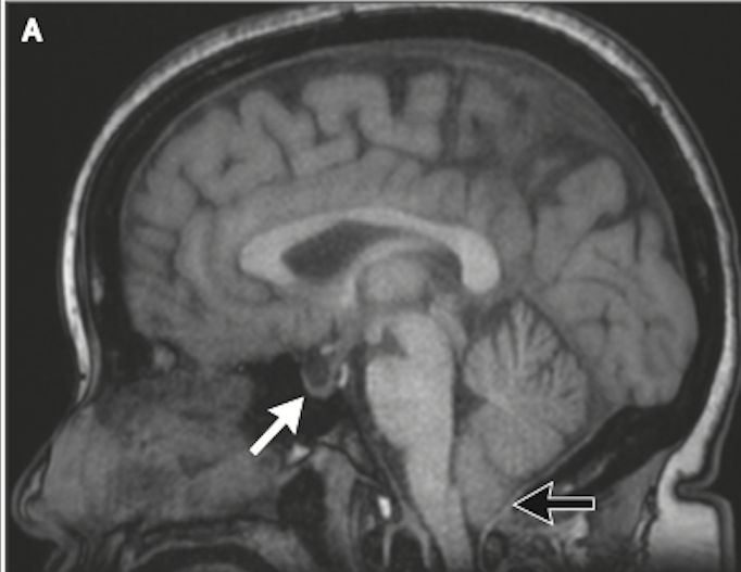
    • This MRI shows (hint: pt w/IIH)
      • white arrow: partially empty sella[…]
      • black arrow: cerebellar tonsils descent[…]

    source: dr mehendale article on IIH shared w/residents
    Rapid Review 
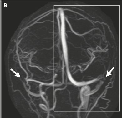
    • This MRI shows (hint: pt w/IIH)
      • white arrows: BL transverse venous sinus stenosis[…]

    source: dr mehendale article on IIH shared w/residents
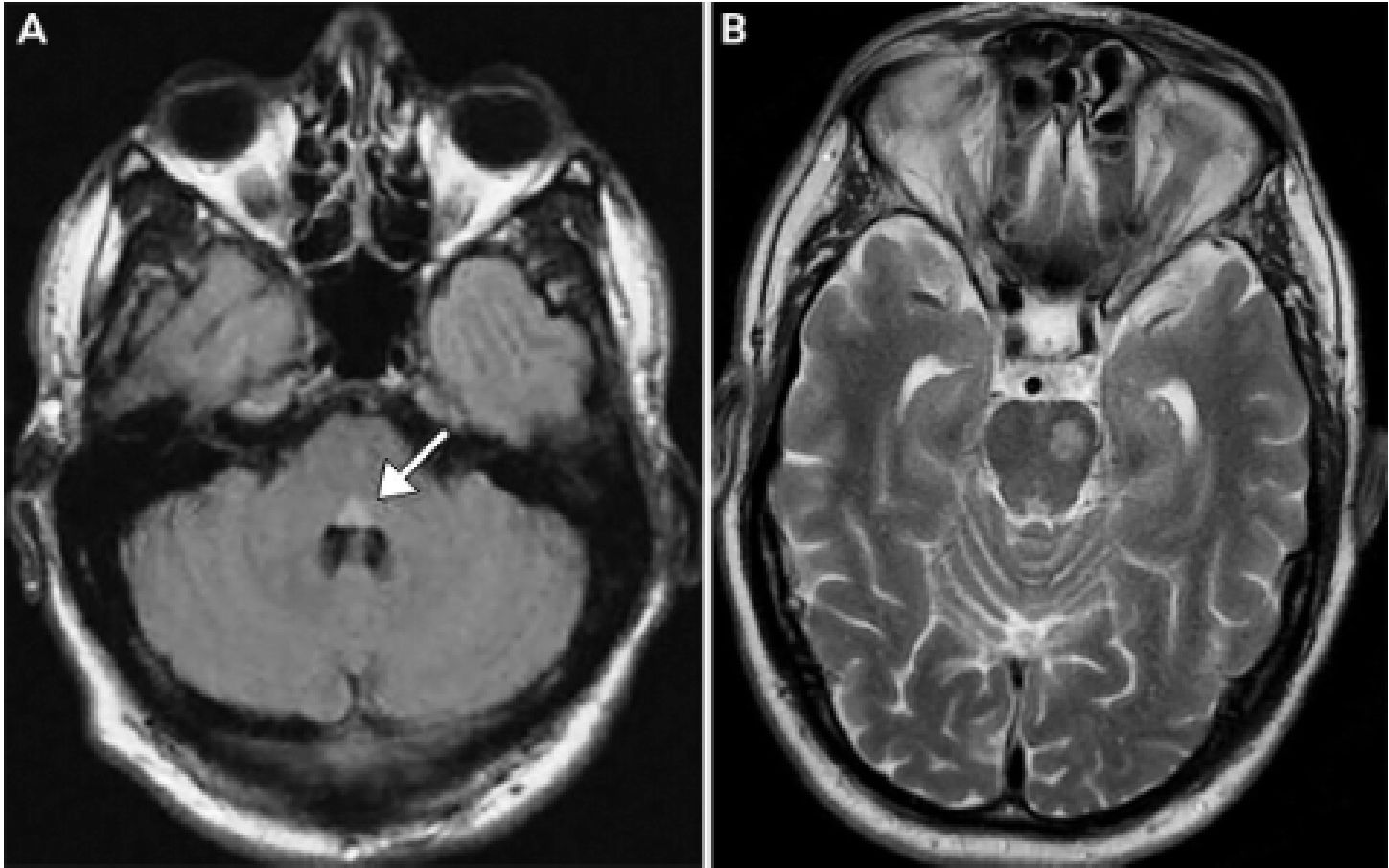
    • A: MRIb axial FLAIR sequence: shows increased signal in the dorsal pons[…]
    • B: MRIb axial T2W sequence: shows increased signal in the ventral L pons[…]