Neuro Ophthalmology

Neuro Ophthalmology

Subspecialties Β· 120 cards Β· 5 labeled figures

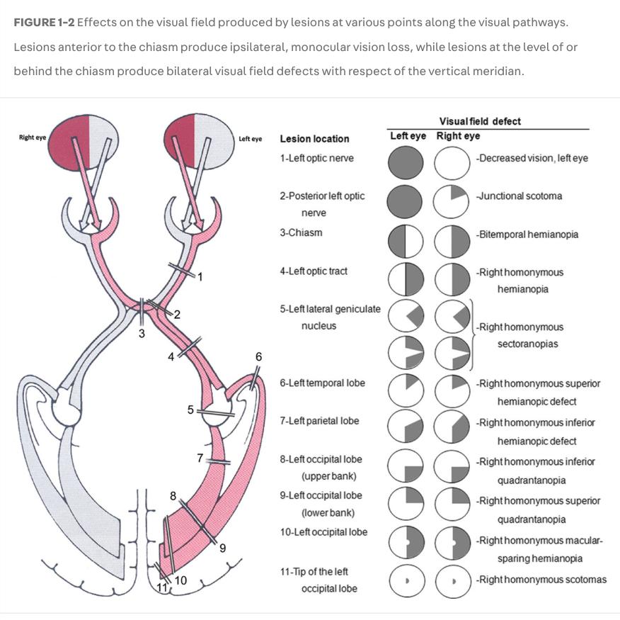
Visual Pathway Lesions - note the defect noted at each step of the pathway
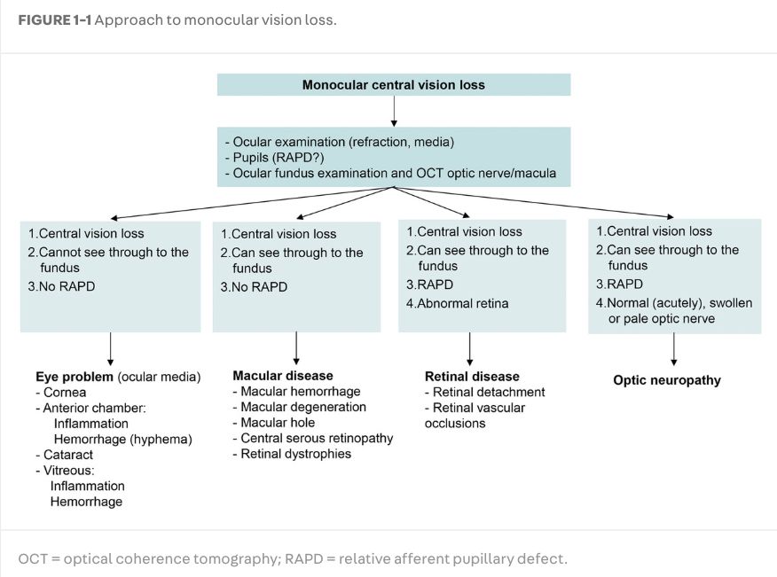
Patient with monocular vision loss.
  • First step: rule out a RAPD[…] -- if there is none: the problem is likely intra-ocular[…]
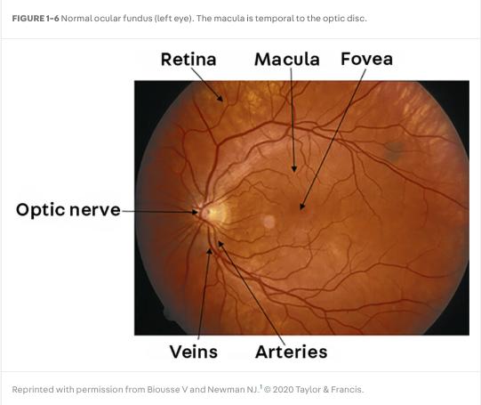
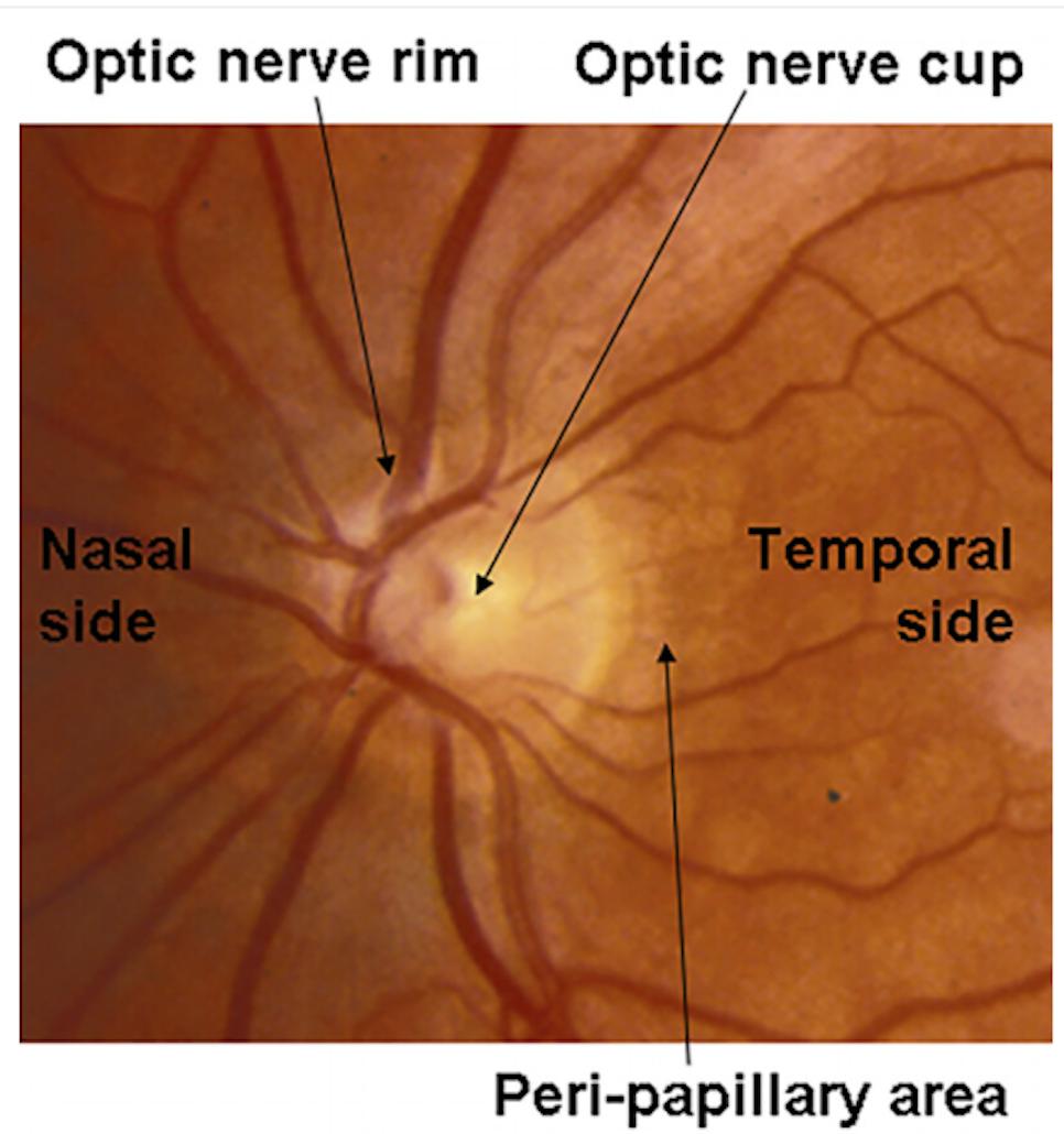
  • Second step: if there is an RAPD[…]: determine if the retina[…] versus the optic nerve[…] looks abnormal
    • if retina[…]: this is more likely retinal[…] issue (detachment, vascular occlusion, etc)
    • if optic nerve[…]: this is more likely optic neuropathy[…] (could be normal acutely, and swollen/pale later)
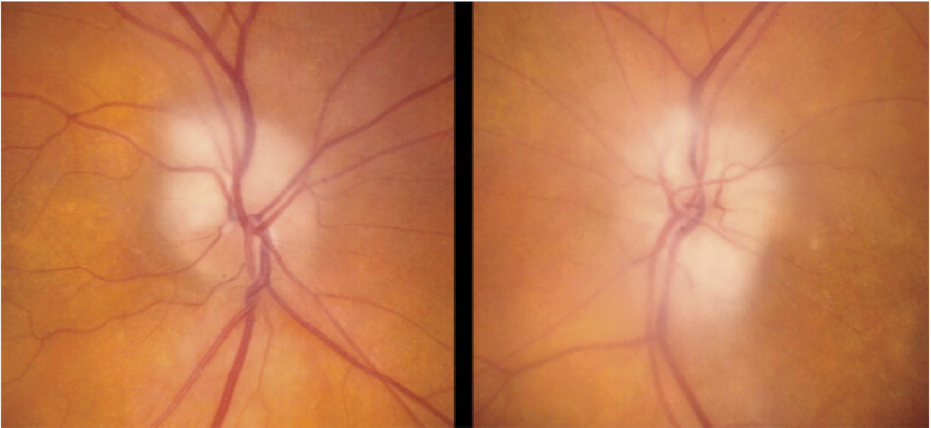
Patient with AION from GCA
This image shows pallid bilateral optic disc edema[…] in both eyes


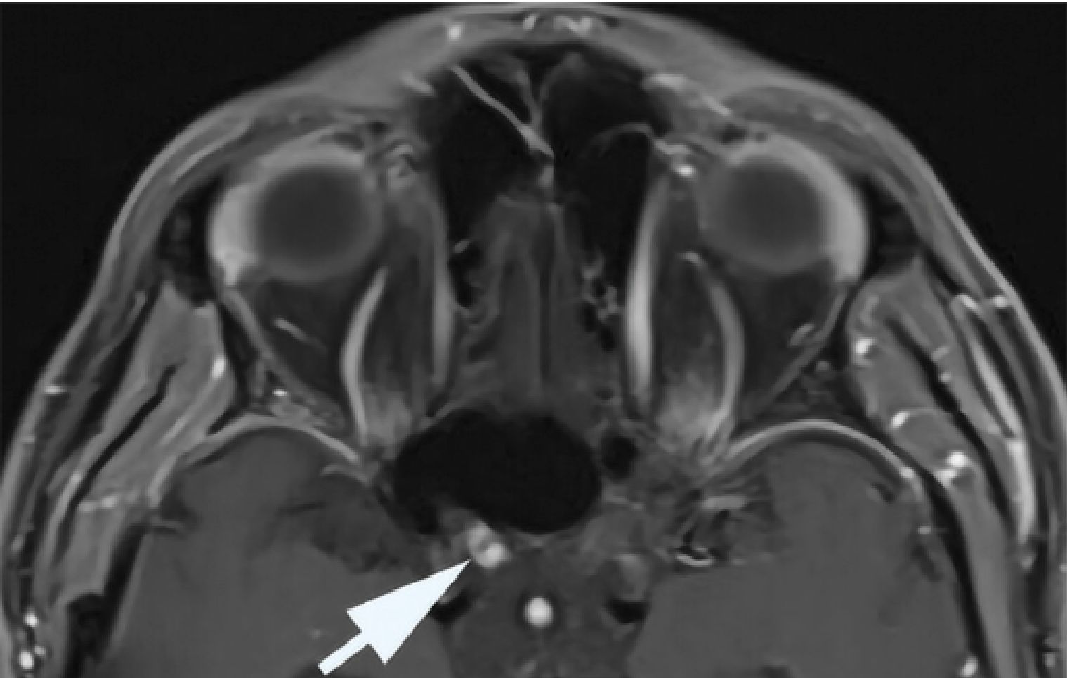
patient with radiation-induced optic neuropathy
  • MRIb/orbits axial T1W postcontrast fat suppresed sequence: shows characteristic juxtachiasmal[…] enhancement of the right optic nerve 


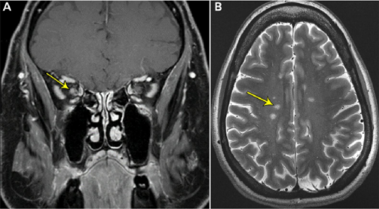
Patient with MS
  • A: MRIb coronal T1W postcontrast fat-sat: shows abnormal enhancement of the optic nerve[…]
  • B: MRIb axial T2W: shows periventricular hyperintensities many with a central vein[…] sign

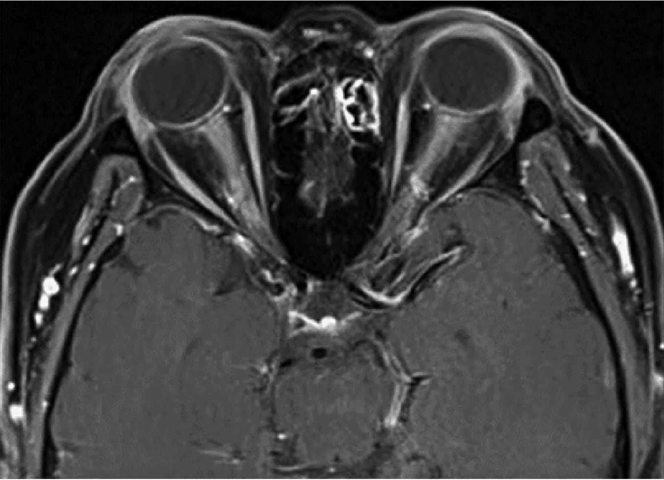
  • MRIb/orbits axial T1W postcontrast fat-suppressed sequence: shows diffuse enhancement of the optic nerves[…] bilaterally along with the perineural[…] optic nerve sheath


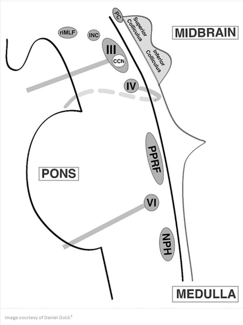
Brainstem Ocular Motor Structures (Sagittal Brainstem View)
  • Cranial Nerves:
    • CN III[…] (with midline central caudal nucleus CCN) > located in midbrain[which part of brainstem?]
    • CN IV[…] > located at midbrain-pons[which part of brainstem?] junction
    • CN VI[…] > located in pons[which part of brainstem?]
  • Neural Integrators:
    • Interstitial nucleus of Cajal[…] > for vertical[…] and torsional[…] gaze holding; located in midbrain[which part of brainstem?]
    • Nucleus prepositus hypoglossi[…] > for horizontal[…] gaze holding; located in medulla[which part of brainstem?]
  • Saccadic Burst Neurons:
    • Rostral interstitial medial longitudinal fasciculus (riMLF)[…] > for vertical[…] and torsional[…] saccades
    • Paramedian pontine reticular formation (PPRF)[…] > for horizontal[…] saccades
  • Other:
    • Posterior commissure (PC)[…] > plays a role in upward[…] gaze; located in midbrain[which part of brainstem?]

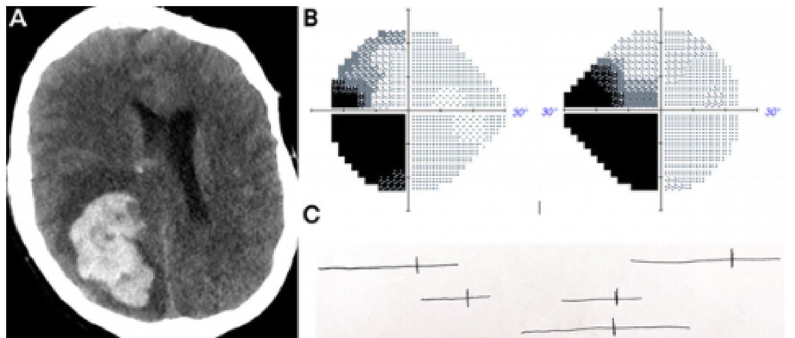
  • A: CTHwo axial sequence: shows a large R parietal[…] lobe ICH
  • B: Humphrey VIsual Field Testing: shows a left homonymous hemianopia[…] worse in the left inferior[…]>superior[…] quadrants 
  • skip C (bisection line test)
  • Diagnosis: L homonymous inferior quadrantanopia[…] with some L superior field involvement

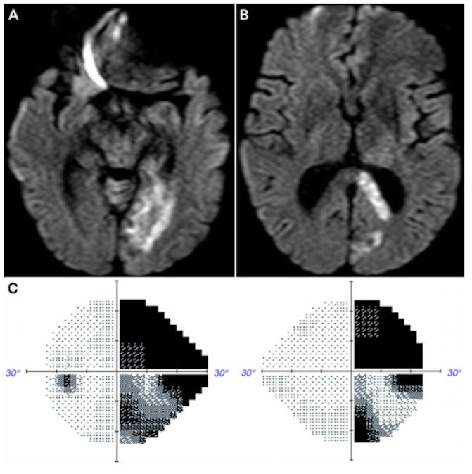
53M, hx complex migraines, p/w x1d of peripheral vision loss, dificulty concentrating, word-finding issues, dizziness
  • A/B MRIb axial DWI: shows L PCA[arterial territory?] stroke involving the L medial temporal[…] and occipital[…] lobes (A) along with the L corpus callosum[…] and forceps major[…] (B)
  • C/D: Humphrey visual field shows a homonymous hemianopia[…] worse in the R superior[…]>R inferior[…] quadrants

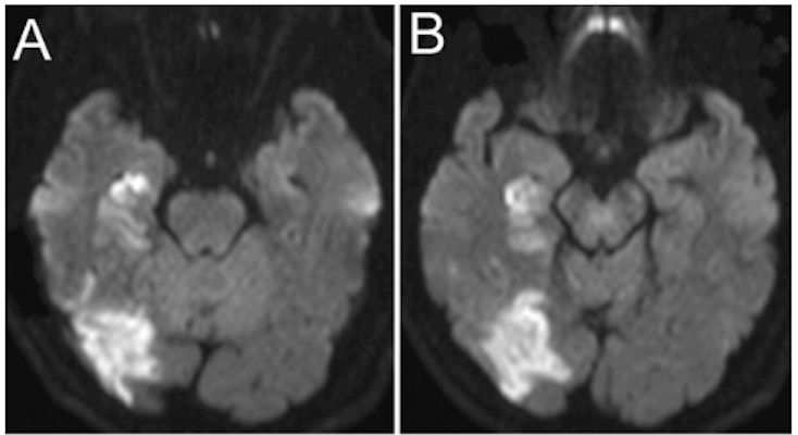
68M p/w x3h dizziness, LUE weakness, L homonymous hemianopia superior>inferior quadrants, and prosopagnosia. CTA: R P2 occlusion. s/p IVT. 
  • A/B: MRIb axial DWI sequence: shows AIS in the R occipitotemporal region involving: parahippocampal[…] gyrus, fusiform[…] face area (area V4), and occipital[…] face area 

Approach to Vision Loss - History and Exam

Most important first questions: monocular vs binocular[…] and transient vs persistent[…].

Monocular[…] β†’ problem is in the eye itself or optic nerve anterior to the optic chiasm[…]; Binocular[…] β†’ bilateral eyes/optic nerves, optic chiasm, or retrochiasm. Additional history: acuity? pain with EOM? headache?

Visual field rules: The retina and optic nerve are organized by the horizontal[…] meridian β€” lesions respecting horizontal meridian β†’ optic nerve head or RNFL[…]; The post-chiasmal visual pathway is organized by the vertical[…] meridian β€” lesions respecting vertical meridian β†’ optic chiasm or retrochiasm. Color and red saturation: compare across meridians.

Pupils: Anisocoria worse in light[…] β†’ large pupil abnormal; Anisocoria worse in dark[…] β†’ small pupil abnormal; Test for RAPD[…] with the swinging-flashlight test β€” relative difference in light transmission to both EW nuclei; positive = unilateral or asymmetric CN II dysfunction.

OCT: cross-sectional retinal layers β€” quantifies RNFL and ganglion cell[…] loss.
Transient Monocular Vision Loss
  • Vascular (most important ophthalmic symptom of carotid[…] artery disease): Transient, monocular, painless[…] partial or complete vision loss lasting minutes ("blackout" or "curtain").
    • Mechanism: thromboembolism[…] (transient BRAO/CRAO) vs hypoperfusion through stenosed carotid.
    • Workup like TIA[…]; rule out GCA[…] if >50yo.
    • If monocular vision loss + disc edema β†’ rule out AION[…].
  • TVOs (transient visual obscurations): Seconds-long blackouts/grayouts precipitated by posture changes[…]; Mechanism: high ICP β†’ papilledema β†’ disc edema β†’ pressure at optic nerve head; Workup like IIH[…] (MRI/MRV β†’ LP).
  • Painful: think (3) angle-closure glaucoma, carotid dissection, or GCA[…].
  • Uhthoff phenomenon[…]: transient vision worsening with heat or exercise in patients with prior optic neuropathy.
Persistent Monocular Vision Loss

Typical features: central[…] vision loss + RAPD Β± red color desaturation + swollen or pale optic nerve head. Exceptions: many cases of AION and ON spare central acuity.

Optic neuropathy patterns: Optic neuritis[…]: + pain with EOM, acute-subacute central vision loss; AION[…]: acute, painless (unless GCA); Compressive[…]: slowly progressive.

Maculopathy vs Optic neuropathy β€” differentiate with OCT[…]: Maculopathy: no RAPD, but metamorphopsia[…] (distortion); Optic neuropathy: no metamorphopsia, but RAPD[…].

Retinal artery occlusion (BRAO/CRAO): acute, painless, with RAPD[…].

Triage as a stroke[…].

CRVO: dilated veins, diffuse hemorrhages, cotton wool[…] spots, optic nerve head swelling.
Transient Binocular Vision Loss

Migraine with aura (most common): gradual[…] onset with scotoma[…] β†’ jagged shimmering lights β†’ headache.

Positive[…] phenomena.

Vertebrobasilar TIA (older patients): acute[…] onset, negative[…] phenomena. Mechanism: basilar + bilateral PCA ischemia.

May have headache over eyebrow contralateral[…] to visual field loss (referred via CN V1[…]). Occipital seizures: very brief (seconds) binocular visual issues.

PRES: cerebral blindness from HTN, eclampsia, or tacrolimus[…].
Persistent Binocular Vision Loss

Bilateral optic neuropathy: same as monocular except no RAPD[…].

Compressive lesion in chiasm (aneurysm, adenoma, meningioma, craniopharyngioma): typically bitemporal hemianopsia[…].

Can extend to cavernous sinus[…] (diplopia, ptosis, anisocoria, facial numbness) or cause endocrine effects (hypothalamus-pituitary axis) or obstructive hydrocephalus[…].

Retrochiasmal lesion (tract, LGN, optic radiations, occipital cortex): contralateral homonymous hemianopia[…], often PCA stroke[…]. Papilledema: disc edema from elevated ICP β€” central acuity spared until late.
Optic Neuropathies - Signs and Vasculature

Signs of optic nerve dysfunction: decreased acuity[…], visual field deficits, color[…] vision issues, RAPD[…] in affected eye.

Exam: disc edema[…] acutely β†’ pallor chronically (>6 weeks).

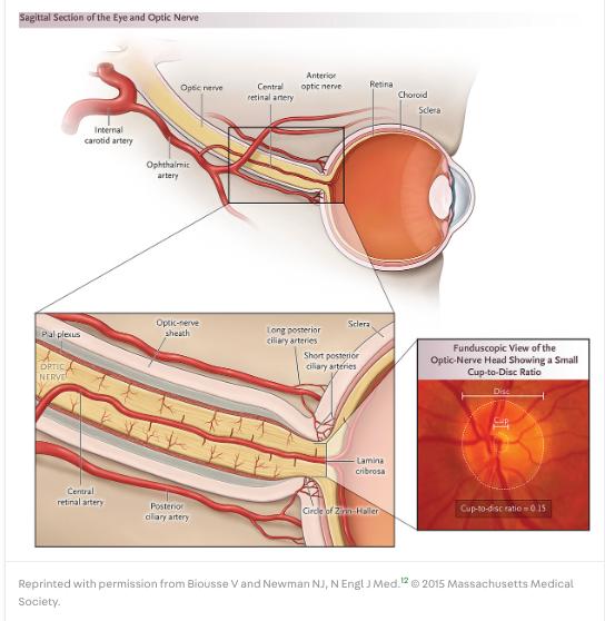
Eye blood supply: ICA β†’ cavernous sinus β†’ ophthalmic artery[…].: 1st branch: central retinal artery[…] β†’ enters optic nerve ~10mm behind eye β†’ supplies inner retina and anterior intraorbital nerve; Other branches: short posterior ciliary arteries β†’ Circle of Zinn-Haller[…] β†’ supplies optic disc in centripetal/segmental pattern (hence altitudinal field loss with infarcts).
Anterior Ischemic Optic Neuropathy (AION)

Anterior (AION): infarct at optic disc[…] level β†’ always has disc edema.

Arteritic AION (GCA): Like NAION but more severe[…]. Systemic symptoms (myalgias, weight loss, fatigue) and cranial symptoms (HA, jaw claudication, scalp tenderness). Pallid disc edema is the hallmark.

Labs: ESR, CRP, platelets[…] elevated.

Temporal artery biopsy[…] confirms; if negative but suspicion high β†’ TCD.

Treatment: immediate IVMP 1g daily[…] even before biopsy to prevent vision loss within days-weeks.

Consider tocilizumab[…] (or MTX) maintenance.

Nonarteritic AION (NAION): Acute, unilateral, painless[…] vision loss + RAPD + segmental disc edema β†’ pallor >6 weeks.

Edema/pallor resolves by 8 weeks[…]. OCT shows RNFL defects.

Rule out GCA[…] if >50yo; look for vascular risk factors and meds (sympathomimetics, PDE-5[…] inhibitors).

Treatment: none[…] restores vision β€” focus on risk factor management. Posterior (PION): infarct behind eye β†’ normal disc at presentation.

Common causes: dissection, hypotension, or vasculitis[…]. Rule out GCA if >50yo; MRI to exclude other retrobulbar causes.
Optic Neuritis - MS vs NMOSD vs MOGAD

Presentation: decreased acuity, dyschromatopsia (red desaturation)[…], RAPD, pain with EOM, MRI showing optic nerve enhancement[…].

May have Uhthoff phenomenon[…].

MS optic neuritis: typically <50%[…] optic nerve length enhancement, retrobulbar[…] location.

AQP4+ NMOSD optic neuritis: typically longitudinally extensive (>50%)[…] enhancement, posterior[…] predominant. Vision typically worse than MS, less disc edema, poor prognosis.

Often with longitudinally-extensive transverse myelitis or area postrema[…] syndrome (n/v/hiccups).

MOGAD: typically bilateral with longitudinally-extensive anterior[…] optic nerve involvement, often with perineural tissue[…] enhancement and disc edema (unlike MS/NMOSD); rare chiasm involvement. Lesions more likely to resolve. MOG titer often reverts to undetectable after the initial attack. Diagnostics: serum AQP4/MOG antibody in all first-presentation ON patients. MRI brain/orbits Β± spine.

LP for kappa free light chains[…] >0.1 mg/dL or OCBs.
Optic Neuritis - Treatment

MS optic neuritis (per the Optic Neuritis Treatment Trial (ONTT)[…]): IVMP[…] followed by PO prednisone taper β€” accelerates vision recovery but does not change ultimate acuity.

Clinically dose 1g daily Γ— 3-5 days[…] β†’ taper.

AQP4/NMOSD: IVMP Γ— 5 days[…] β†’ longer PO prednisone taper (~6 weeks); If minimal IVMP response β†’ PLEX[…] early or first-line (common need given aggressive nature); Maintenance: rituximab (off-label), complement inhibitors (eculizumab, ravulizumab, satralizumab, inebilizumab[…]), or off-label tocilizumab; Note: some MS DMTs (IFNΞ², fingolimod, natalizumab[…]) worsen[…] NMOSD. MOGAD: IVMP 1g Γ— 3-5 days β†’ taper similar to NMOSD.

Most cases resolve, often monophasic[…]. PLEX/IVIG reserved for non-responders. Long-term PPX in relapsing cases: monthly IVIG, rituximab, or other immunotherapies.
Optic Neuritis - Other Inflammatory and Infectious

Sarcoidosis: multisystem disease with noncaseating[…] granulomas. PET to identify accessible LN biopsy targets (cervical, mediastinal). Treat steroids β†’ TNF inhibitors.

GPA (granulomatosis with polyangiitis): small-vessel PR3/c-ANCA[…] vasculitis + necrotizing[…] granulomas. Triad: upper resp + lower resp + kidney involvement, Β± ocular (uveitis/scleritis + optic neuropathy), Β± mononeuritis multiplex. Treat steroids β†’ immunotherapy. GFAP astrocytopathy: meningoencephalitis Β± optic neuropathy.

Diagnose GFAP-IgG[…] in serum + CSF.

MRI: linear, perivascular radial[…] enhancement.

CRMP-5 (CV2) optic neuropathy: paraneoplastic, associated with SCLC or thymoma[…]. Treat steroids + tumor.

Ocular TB: usually bilateral[…], spreads contiguously dura β†’ optic nerve. Can cause meningitis with papilledema. Diagnose TST + IFN-Ξ³ assay.

Treat RIPE[…]; ethambutol is oculotoxic.

Bartonella neuroretinitis: cat-scratch with fever, lymphadenopathy, possible disc edema and macular star[…]. Often self-resolves. Ocular syphilis (any stage): lymphocytic meningitis + radiculitis + cranial neuropathy + ocular symptoms.

Treat per ID (IV PCN G[…]). Rhino-orbital-cerebral fungal (Mucor, Aspergillus): immunocompromised β†’ paranasal sinus β†’ orbital apex β†’ skull base β†’ cavernous sinus β†’ brain.

Early ENT[…] consult for biopsy.
Hereditary, Toxic, and Compressive Optic Neuropathies

Hereditary optic neuropathies: mitochondrial[…] dysfunction β†’ retinal ganglion cell injury.

LHON (most common): acute-subacute unilateral[…] central vision loss β†’ eventually involves the other eye. Often initially hyperemic pseudoedematous discs β†’ temporal pallor, RNFL/GCL loss. 3 common mtDNA variants (e.g., m.11778G>A, m.14484T>C).

OPA-1 (Dominant Optic Atrophy): chronic bilateral[…] central vision loss. No treatment.

Toxic/nutritional optic neuropathy: chronic bilateral central vision loss, scotomas[…], rare disc edema. Treat by removing toxin and treating deficiencies.

Classic causes include B12 deficiency, ethambutol, methanol[…]. Compressive/infiltrative: meningiomas, craniopharyngiomas, pituitary adenomas, gliomas, mets.

Chronic painless vision loss; can be acute if aneurysm, pituitary apoplexy, or aspergilloma[…].

Radiation-induced optic neuropathy: total dose >50[…] Gy, progressive irreversible vision loss 3 months to 9 years post-treatment.

Traumatic optic neuropathy: get CTH/Max-Face[…] to rule out fractures.
Papilledema - Pathophysiology and Symptoms

Pathophys: high ICP β†’ transmitted from subarachnoid space via optic nerve sheath[…] (axoplasmic stasis[…]) β†’ optic nerve head edema.

Other causes of high ICP: malignant HTN, severe anemia, drugs (vitamin A derivatives, tetracyclines, lithium[…]).

Symptoms: Headache worse in AM, lying flat, or Valsalva[…] (migraine-like features make this a mimic); Tinnitus (nonspecific) from transverse sinus stenosis[…] β†’ turbulent flow ("whoosh"); Bilateral TVOs[…] precipitated by posture/strain; Horizontal binocular diplopia from CN VI[…] compression; Absence of spontaneous venous pulsations[…]; Dyschromatopsia[…] = sign of vision-threatening severity.

Grading: use the FrisΓ©n[…] scale (0 = normal, 5 = complete obscuration of vessels). Moderate-to-severe β‰₯3. Pseudopapilledema: optic disc edema not from high ICP (drusen, masses, myopia, myelinated fibers, congenital).

Differentiate with OCT[…].
Papilledema - Diagnostics and Treatment

Imaging: MRI brain + MRV + MR orbits with and without contrast[…] β†’ CT if MRI unavailable.

Exclude CVST, masses, hydrocephalus, meningeal process[…].

MR orbits signs: flattening of posterior globes, optic nerve tortuosity, large optic nerve sheath, optic nerve enhancement, intraocular protrusion of optic nerve head[…].

MR brain signs: empty/partially empty sella, decreased pituitary height, enlarged Meckel's cave, tonsillar ectopia, slitlike ventricles[…].

LP (lateral decubitus or fluoro prone with legs straight): >25[…] cm H2O abnormal in adults; >28 in kids.

IIH treatment (per the IIH Treatment Trial): Reduce CSF secretion: acetazolamide (Diamox) or topiramate[…] (carbonic anhydrase inhibitors); off-label furosemide, amiloride, spironolactone; Weight loss[…] Β± GLP-1 agonists; Lumbar drain as temporizing measure; Surgical options: optic nerve sheath fenestration (ONSF)[…] for vision-threatening cases without HA; CSF diversion (LP shunt or VPS)[…]; venous sinus stenting (requires DAPT[…] postprocedurally). CVST treatment: AC with LMWH Β± endovascular (fibrinolysis/thrombectomy) β†’ long-term DOAC or warfarin.

Avoid steroids[…].
Diplopia - Terminology

Phoria vs tropia: "Phoria[…]" present only when one eye is covered (latent); "Tropia[…]" present with both eyes open; Eso/exo/hyper/hypotropia: eyes deviate toward nose, away from nose, upward, downward.

Monocular vs binocular: Monocular[…] diplopia persists with one eye closed β†’ usually ocular (refractive)[…] cause; Binocular[…] diplopia resolves with one eye closed β†’ usually neurologic[…] cause (eyes misaligned, brain sees two images). Saccades vs smooth pursuit: saccades are very fast.

Distinguish true INO[…] (adducting saccade slow throughout) vs pseudo-INO[…] (slow only at the end) β€” pseudo-INO is classic for MG[…].
Myasthenia Gravis Ocular Signs

MG ocular signs: Fatigueable ptosis[…] with prolonged upgaze; Cogan eyelid twitch[…]: brief upward twitch on returning to primary from 5-10s of downgaze; Enhancement of ptosis[…] with contralateral manual eyelid elevation; Improvement with ice pack[…] application for ~2 minutes. MG can mimic INO (pseudo-INO) including bilateral; differentiate by: Convergence preserved in INO (bypasses MLF); Adducting saccade slow at the end only in MG.
Diplopia - Localization Framework

Supranuclear (projections to CN nuclei): toxic/metabolic or neurodegenerative. Skew deviation[…] (vertical misalignment from utricle/vestibular asymmetry) typically from brainstem or cerebellar insult.

Internuclear (INO): MLF[…] lesion strips myelin β†’ can't coordinate ipsilateral CN VI with contralateral CN III.

When looking to side of lesion β†’ can't adduct[…] the contralateral eye + horizontal nystagmus[…] in abducting eye.

Convergence[…] is intact

Nuclear: CN III nucleus β†’ ipsilateral CN III palsy + contralateral supraduction deficit[…] (SR subnucleus decussates; central caudal subnucleus projects to both LPS); CN IV nucleus (dorsal brainstem) β†’ contralateral CN IV palsy[…], Β± ipsilateral Horner[…] from adjacent oculosympathetic fibers; CN VI nucleus β†’ ipsilateral abduction weakness Β± ipsilateral gaze palsy (PPRF nearby) Β± ipsilateral peripheral CN VII palsy[…] (facial colliculus).
CN III Palsy

Findings: ipsilateral ptosis, adduction, elevation, and depression[…] weakness Β± dilated, nonreactive pupil if postganglionic PS fibers hit.

Combined: "down-and-out, fixed/dilated[…]" pupil.

Pupil-sparing vs pupil-involving: PS fibers travel along the outer edge[…] of CN III; Compressive[…] lesions (tumor, aneurysm) β†’ pupil-involving; Microvascular ischemia[…] (DM, HTN) β†’ affects central portion β†’ pupil-sparing.

Diagnostics: CTA/MRA[…] to rule out Pcomm aneurysm[…]; MRI to rule out stroke/tumor/inflammation; DSA case-by-case.
CN IV and CN VI Palsies

CN IV palsy: SO knockout β†’ can't fully intort or depress[…] eye.

Eye drifts up[…] and out[…], needs head tilt[…] to compensate for lost intorsion.

Mnemonic β€” "CN IV GOTS[…] worse": Gaze Opposite, Tilt Same.

Hypertropia worse on gaze toward opposite[…] side and on head tilt toward[…] affected side (excyclotorsion = outward rotation at baseline).

Parks-Bielschowsky 3-step test: affected eye hypertropic[…], worsened on gaze toward opposite side, and worsened on head tilt toward affected side.

CN VI palsy: can't abduct[…] affected eye.

Long course (pontomedullary jxn β†’ prepontine cistern β†’ over petrous ridge β†’ Dorello canal β†’ cavernous sinus β†’ SOF/orbital apex) makes it a falsely localizing[…] sign from elevated ICP or trauma.

Microvascular[…] ischemia (HTN/DM) can cause this, usually self-resolves; MRI brain/orbits for workup.
Multiple CN Palsies and NMJ/Orbital Disorders

Multiple CN palsy DDX: neurosarcoidosis, LMM, lymphoma, Tolosa-Hunt (cavernous sinus, painful), MFS (anti-GQ1b), or orbital apex syndrome[…] (CN II/III/IV/V1/VI).

NMJ disorders: MG[…]: pseudo-INO; many ocular cases are seronegative β†’ get single-fiber EMG[…] of frontalis or orbicularis oculi; LEMS[…]: rarely causes diplopia; Tick-bite disorders or botulism[…].

Orbital diseases (thyroid eye disease, IgG4 disease, GCA): in TED rule out EOM involvement with acronym IM SLO[…] (inferior > medial > superior > lateral > obliques).

Myopathies: oculopharyngeal MD (PABPN1), CPEO[…] (mitochondrial).
Supranuclear Gaze - Cell Types and Pathways

Cell types: Omnipause[…] neurons (e.g., nucleus raphe interpositus): tonically inhibit burst neurons to prevent unwanted saccades.

Dysfunction β†’ saccadic intrusions[…] (square-wave jerks, opsoclonus).

Burst[…] neurons in PPRF (horizontal) and riMLF[…] (vertical/torsional): generate saccade pulse. Motor neurons of CN VI, III, IV project to EOM.

Interneurons connect via the MLF[…] for conjugate movements.

Hemispheric control: saccades are contralateral[…]; smooth pursuit is ipsilateral[…].

Functional consequences: Omnipause dysfunction β†’ square-wave jerks/opsoclonus; Neural integrator dysfunction (nucleus prepositus hypoglossi, medial vestibular) β†’ gaze-evoked nystagmus[…]; Cerebellar flocculus lesions β†’ downbeat[…] nystagmus and gaze-evoked nystagmus; Cerebellar nodulus β†’ periodic alternating[…] nystagmus.
Horizontal Saccade Pathway

Pathway: FEF[…] (frontal eye fields) β†’ contralateral PPRF[…] in dorsal pons β†’ ipsilateral CN VI nucleus β†’ ipsilateral MLF β†’ contralateral CN III nucleus. Alternative route: FEF β†’ caudate β†’ SN β†’ superior colliculus β†’ PPRF. PEF projects through superior colliculus β†’ PPRF. Basal ganglia maintains balance between voluntary and reflexive saccades.

Neural integrator for horizontal gaze: nucleus prepositus hypoglossi[…] + medial vestibular nuclei.

Disorders by site: Cortex (FEF, PEF)[…]: eyes deviate ipsilaterally; overcome with horizontal VOR; Β± cortical signs; PPRF[…]: eyes deviate contralaterally; selective loss of ipsilateral horizontal saccades; preserved horizontal VOR (vestibular fibers bypass PPRF); CN VI[…]: eyes deviate ipsilaterally; all classes affected (saccades, smooth pursuit, VOR); may see ipsilateral peripheral CN VII palsy; INO[…]: MLF lesion β†’ impaired adduction on contralateral gaze + dissociated nystagmus in abducting eye; convergence spared; Bilateral INO β†’ large exotropia[…] β†’ horizontal diplopia in all directions; convergence spared; One-and-a-Half[…] syndrome: ipsilateral CN VI nucleus + ipsilateral MLF β†’ only contralateral eye abduction preserved; Β± "Eight-and-a-Half[…]" with CN VII fascicle.

Hypometria (undershoot): cortex[…].

Hypermetria (overshoot): cerebellum[…].

Acute thalamic ICH β†’ "wrong-way deviation[…]" β€” contralateral gaze deviation from transient MR disruption (pseudoabducens palsy).
Vertical Saccade Pathway and Parinaud
  • Pathway: vertical saccade signals from both hemispheres β†’ riMLF[…] at mesodiencephalic junction β†’ bilateral elevator (SR, IO) + ipsilateral depressor (IR, SO) nuclei.
    • Bilateral riMLF lesions (e.g., from posterior thalamic/subthalamic artery stroke[…]) β†’ abnormal vertical saccades.
  • Neural integrator for vertical gaze: interstitial nucleus of Cajal (INC)[…] in the rostral midbrain.
    • Output through posterior commissure[…] to CN III/IV and contralateral INC. INC lesions β†’ impaired vertical gaze holding and torsional nystagmus.
  • Parinaud (Dorsal Midbrain) Syndrome: damage to posterior commissure[…] from pineal tumors, midbrain stroke, or noncommunicating hydrocephalus from a dilated 3rd ventricle.: Limited upward eye movements + tonic sustained downgaze ("setting sun[…]" sign); Mid-dilated pupils with light-near dissociation[…] (pretectal nuclei); Collier sign[…] (eyelid retraction in primary gaze); Convergence-retraction nystagmus[…] with attempted upgaze.
  • PD vs PSP ocular: PD: blepharospasm (eyelid spasms)[…], decreased blink rate, eyelid opening apraxia, dry eyes, convergence insufficiency; PSP: early ocular involvement β€” slowing of vertical saccades[…] β†’ supranuclear vertical[…] gaze paresis (eventually complete ophthalmoplegia), square-wave jerks.
Smooth Pursuit and VOR

Smooth pursuit pathway: retina β†’ ipsilateral[…] temporo-parietal-occipital junction & FEF β†’ ipsilateral dorsolateral pontine nucleus β†’ decussates to contralateral cerebellum (flocculus[…]) β†’ CN VIII vestibular nuclei β†’ decussates again β†’ ipsilateral CN VI nucleus.

End result: smooth pursuit control is ipsilateral[…] (vs saccades which are contralateral).

Disorders: Symmetric smooth pursuit loss: nonlocalizing[…] (toxic-metabolic, brainstem/cerebellar dysfunction, BG disorders); Asymmetric[…] smooth pursuit loss: localizes to ipsilateral pathway dysfunction.

VOR: keeps gaze stable during head motion.: Horizontal: semicircular canals β†’ CN VIII nuclei via MLF β†’ ipsilateral CN III + contralateral CN VI; Test with the head impulse[…] test: if gaze stays on target = normal; if catch-up saccade[…] back to target = abnormal.
Skew Deviation and HINTS

Skew deviation: disrupted otolith (utricle)[…] signal (labyrinthitis, demyelinating/ischemic disease) β†’ through dorsal brainstem β†’ to INC[…] β†’ vertical binocular misalignment.

Unlike CN IV lesions, skew deviation does not[…] have excyclotorsion (outward eye rotation).

HINTS exam (for AVS β€” acute vestibular syndrome): Head Impulse[…] Test: peripheral[…] lesion shows catch-up saccade; no catch-up saccade implies central[…] lesion; Nystagmus[…]: unidirectional typically peripheral; multidirectional usually central; Test of skew[…]: skew deviation present usually central.
Nystagmus Subtypes

Oscillopsia: stationary objects appear to move (resolves with eyelid closure); vertigo[…] persists despite closure.

Acute vestibular disorders can have both.: Sitting oscillopsia: spontaneous nystagmus independent of head movement; Walking oscillopsia: dependent on head movements (bilateral vestibular nuclei loss[…] β†’ no VOR β†’ world "bounces"). Ocular bobbing: downward/upward jerk with slow return to baseline.

Coma, often with impaired horizontal movements due to toxic/metabolic/medication encephalopathy affecting the pons[…].

Saccade slowing: horizontal β†’ PPRF or pons[…]; vertical β†’ riMLF or midbrain[…].

Saccadic intrusions: Square-wave jerks[…]: have intersaccadic intervals; common in asymptomatic aging but in young + gait ataxia + gaze-evoked/downbeat nystagmus β†’ posterior fossa lesion or Friedreich ataxia[…].

In older patient with bradykinesia β†’ PD[…]. Ocular flutter: horizontal plane only.

Opsoclonus[…]: all planes β€” infection or paraneoplastic in adults; neuroblastoma[…] (opsoclonus-myoclonus) in infants.
Nystagmus - Specific Types

BPPV: triggered by positional changes.: Posterior canal[…] (most common): Dix-Hallpike β†’ mixed upbeat/torsional nystagmus toward affected ear; Horizontal canal[…]: supine roll test β†’ horizontal nystagmus.

Vestibular neuritis: acute onset, beats contralateral[…] to lesion side, unidirectional, intensifies as patient looks toward fast-phase side. Physiologic endpoint nystagmus: normal in far lateral gaze, small amplitude, fatigues quickly; disappears at 75% mark.

Downbeat[…] nystagmus: cerebellar flocculus/tonsils[…] dysfunction. Progressive imbalance, vertical oscillopsia, gaze-evoked nystagmus in lateral gaze.

Treatment: trial dalfampridine (4-AP)[…]; avoid bifocals (forces downgaze).

Periodic alternating[…] nystagmus: spontaneous horizontal nystagmus reversing direction every minute or two ("null period").

Cerebellar nodulus/uvula[…].

Treatment: baclofen[…].

Pendular nystagmus: often seen with MS[…]; heavy posterior fossa/brainstem paramedian involvement.

Seesaw[…] nystagmus: one eye rises and incycloducts while the other depresses and excycloducts. Mesodiencephalic junction or optic chiasm area lesions.

Upbeat nystagmus: midline brainstem (e.g., Wernicke's encephalopathy[…]).

Treat empirically with IV thiamine[…]. Oculopalatal tremor: presents with oscillopsia or imbalance months after a posterior fossa insult (cavernoma, surgery).

Hits the Guillain-Mollaret triangle[…]. Pathway: inferior olive β†’ contralateral cerebellar dentate β†’ wraps around red nucleus β†’ down via central tegmental tract β†’ back.

MRI: T2 FLAIR hyperintensity of the inferior olivary nuclei[…].
Mydriasis - Unilateral Causes

Unilateral mydriasis (dilated, sluggishly reactive pupil) β†’ anisocoria worse in light[…].

CN III palsy: injures preganglionic[…] oculoPS fibers from EW nucleus. Poor response to near accommodation too.

Tonic pupil: injures postganglionic[…] oculoPS at ciliary ganglion or short ciliary nerves.

Causes: infectious (VZV, syphilis[…]), autoimmune (GBS, MFS), metabolic (DM), iatrogenic.: Acutely: dilated, sluggishly reactive, poor near response; Eventually: light-near dissociation[…] β€” sluggish to light, good near response; Test with dilute pilocarpine[…] β€” constricts tonic pupil but not normal (cholinergic supersensitivity); Adie syndrome[…]: tonic pupil + diminished muscle stretch reflexes.

Iatrogenic mydriasis: anticholinergics (scopolamine patch, ipratropium DuoNebs[…]) or sympathomimetics (epinephrine, phenylephrine, cocaine).

Mechanical mydriasis: iris sphincter damage β†’ dilated, sluggish, irregular pupil; will not[…] constrict to pilocarpine (unlike neurologic causes).
Bilateral Mydriasis and Light-Near Dissociation

Bilateral mydriasis causes: Bilateral tonic pupil[…]: autoimmune autonomic ganglionopathy or paraneoplastic; Bumke[…] pupils: high anxiety in a young, healthy person; Parinaud syndrome[…]: bilateral mydriasis + light-near dissociation + upgaze palsy + convergence-retraction nystagmus + Collier sign; Miller Fisher syndrome[…]: post-infectious anti-GQ1b[…] β†’ bilateral mydriasis + light-near dissociation.

Light-near dissociation DDX: Optic neuropathy[…]: hits CN II input to pretectal nucleus; vision loss accompanies; Parinaud[…]: hits pretectal nucleus in dorsal midbrain; Tonic pupil[…] (ciliary ganglion / short ciliary nerves): aberrant reinnervation; Argyll-Robertson[…] pupils from neurosyphilis: ciliary ganglion or PAG β†’ bilateral, small pupils with brisk near response; Chronic CN III palsy: pseudo-light-near dissociation; adduction weakness.
Horner Syndrome

Triad: ptosis, miosis, anhidrosis (variable by lesion level). Mechanisms: Ptosis: sympathetic innervation to MΓΌller[…] muscle cut β†’ upper/lower lid ptosis with apparent enophthalmos[…].

Miosis: impaired dilation β†’ anisocoria worse in dark[…].

Anhidrosis: preganglionic (2nd order)[…] lesion β†’ hemifacial anhidrosis ("harlequin sign").

Postganglionic (3rd order)[…] lesion β†’ medial forehead only (fibers travel with ICA).

Pharmacologic confirmation: Cocaine[…] drops (NE reuptake blocker): dilate normal eye but not Horner's eye; Apraclonidine[…] (Ξ±1 agonist): dilates Horner's eye and reverses ptosis (denervation supersensitivity).

3-neuron pathway: 1st order: hypothalamus β†’ ciliospinal center of Budge at C8-T2[…] β†’ exits via T2-T3 roots β†’ sympathetic chain; 2nd order: superior cervical ganglion[…] (preganglionic); 3rd order: postganglionic β€” follows ICA[…] through cavernous sinus β†’ via nasociliary nerve to eye.

Painful Horner + headache β†’ carotid dissection[…]. Pediatric Horner after subclavian line β†’ image neck/mediastinum.
RAPD

Cause: most commonly optic neuropathy[…] β†’ asymmetric pupil response on alternating-light test.

DDX of RAPD: Retinal disease; Optic nerve[…]: ipsilateral vision loss; Optic chiasm[…]: asymmetric bilateral vision loss; Optic tract[…]: contralateral[…] eye has the RAPD, with homonymous hemianopia contralaterally; Dorsal midbrain (Parinaud)[…]: no vision loss but RAPD because light pathway disrupted at pretectal nucleus.
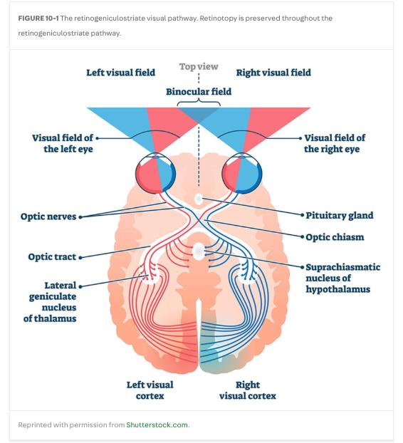
Visual Pathways - Standard and Specialized

Retinogeniculate pathway (standard light pathway): Color, fine details, stationary perception β†’ parvocellular[…] ganglion cells (cone-rich) β†’ optic nerve β†’ chiasm β†’ LGN layers 3-6 β†’ striate cortex (V1) above/below calcarine[…] sulcus; Motion, depth, contrast perception β†’ magnocellular[…] ganglion cells β†’ LGN layers 1-2 β†’ optic radiations β†’ V1.

Tectopulvinar[…] pathway (visuospatial, blindsight): optic tract gives off fibers before LGN β†’ superior colliculus[…] β†’ pulvinar nucleus of thalamus β†’ V1.

Retinohypothalamic[…] pathway (circadian rhythm): intrinsically photosensitive retinal ganglion cells with melanopsin β†’ SCN[…] of hypothalamus β†’ pineal gland for melatonin release.

Higher-order streams: Dorsal[…] stream ("where[…]/action"): occipital β†’ posterior parietal; Ventral[…] stream ("what[…]"): occipital β†’ posterior temporal.
Higher-Order Visual Syndromes

Anton syndrome: bilateral V1[…] damage β†’ cortical blindness + visual anosognosia[…] (blindsight from tectopulvinar). Patient confabulates; light pathway preserved.

Common cause: bilateral PCA[…] stroke. Balint syndrome: bilateral occipitoparietal damage.

Triad: simultanagnosia + optic ataxia + oculomotor apraxia[…].

Causes: bilateral posterior strokes, PML[…], neurodegenerative (Alzheimer, DLB).

Visual neglect: parietal[…] (often right[…]) damage.

Inattention to contralateral[…] hemifield + visual anosognosia.

Akinetopsia: damage to V5[…] (occipitotemporoparietal junction). Moving objects "jump"; liquids appear "frozen."

Pure alexia without agraphia: left/dominant occipital cortex + corpus callosum (splenium) β€” disconnects left fusiform[…] gyrus (visual word form area) from visual processing. Can write a sentence but cannot read it.

Prosopagnosia: occipitotemporal (R or BL) damage to fusiform[…] gyrus. Can't recognize previously learned faces. Causes: PCA stroke, FTD/AD, encephalitis, temporal surgery, tumor.

Central achromatopsia: damage to V4[…] (lingual + fusiform gyri). Cortical loss of color perception with intact optic pathway.

Bilateral β†’ achromatopsia; unilateral β†’ hemiachromatopsia. Β± co-occurring prosopagnosia, superior quadrantanopia[…]. Causes: PCA stroke, CO poisoning.
Practice Q - INO vs MG Differentiation

MLF lesion = adduction deficit + abducting nystagmus + preserved convergence[…].

Slow adducting saccades[…] distinguish INO from MG.

One-and-a-half[…] = dorsal pontine lesion of CN VI nucleus + ipsilateral MLF β€” only contralateral abduction preserved.
Practice Q - Decompensated Congenital CN IV Palsy

Decompensated congenital CN IV palsy: head tilt away[…] from affected eye, hyperdeviation worse in contralateral gaze + ipsilateral head tilt (Park-Bielschowsky 3-step test).
Practice Q - CN VI Nuclear Lesion

A CN VI nuclear/fascicular lesion in the dorsal pons often co-affects the CN VII[…] fascicle as it wraps around the abducens nucleus (facial colliculus).
Practice Q - Pharmacologic Confirmation of Horner

Apraclonidine[…] drops cause pupillary dilation and reverse ptosis in Horner's syndrome via denervation supersensitivity. Cocaine drops fail to dilate the Horner pupil.
Practice Q - Pediatric Horner After Subclavian Line

Pediatric Horner syndrome after subclavian line placement should prompt imaging of the neck and mediastinum[…] to rule out lesions along the sympathetic chain (including neuroblastoma[…]).
Practice Q - Anticholinergic Iatrogenic Mydriasis

Topical anticholinergic exposure causes a fixed dilated[…] pupil unresponsive to light or near accommodation and no response[…] to pilocarpine drops.
Practice Q - Tonic Pupil Cholinergic Supersensitivity

Ciliary ganglion / short ciliary nerve PS lesion produces a tonic pupil β€” poorly light-reactive but tonic near response, with cholinergic supersensitivity (dilute pilocarpine[…] constricts the affected pupil).
Practice Q - Young Woman with Painful Monocular Vision Loss
  • A young woman with painful subacute monocular vision loss, central scotoma, and posterior optic nerve enhancement = optic neuritis. IVMP speeds recovery[…] only (does not change ultimate acuity per ONTT)
  • Retrobulbar ON has a normal disc[…] acutely
Practice Q - MOGAD Optic Neuritis MRI Features

MOGAD optic neuritis classically shows bilateral[…] disc edema and perineural sheath[…] enhancement, with frequent complete MRI resolution.

Practice Q - AQP4-NMOSD ON Treatment

AQP4-NMOSD optic neuritis needs PLEX[…] early or first-line plus rituximab[…] (or complement inhibitors) for long-term prevention.

Avoid standard MS DMTs[…] (IFN-Ξ², fingolimod, natalizumab) β€” they worsen NMOSD.
Practice Q - GCA Emergency Treatment

Suspected GCA with elevated ESR/CRP and systemic symptoms β†’ start immediate IV methylprednisolone[…] before temporal artery biopsy[…].

Pallid disc edema[…] is the GCA hallmark on fundoscopy.
Practice Q - NAION 'Disc at Risk'

NAION arises in patients with a crowded fellow disc[…] ("disc-at-risk"). Treatment does not restore vision β€” focus on vascular risk factor management.
Practice Q - PION Distinguishing Features

Posterior ischemic optic neuropathy presents with acute altitudinal loss + RAPD + normal-appearing disc[…] + recent hypotension[…] (vs AION which has acute disc edema).
Practice Q - CRAO as Stroke

CRAO produces sudden painless severe monocular vision loss with RAPD[…] β€” treat as an anterior-circulation stroke[…], eligible for IV thrombolysis within 4.5 hours.
Practice Q - Compressive Optic Neuropathy

Chronic painless vision loss + dyschromatopsia + RAPD + temporal field defect + disc pallor[…] β†’ image for a compressive[…] lesion (e.g., tuberculum sellae meningioma). Skull-base lesions cause atrophy without preceding edema; orbital lesions go edema β†’ pallor.
Practice Q - Radiation Optic Neuropathy MRI

Radiation-induced optic neuropathy shows juxtachiasmal enhancement[…] on fat-suppressed post-contrast T1 MRI.
Practice Q - LHON Presentation

LHON: painless sequential bilateral[…] central scotomas in a young man; initially hyperemic pseudoedematous[…] discs β†’ temporal pallor with RNFL/GCL loss on OCT.
Practice Q - Thiamine Toxic-Nutritional Optic Neuropathy

Thiamine deficiency causes bilateral symmetric central scotomas with initially normal discs; treat empirically with IV thiamine[…] to prevent irreversible axonal loss.
Practice Q - IIH Visual Field Testing
  • IIH monitoring requires visual automated perimetry[…] as visual confrontation testing is insufficient.
  • New dyschromatopsia[…] is a red flag for vision-threatening disease.
Practice Q - Drug-Induced IIH

Medications that can cause IIH/papilledema include lithium[…], vitamin A and retinoids, steroids withdrawal, growth hormone, and tetracyclines[…]/fluoroquinolones
Practice Q - CVST Papilledema

CVST-related papilledema initially has normal acuity and visual fields[…], and avoid steroids[…]. Treat with LMWH + endovascular options as needed.
Practice Q - Superior Quadrantanopia (Meyer's Loop)

A left superior quadrantanopia is most often due to a lesion of the right temporal[…] lobe, disrupting Meyer's loop[…].

Lesions in the superior parietal or occipital lobes typically produce inferior[…] quadrantanopias.
Practice Q - Right Occipital Stroke Hemianopia

A right occipital (PCA territory) stroke causes a left homonymous hemianopia[…] with difficulty finding line starts; reading rehabilitation uses tracking strategies.
Practice Q - Parinaud Urgent Workup

Parinaud syndrome (upgaze palsy + convergence-retraction nystagmus + light-near dissociation) needs urgent MRI[…] to evaluate for a pineal germ cell tumor[…].

Vertical VOR[…] overcomes the supranuclear palsy.
Practice Q - HINTS Central vs Peripheral

HINTS findings supporting a central[…] lesion include non-horizontal nystagmus, no catch-up saccade[…] on Head Impulse test, and skew deviation[…] on test of skew.
Practice Q - Vestibular Neuritis Nystagmus Direction

Right vestibular neuritis produces a horizontal-torsional[…] nystagmus with slow phase toward the affected (right) ear and fast phase toward the healthy (left) ear.
Practice Q - 4-Aminopyridine for Nystagmus

4-aminopyridine (dalfampridine)[…] is used for downbeat and gaze-evoked nystagmus and the oscillopsia of cerebellar ataxia.
Practice Q - PSP Ocular Features

Progressive supranuclear palsy: vertical supranuclear gaze palsy[…] (downgaze predominant), axial rigidity, early falls, hummingbird[…] midbrain atrophy, and square-wave jerks[…]. Manage downgaze palsy by placing reading material at eye level.
Practice Q - Posterior Cortical Atrophy

Posterior cortical atrophy (an Alzheimer variant) presents with simultanagnosia[…], dyscalculia, and constructional apraxia.
Practice Q - V1 Spares Pupillary Reflex

Bilateral V1 lesions cause cortical blindness but spare[…] the pupillary light reflex via the pretectal[…] pathway (retinotectal route bypasses cortex).
Practice Q - Monocular vs Binocular Diplopia

Monocular[…] diplopia (persists with one eye closed) = intraocular cause (refractive, astigmatism).

Binocular[…] diplopia (resolves with one eye closed) = eye misalignment with a neurologic cause.

An esophoria[…] is a latent inward deviation that fuses with both eyes open.
Practice Q - Ocular MG and Pseudo-INO

Ocular myasthenia gravis can mimic INO[…] (pseudo-INO with bilateral adduction deficits) and can coexist with thyroid eye disease[…], where EOM enlargement and proptosis complicate the picture.
Practice Q - Migraine vs Occipital Seizure vs TIA

Bilateral gradual[…] visual aura over minutes suggests migraine.

Abrupt static[…] onset suggests occipital seizure or vertebrobasilar TIA. Post-CVST chronic headache may be prevented with CGRP-targeted therapy or onabotulinumtoxinA.
Practice Q - GCA Retinal Angiography

Retinal angiography can detect choroidal nonperfusion[…] in GCA and characterize retinal arteriolar branch involvement in Susac[…] syndrome.
Practice Q - PD and Dry Eyes

Dry eye prevalence in Parkinson disease is 60-70[…]%, related to decreased blink rate and autonomic dysfunction.
Practice Q - Visual Impairment and Dementia Risk

Visual impairment is associated with ~47[…]% higher risk of dementia.