Cranial / Nerves

Cranial / Nerves

Anatomy · 16 cards

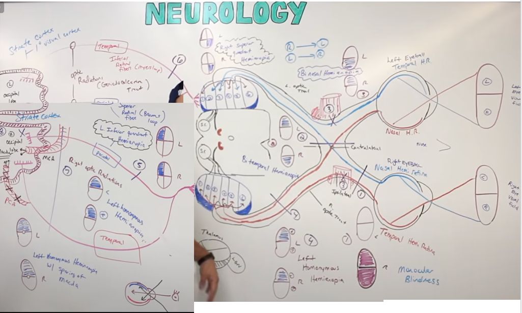
CN II Optic :: Visual Pathway

Light from the left[…] visual field hits the right[…] hemiretina of each eye.

From the right eye, fibers travel via optic nerve → do not cross[…] the optic chiasm → right optic tract. From the left eye, fibers travel via optic nerve → cross[…] the optic chiasm → right optic tract.

Both tracts synapse at the lateral geniculate nucleus (LGN)[…] of the thalamus: ipsilateral fibers go to layers 2, 3, 5[…]; contralateral fibers go to layers 1, 4, 6[…].

Some LGN fibers project to the superior colliculus and pretectal nucleus[…] in the midbrain (for the light reflex and saccades).

The rest project as the optic radiations through the parietal lobe (Baum's loop, superior fibers)[…] and temporal lobe (Meyer's loop, inferior fibers)[…], arriving at the primary visual cortex (V1)[…] along the calcarine sulcus.
CN III Oculomotor :: Nucleus and Course

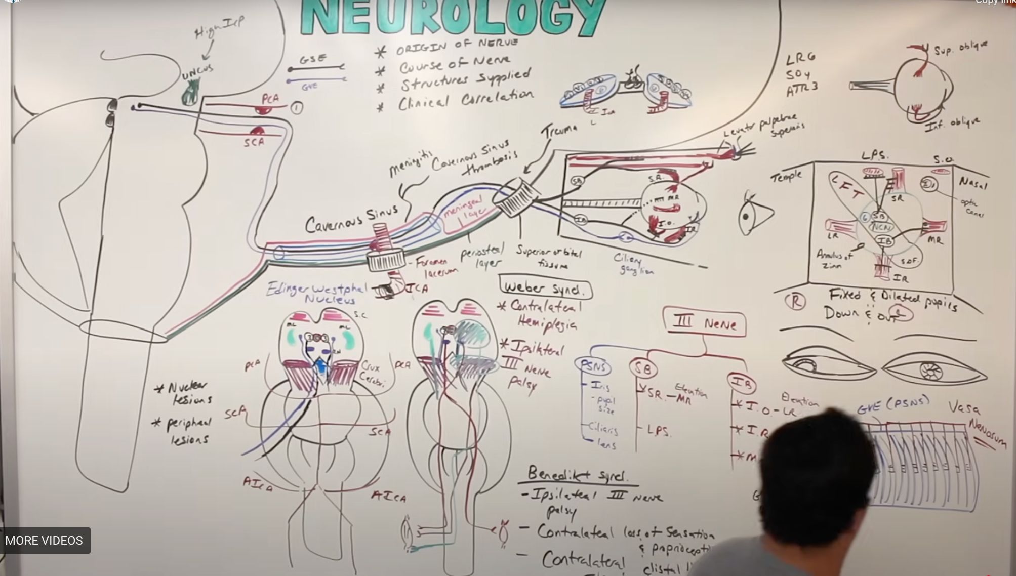
The CN III nucleus sits in the midbrain at the level of the superior colliculus[…], just lateral to the cerebral aqueduct[…] and PAG.

Lateral to CN III is the Edinger-Westphal nucleus[…], which provides parasympathetic[…] fibers (pupil constriction, accommodation).

Fibers exit anteriorly into the interpeduncular fossa[…] between the cerebral peduncles, passing under the PCA[…] and above the SCA[…].

CN III then runs through the lateral wall of the cavernous sinus[…] along with CN IV, V1, V2, and CN VI (the abducens runs free within the sinus near the ICA).

It enters the orbit through the superior orbital fissure[…].
CN III Oculomotor :: Branches and Targets

Within the orbit, CN III splits into a superior[…] branch supplying superior rectus and levator palpebrae superioris, and an inferior[…] branch supplying inferior rectus, medial rectus, and inferior oblique.

Parasympathetic fibers travel with the inferior[…] branch to synapse in the ciliary ganglion[…], then send short ciliary nerves to the sphincter pupillae[…] (pupil constriction) and ciliary muscle (accommodation).

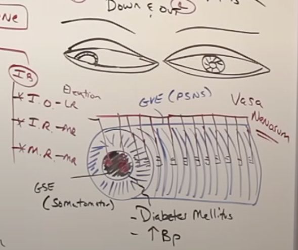
Within the nerve, motor fibers run medially/centrally[…] while parasympathetic fibers run superficially/peripherally[…] — explaining why compression (uncal herniation) hits the pupil FIRST while ischemia (diabetes) spares it.
CN III Oculomotor Lesion Syndromes
  • Damage to motor fibers causes the eye to deviate down and out[…] d/t unopposed action of cranial nerves (2) (IV (superior oblique) and VI (lateral rectus)[…]).
  • Damage to parasympathetic fibers causes a fixed, dilated[…] pupil.
  • High ICP with uncal[…] herniation compresses parasympathetic fibers first (blown pupil), then motor fibers.
  • Benedikt[…] syndrome: ipsilateral CN III palsy + red nucleus tremor + medial lemniscus sensory loss.
  • Weber[…] syndrome: ipsilateral CN III palsy + contralateral hemiparesis from cerebral peduncle (CST) involvement.
  • Claude[…] syndrome: ipsilateral CN III palsy + contralateral ataxia (SCP involvement).
"pope has a tremor" 
"webby" arm goes weak
"learning how to use claude makes me go dizzy"
CN IV Trochlear :: Course and Function

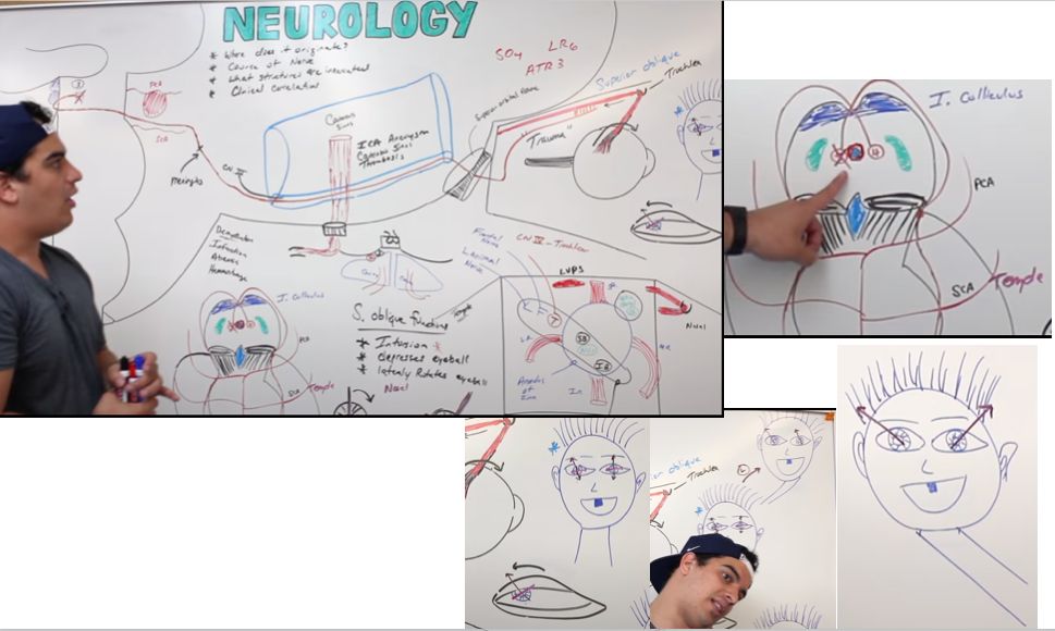
The CN IV nucleus lies lateral to the cerebral aqueduct/PAG[…] at the level of the inferior colliculus[…].

It is the only CN to exit dorsally[…] and the only one to decussate before exiting[…] — so each nucleus supplies the contralateral eye.

Course: exits dorsally → crosses → wraps around the midbrain → passes under PCA[…] above SCA → through the lateral wall of the cavernous sinus[…] → through the superior orbital fissure[…] → supplies the superior oblique[…] muscle.

Superior oblique pulls the eye down, in, and intorts[…] it.

Lesion: the affected eye is extorted[…] at baseline and pulled up and out. Patients compensate by tilting the head to the contralateral[…] (uninvolved) shoulder.

A nucleus[…] lesion produces a CONTRALATERAL palsy; a peripheral nerve[…] lesion produces an IPSILATERAL palsy.
CN V Trigeminal :: Sensory Nuclei

Midbrain mesencephalic[…] nucleus: DCML-like proprioception from face/muscles of mastication.

Pons principal (chief) sensory[…] nucleus: DCML-like fine touch from face.

Medulla spinal nucleus of CN V[…]: spinothalamic-like pain and temperature from the entire face (extends down to upper cervical cord).

Pons motor nucleus of CN V[…]: motor to muscles of mastication (travels with V3).
CN V Trigeminal :: V1 Ophthalmic Division

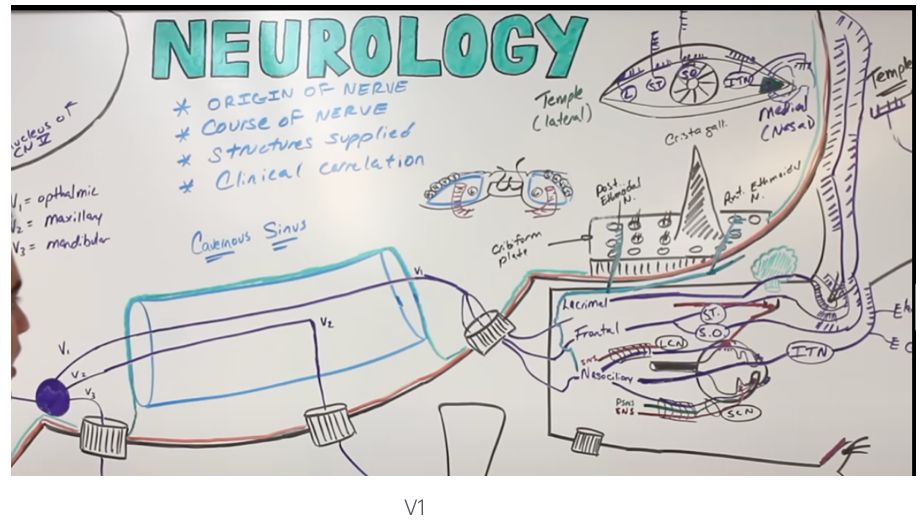
V1 leaves the trigeminal ganglion[…], runs through the lateral wall of the cavernous sinus[…], then enters the orbit via the superior orbital fissure[…].

Three main branches: lacrimal[…] (skin and conjunctiva at temple-facing upper eye, pierces lacrimal gland); frontal[…] (gives supratrochlear + supraorbital nerves to forehead and scalp); and nasociliary[…] (long + short ciliary nerves to cornea, plus ethmoidal and infratrochlear branches to nasal mucosa).

V1 mediates the corneal reflex[…] afferent limb (efferent is CN VII).
CN V Trigeminal :: V2 Maxillary and V3 Mandibular Divisions

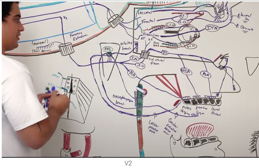
V2 exits the cavernous sinus via the foramen rotundum[…] into the pterygopalatine fossa, supplying sensation to the maxillary skin, upper teeth, palate, and nasal cavity.

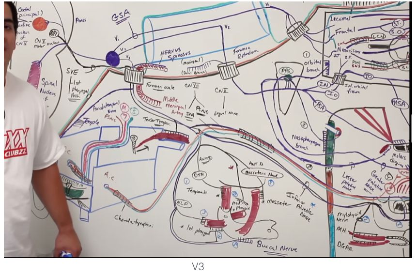
V3 exits via the foramen ovale[…]. Sensory branches supply the mandibular skin, anterior 2/3 tongue (general somatic afferent — NOT taste), lower teeth, and TMJ.

V3 carries the motor[…] fibers from the CN V motor nucleus to muscles of mastication: masseter, temporalis, medial and lateral pterygoids, plus tensor tympani, tensor veli palatini, mylohyoid, and anterior digastric.

A V3 motor lesion causes the jaw to deviate toward the affected side[…] (unopposed pterygoid action on contralateral side).
CN VI Abducens :: Course and Lesions

The CN VI nucleus is in the pons[…], just anterior to the 4th ventricle. The CN VII[…] fibers wrap posteriorly around this nucleus, creating an indent in the 4th ventricle called the facial colliculus[…].

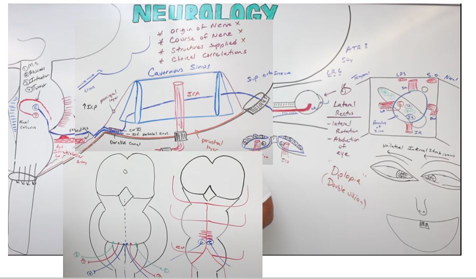
Pathway: fibers exit at the pontomedullary junction[…] → ascend along the clivus[…] → enter Dorello's canal[…] (with the inferior petrosal sinus) → cavernous sinus (running FREE within the sinus near the ICA, not in the lateral wall) → superior orbital fissure[…] → supplies the lateral rectus[…] (abducts the eye).

Lesion: the affected eye deviates inward (medial)[…] from unopposed medial rectus pull.

CN VI is uniquely vulnerable to elevated ICP[…] (long intracranial course) — making it a non-localizing sign in IIH or any increased-ICP state.

Other peripheral causes: AICA aneurysm[…], cavernous sinus disease (CVST, meningitis), and basilar skull base lesions.
CN VII Facial :: Motor Branch and Hyperacusis Prevention

CN VII nucleus is at the level of CN VI[…] in the pons. Fibers wrap around the CN VI nucleus (forming the facial colliculus), then exit at the pontomedullary junction[…].

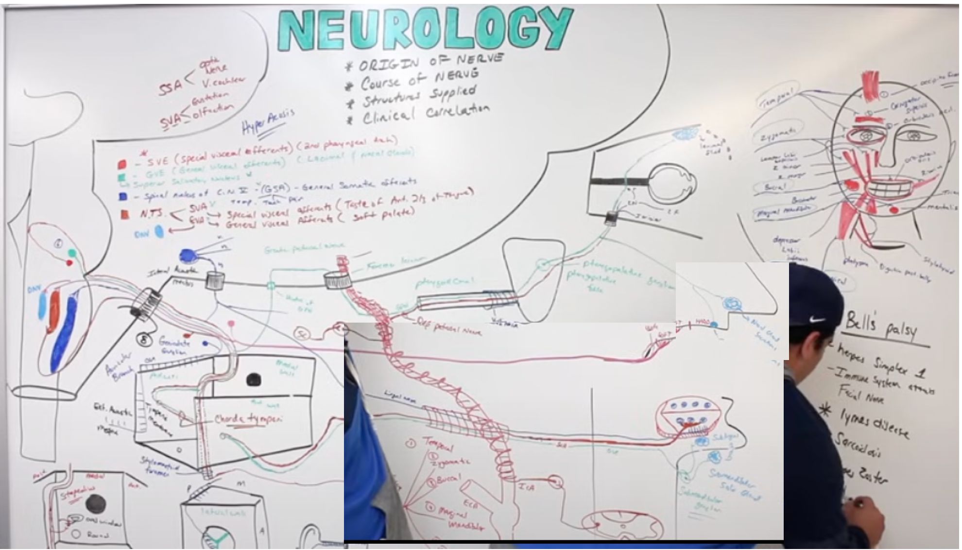
It travels through the internal auditory canal[…] (with CN VIII), enters the facial canal[…], gives off a branch to the stapedius[…] muscle (which dampens stapes vibration to prevent hyperacusis[…]), then exits via the stylomastoid foramen[…].

Five motor branches supply facial expression: temporal[…] (frontalis, orbicularis oculi), zygomatic[…] (zygomaticus), buccal[…] (buccinator, orbicularis oris), marginal mandibular[…] (mentalis), and cervical[…] (platysma).
red = motor (SVE); green = lacrimation/salivation (GVE); dark blue = anterior 2/3 tongue touch/pain/T/proprioception (GSA); brown = taste anterior 2/3 (SVA); light blue = soft palate sensation (GVA)
CN VII Facial :: Parasympathetic, Taste, and Lesion Localization

The superior salivatory nucleus[…] provides parasympathetic fibers that travel via:
- Greater petrosal nerve → pterygopalatine ganglion[…] → lacrimal gland and nasal/palatine mucosa.
- Chorda tympani → submandibular ganglion[…] → submandibular and sublingual salivary glands.

Taste from the anterior 2/3 tongue[…] travels via chorda tympani → geniculate ganglion → nucleus tractus solitarius[…].

Lesion localization:
- Upper motor neuron (cortical/CST): contralateral lower face only[…] (forehead spared by bilateral innervation).
- Lower motor neuron (nucleus or distal to it): ipsilateral entire face[…] weakness (forehead included).

Bell's palsy[…]: peripheral LMN CN VII palsy — entire ipsilateral face affected, often with hyperacusis (stapedius) and loss of taste (chorda tympani) if proximal to the lesion.
red = motor (SVE); green = lacrimation/salivation (GVE); dark blue = anterior 2/3 tongue touch/pain/T/proprioception (GSA); brown = taste anterior 2/3 (SVA); light blue = soft palate sensation (GVA)
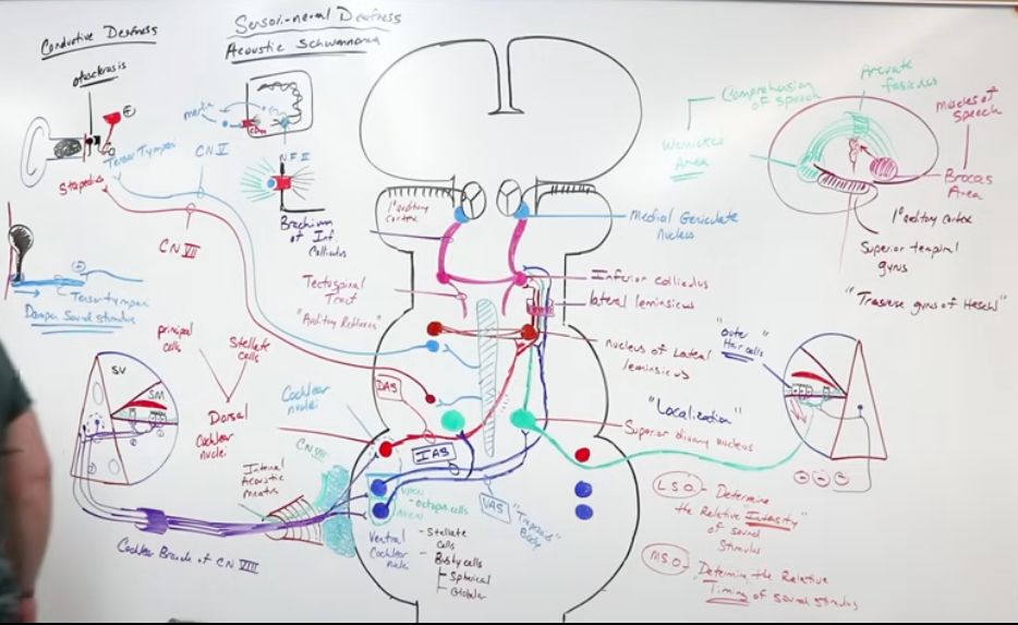
CN VIII Auditory Pathway

Sound → external acoustic meatus → vibrates the tympanic membrane[…] → ossicles (malleus → incus → stapes[…]) → stapes taps the oval window[…] → fluid waves in the cochlea vibrate hair cell cilia[…] → spiral ganglion → cochlear branch of CN VIII.

CN VIII travels through the internal auditory canal[…] (with CN VII) to the pontomedullary junction[…], synapsing on the ventral and dorsal cochlear nuclei.

Cochlear output crosses (mostly) via the trapezoid body[…] → bilateral superior olivary nucleus[…] (sound localization) → lateral lemniscus[…]inferior colliculus[…]medial geniculate nucleus (MGN)[…] of thalamus → primary auditory cortex[…] (Heschl's gyrus).

Branches activate CN V (tensor tympani) and CN VII (stapedius) to dampen loud sounds — the stapedial reflex[…].
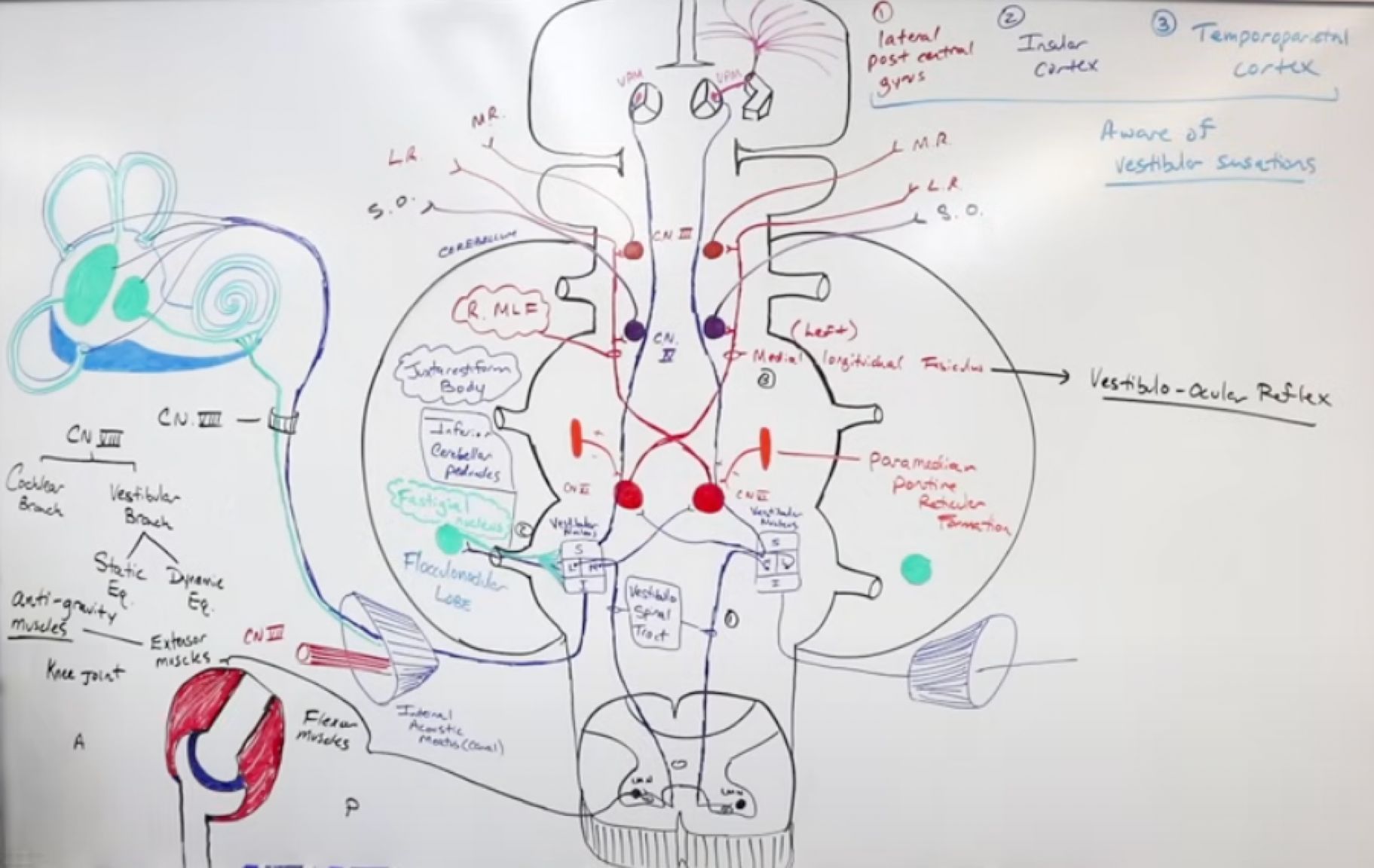
CN VIII Vestibular Pathway

Vestibular branch travels through the IAC, synapsing on the vestibular nuclear complex[…] (superior, lateral, medial, inferior) in the pontomedullary junction.

Four output pathways:
1. Vestibulospinal[…] (descending): medial nucleus → head/neck postural control; lateral nucleus → trunk/limb antigravity tone.

2. Vestibulocerebellar[…] (posterior): inferior nucleus → inferior cerebellar peduncle → flocculonodular lobe[…] → fastigial nucleus (2-way modulation).

3. Vestibulocortical[…] (ascending): superior nucleus → VPM thalamus[…] → postcentral gyrus + insula (conscious vestibular sense).

4. MLF (anterior)[…]: vestibular nuclei → CN III, IV, VI nuclei for the vestibulo-ocular reflex (VOR).
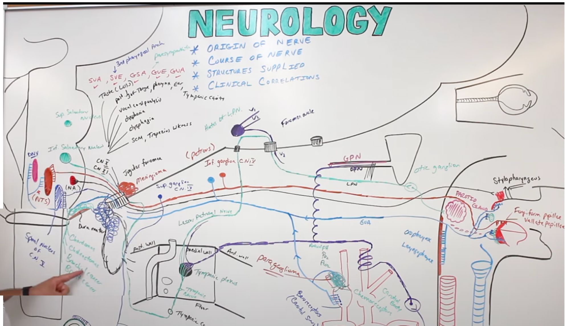
CN IX Glossopharyngeal :: Functions

All CN IX fibers exit/enter the skull via the jugular foramen[…].

Sensory:
- Taste from posterior 1/3 tongue[…] (fungiform/vallate papillae) → inferior (petrous) ganglion → nucleus tractus solitarius[…].
- General sensation (touch, pain, T) from posterior 1/3 tongue, pharynx, soft palate, tonsils, middle ear, and external auditory meatus → superior ganglion → spinal nucleus of CN V[…].

Motor (from nucleus ambiguus): stylopharyngeus[…] muscle (elevates pharynx during swallowing).

Parasympathetic (from inferior salivatory nucleus[…]): via lesser petrosal → otic ganglion → parotid gland[…].

Visceral afferents: from carotid sinus (baroreceptors → BP) and carotid body (chemoreceptors → O2/CO2) → NTS.

CN IX is the afferent limb of the gag reflex[…] (efferent is CN X).
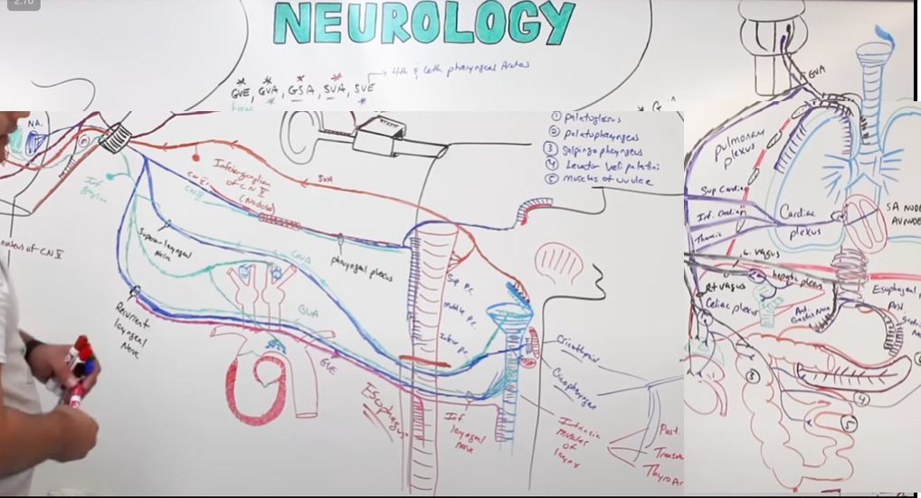
CN X Vagus :: Motor and Parasympathetic

All CN X fibers exit/enter via the jugular foramen[…].

Motor (from nucleus ambiguus[…]): joins CN IX and XI cranial part to form the pharyngeal plexus[…], supplying pharyngeal constrictors, soft palate, and uvula. Branches:
- Superior laryngeal nerve → external[…] branch to cricothyroid (phonation).
- Recurrent laryngeal nerve[…] → all other intrinsic laryngeal muscles (phonation, vocal cords).

Parasympathetic (from dorsal motor nucleus of CN X[…]): supplies esophagus (peristalsis), heart (slows HR), lungs/bronchi (bronchoconstriction), GI tract through splenic flexure[…] (the "vagal break"), and other thoracoabdominal organs via celiac and hepatic plexuses.

Sensory: visceral and mucosal sensation from epiglottis, larynx, and pharynx → NTS + dorsal nucleus of CN X[…].

CN X is the efferent limb of the gag reflex[…].
CN XII Hypoglossal :: Course and Lesions

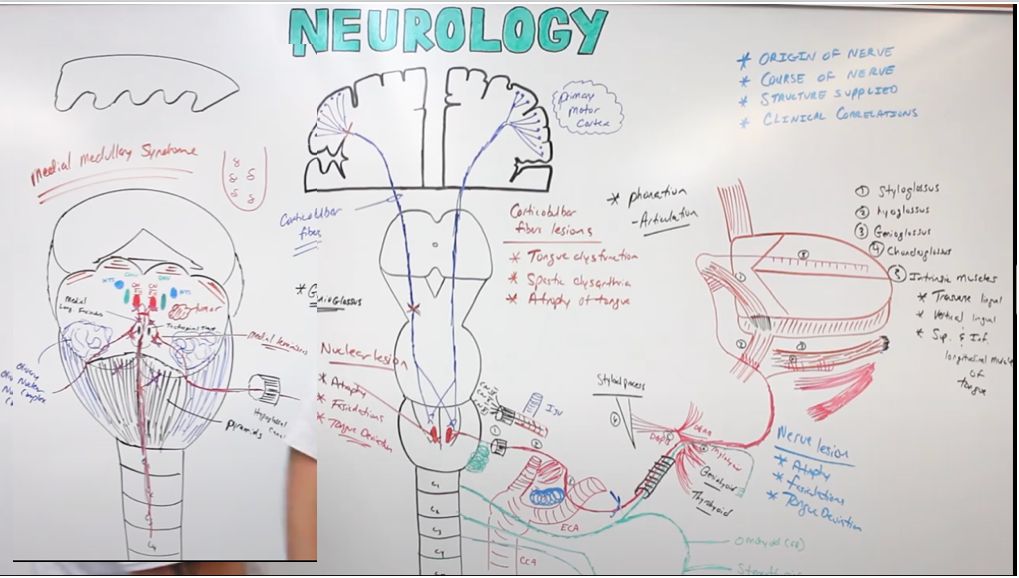
The CN XII[…] nucleus lies in the medial medulla just lateral to the midline, with the dorsal motor nucleus of CN X just lateral.

Fibers travel ventrally through the reticular formation, MLF, medial lemniscus, and the olive[…], exiting in the sulcus between the pyramids and olives[…].

Course: through the hypoglossal canal[…] → below the IJ vein → between ICA and ECA → below the styloid process → pierces tongue muscles.

Central innervation: primary motor cortex (face area) → corticobulbar tract → synapses bilaterally[…] on CN XII nuclei (UMN lesions cause MILD contralateral tongue weakness).

Lesion patterns:
- LMN lesion (nucleus or peripheral nerve): tongue deviates TOWARD[…] the affected side, with ipsilateral atrophy and fasciculations[…].
- UMN lesion (cortex/CBT): tongue deviates AWAY[…] from the lesion (toward the weak side), no atrophy or fasciculations.

Bilateral UMN lesions cause spastic dysarthria[…] (e.g., in ALS or bilateral strokes).