Spinal / Tracts

Spinal / Tracts

Anatomy Β· 17 cards

Spinal Cord :: Levels and Nerve Counts

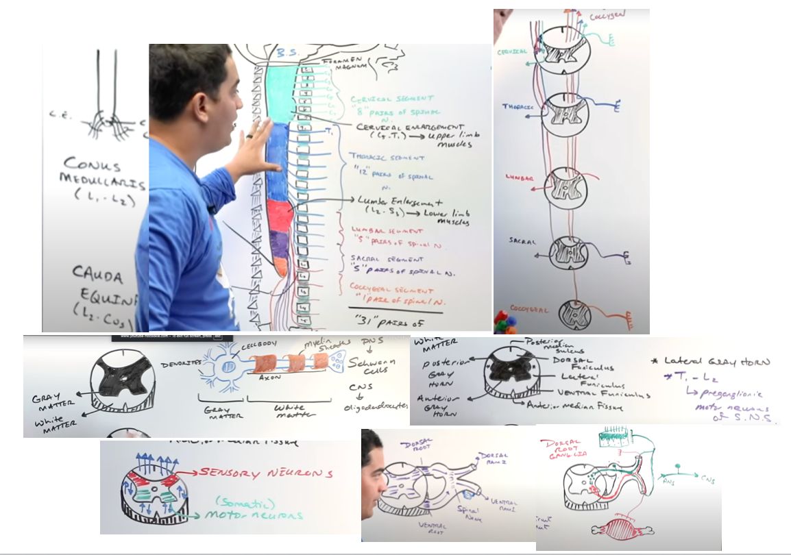
The spinal cord starts at the C1[…] level and ends at L1-L2[…] as the conus medullaris; below this lies the cauda equina[…] ("horse's tail").

Total spinal nerves: 31[…] β€” 8 cervical, 12 thoracic, 5 lumbar, 5 sacral, 1 coccygeal.

The first cervical nerve (C1) exits ABOVE[…] the C1 vertebra. From T1 onward, each spinal nerve exits BELOW its corresponding vertebra.

Enlargements: the cervical[…] enlargement spans C5-T1 (BUE innervation); the lumbar[…] enlargement spans L2-S3 (BLE innervation).

As you descend the cord, gray[…] matter increases (more motor pools needed for limbs) and white[…] matter decreases (fewer ascending/descending fibers remain).
Spinal Cord :: Cross-Section Anatomy

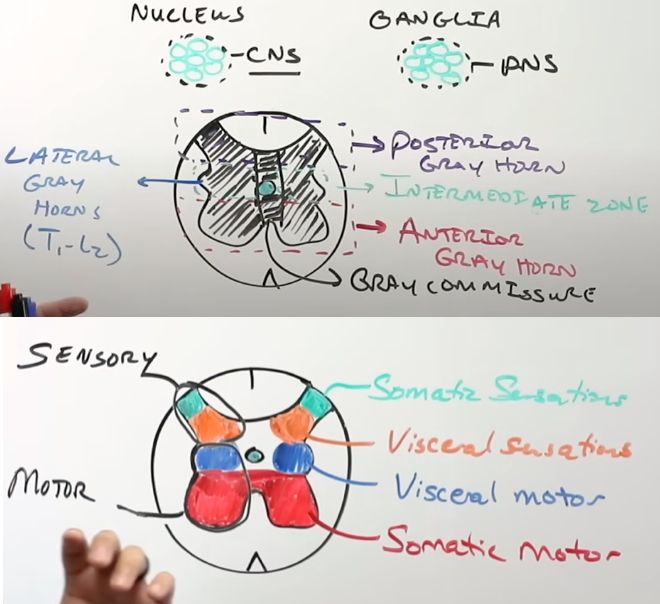
Gray matter forms the inner butterfly/starfish[…] (or H-shape), surrounded by white matter.

Gray matter zones: posterior gray horn[…] (somatic + visceral sensory), anterior gray horn[…] (somatic motor), and intermediate zone[…] containing the lateral gray horn[…] (visceral motor β€” only present at T1-L2 for sympathetic preganglionics, and S2-S4 for parasympathetic preganglionics).

The slab of gray matter connecting both sides is the gray commissure[…], with the central canal[…] running through it.

White matter columns (funiculi): dorsal/posterior, lateral, and anterior/ventral. The anterior white commissure[…] is the crossing point for STT fibers.

A group of neuron cell bodies in the CNS is a nucleus[…]; in the PNS, a ganglion[…].
Spinal Cord :: Spinal Nerve Formation and Rami

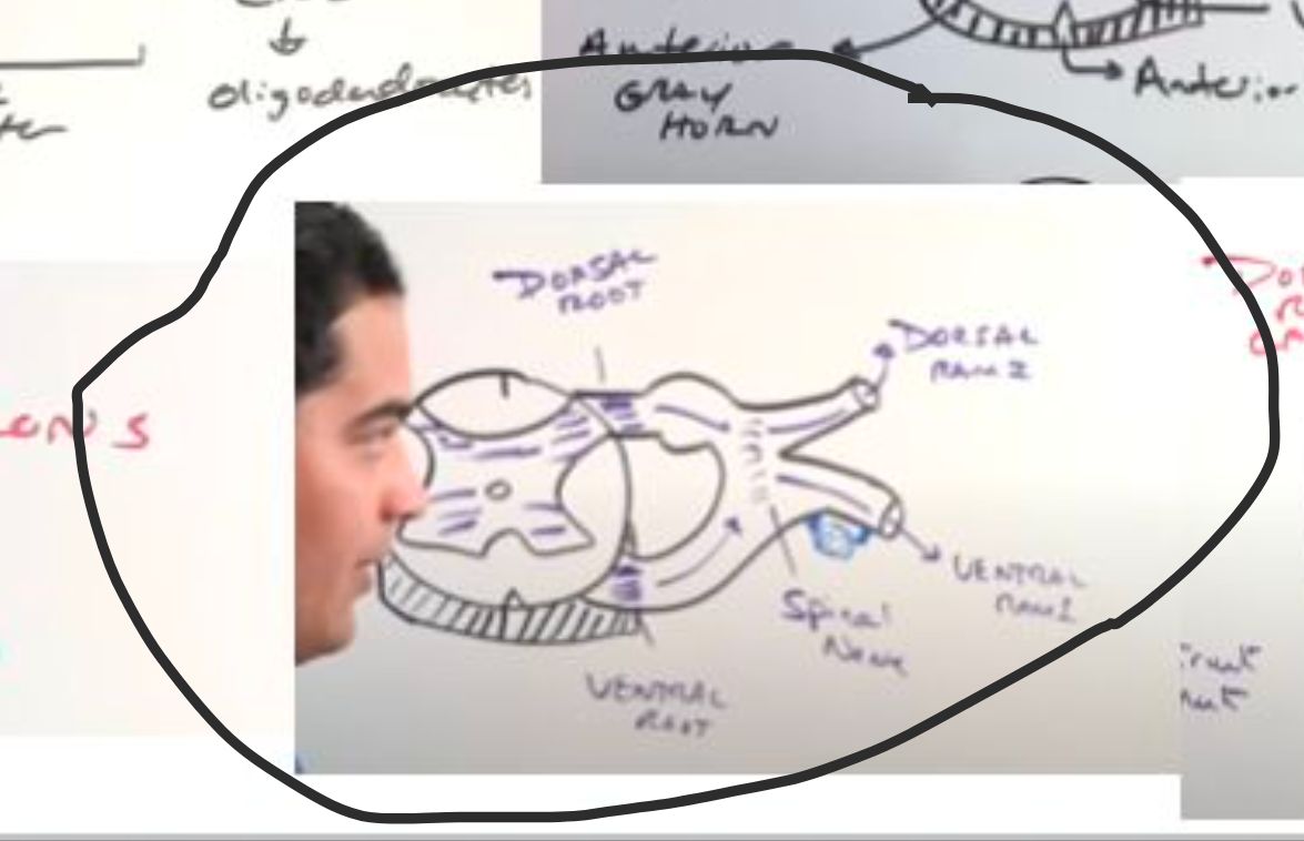
  • Motor output: anterior gray horn Ξ±/Ξ³ motor neurons β†’ ventral root[…]. Sensory input enters via the dorsal root[…] (cell bodies in the dorsal root ganglion).
  • Dorsal + ventral roots fuse into a spinal nerve[…], which then splits into dorsal and ventral rami β€” making spinal nerves mixed sensory and motor[…].
  • Dorsal rami pick up sensation from the neck, back, and vertebrae[…].
  • Ventral rami pick up sensation from the trunk and limbs[…] (and form the plexuses).
  • Communication branches: white and gray rami communicans[…] link spinal nerves to the sympathetic chain ganglion.
Ascending Tracts :: DCML Pathway

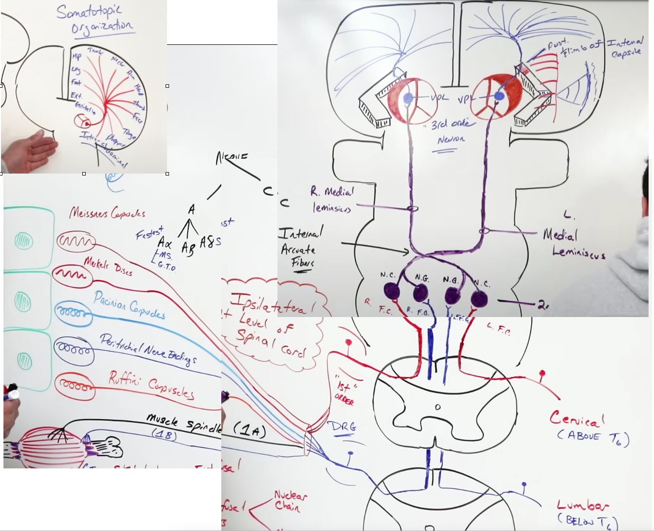
Functions: discriminative touch, pressure, vibration, proprioception, and stretch[…].

PNS receptors: Meissner's corpuscles, Merkel's discs, Pacinian corpuscles, Ruffini corpuscles, peritrichial endings, plus muscle spindles (1a fibers[…]) and Golgi tendon organs (1b fibers[…]).

1st-order neuron: cell body in DRG[…] β†’ posterior gray horn β†’ ascends in the dorsal/posterior white column[…] IPSILATERALLY. Below T6 (legs): fasciculus gracilis[…]; above T6 (arms): fasciculus cuneatus[…].

2nd-order neuron: synapses at the nucleus gracilis or cuneatus in the medulla β†’ fibers DECUSSATE as the internal arcuate fibers[…] β†’ ascend as the medial lemniscus[…].

3rd-order neuron: synapses in the VPL[…] of thalamus β†’ projects via posterior limb of internal capsule β†’ primary somatosensory cortex (postcentral gyrus)[…].
Ascending Tracts :: Spinothalamic Tract (STT)

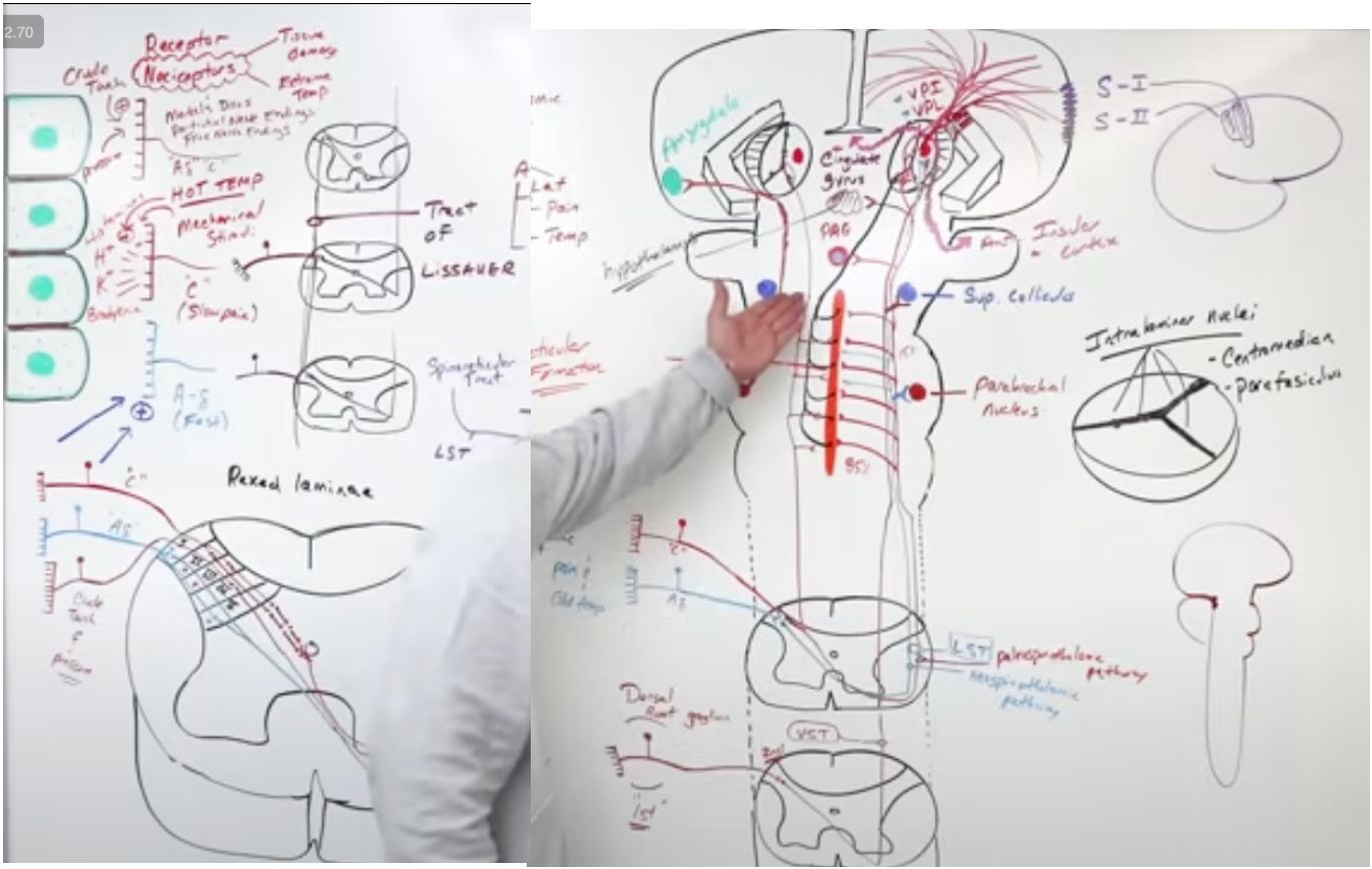
Functions: crude touch, pressure, pain, and temperature[…].

PNS receptors: nociceptors, Merkel's discs, free nerve endings; carried by A-delta[…] fibers (sharp pain/cold) and C[…] fibers (dull pain/crude touch).

1st-order neuron: DRG.

2nd-order neuron: synapses in the posterior gray horn[…] β†’ MOST fibers CROSS via the anterior white commissure[…] β†’ ascend contralaterally. The lateral STT (lateral white column) carries pain/T; the ventral STT (anterior white column) carries crude touch.

STT collaterals project to reticular formation[…] (arousal), intralaminar thalamus (cortex "wake up"), parabrachial nucleus[…] β†’ amygdala (emotional pain), midbrain tectum/superior colliculus, and PAG[…] (descending pain modulation).

3rd-order neuron: synapses in VPL[…] of thalamus β†’ projects to primary somatosensory cortex.
Ascending Tracts :: Spinocerebellar System

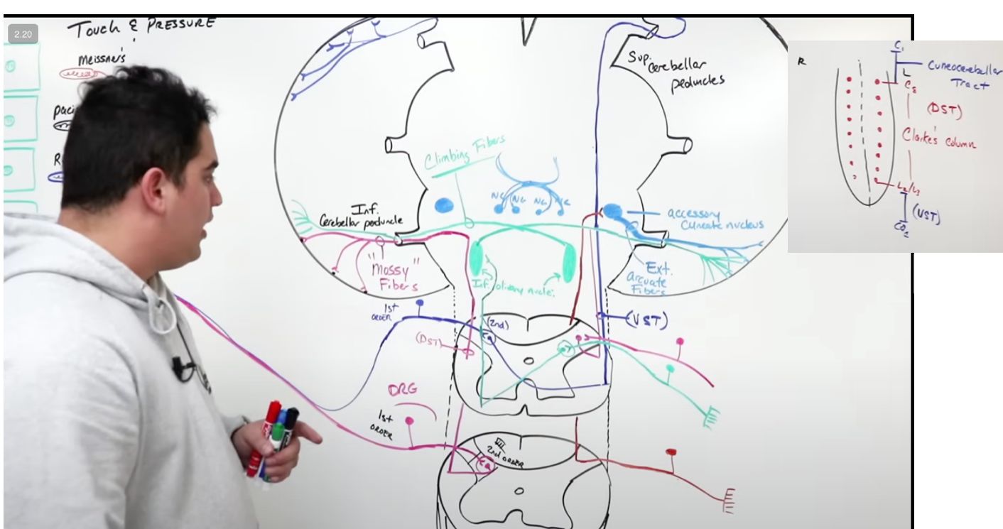
All carry touch, pressure, and proprioception[…] to the cerebellum (unconscious proprioception).

Dorsal spinocerebellar tract (DSCT)[…]: carries IPSILATERAL info from C8-L2/3 (origin = Clarke's column[…] / nucleus dorsalis) β†’ lateral white column β†’ inferior cerebellar peduncle (ICP)[…] β†’ cerebellum as mossy fibers.

Cuneocerebellar tract[…]: carries IPSILATERAL info from ABOVE C8 β†’ ascends to the accessory cuneate nucleus[…] in the medulla β†’ external arcuate fibers through the ICP[…] β†’ cerebellum.

Ventral spinocerebellar tract (VSCT)[…]: carries info from L2/3 and BELOW β†’ crosses via the anterior commissure β†’ ascends in the contralateral lateral white column β†’ enters via the superior cerebellar peduncle (SCP)[…] β†’ CROSSES AGAIN in the cerebellum, ending up ipsilateral.

Rostral spinocerebellar tract[…]: cervical/upper extremity proprioception (analog of VSCT for arms).
Ascending Tracts :: Pain Modulation

Two mechanisms:

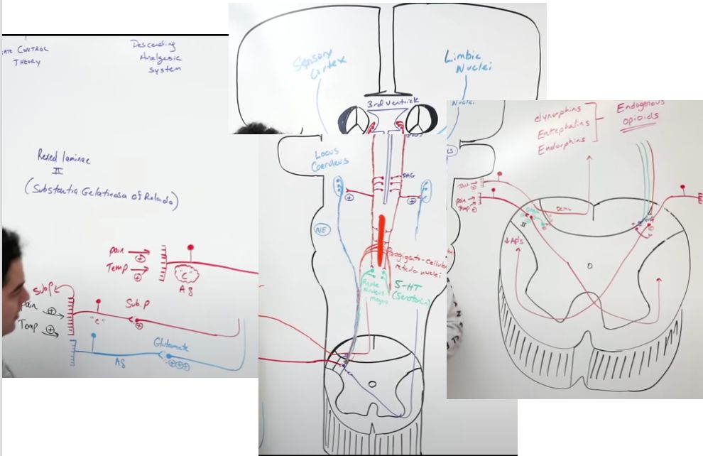
Gate control theory[…]: rubbing an injured area activates the DCML[…] pathway, whose collaterals stimulate inhibitory interneurons in the dorsal gray horn. These release GABA[…] onto the STT synapse β€” reducing pain perception.

Descending analgesic system[…]: the periaqueductal gray (PAG)[…] around the cerebral aqueduct and periventricular gray around the 3rd ventricle stimulate the locus ceruleus[…] (NE), raphe nuclei[…] (serotonin), and reticular formation.

These descend through the posterior white column β†’ dorsal gray horn, where they stimulate inhibitory interneurons to release endorphins and endogenous opioids[…] β€” blocking pain transmission.

Pharmacologic relevance: SSRIs/SNRIs (e.g., duloxetine), TCAs (amitriptyline), and opioids all act on this system.
Descending Tracts :: Corticospinal Tract (CST)

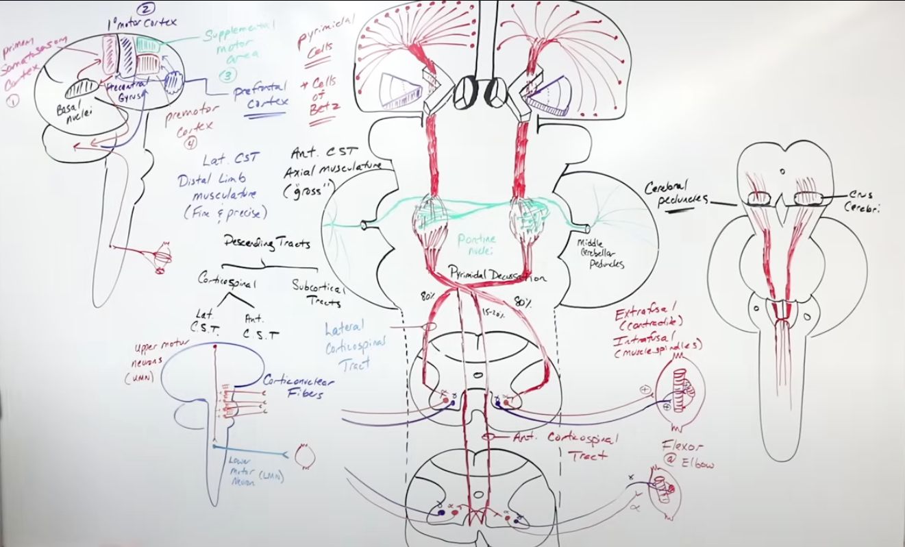
Origin: primary motor cortex (~30%), motor association cortex (~30%), and primary somatosensory cortex (~40%)[…]; cell bodies in the pyramidal (5th) cortical layer[…].

Pathway: centrum semiovale β†’ corona radiata[…] β†’ posterior limb of internal capsule[…] β†’ crus cerebri[…] (cerebral peduncles) β†’ pons (some fibers branch to pontine nuclei β†’ ponto-cerebellar[…]) β†’ pyramids[…] of medulla.

At the pyramidal decussation, ~85%[…] of fibers CROSS to form the lateral CST (limbs), and ~15% stay ipsilateral as the anterior CST (axial muscles).

Lateral CST runs in the lateral white column[…] β†’ synapses on Ξ±/Ξ³ motor neurons in the anterior gray horn[…] β†’ ventral root β†’ muscles.

Anterior CST decussates at its target spinal level via the anterior white commissure[…] before synapsing.
Descending Tracts :: Corticobulbar Tract (CBT)

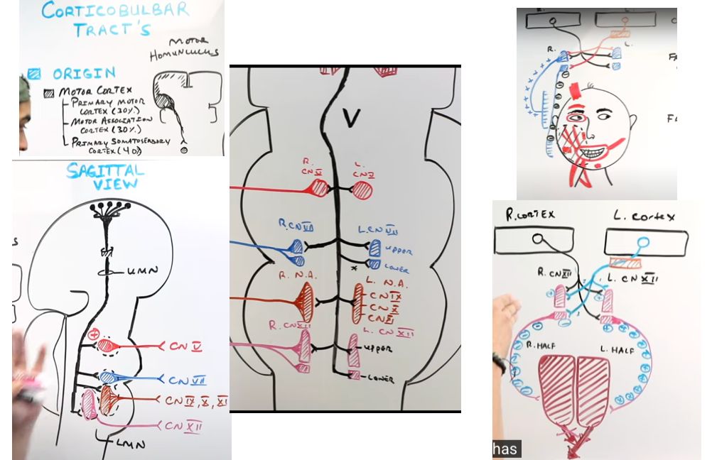
Origin: primary motor cortex, motor association cortex, and primary somatosensory cortex[…]; on the homunculus, it sits laterally[…] (face area).

Pathway: centrum semiovale β†’ corona radiata β†’ genu of the internal capsule[…] β†’ midbrain (medial crus cerebri) β†’ synapses on motor nuclei of CN V, VII, IX, X, XI (cranial), XII[…].

Innervation pattern: MOST CN motor nuclei receive bilateral[…] corticobulbar input β€” explaining why UMN lesions cause only mild contralateral weakness for most CNs.

Exceptions (predominantly contralateral cortical input):
- Lower face (CN VII): UMN lesion β†’ contralateral lower-face[…] weakness only (forehead spared).
- Tongue (CN XII): UMN lesion β†’ tongue deviates AWAY[…] from the cortical lesion.
Descending Cerebellar Tracts: Vestibulospinal Tract
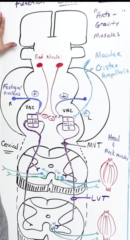
  • Function: activates extensor and antigravity[…] muscles β€” important for posture and balance.
  • Inputs to the vestibular nuclear complex: ear's maculae[…] (linear acceleration) + cristae ampullaris[…] (angular acceleration) via the vestibular branch of CN VIII; cerebellar Purkinje fibers from the flocculonodular lobe (modulatory).
  • Pathway: medial and lateral vestibular nuclei β†’ fibers descend ipsilaterally in the anterior white column β†’ synapse on anterior gray horn β†’ activate Ξ±/Ξ³ motor neurons of extensor muscles.
  • The medial[…] vestibulospinal tract handles head/neck postural reflexes; the lateral[…] vestibulospinal tract handles trunk/limb extensor tone.
Descending Tracts :: Reticulospinal (Pontine vs Medullary)

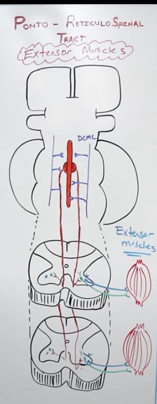
Pontine reticulospinal[…]: arises from pontine reticular formation β†’ descends in the anterior white column[…] β†’ activates extensor[…] muscles (assists the vestibulospinal tract).

Medullary reticulospinal[…]: arises from medullary reticular formation β†’ descends in the lateral white column[…] β†’ activates flexor[…] muscles AND inhibits extensor tone.

These two tracts antagonize each other: pontine = extensor drive; medullary = flexor drive + extensor inhibition.

The CST[…] normally projects collaterals to the medullary reticulospinal nuclei to keep extensor tone in check. Loss of CST in UMN lesions disinhibits the pontine pathway β†’ spasticity[…] with extensor predominance.
Descending Tracts :: Rubrospinal Tract

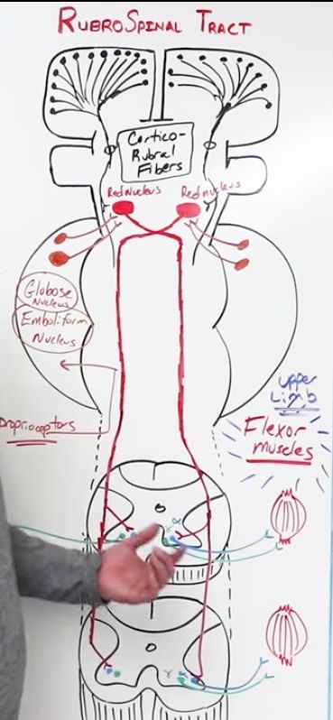
Function: activates flexor[…] muscles, predominantly of the upper extremities (BUE > BLE).

Pathway: cortex sends corticorubral[…] fibers descending to the red nucleus[…] in the midbrain. The red nucleus also receives input from the cerebellum's globose and emboliform (interposed)[…] nuclei.

From the red nucleus, fibers DECUSSATE at the ventral tegmental decussation[…] β†’ descend in the contralateral lateral white column β†’ synapse on Ξ±/Ξ³ motor neurons in the anterior gray horn[…].

In humans, the rubrospinal tract is functionally overshadowed by the CST β€” but it becomes prominent in decorticate[…] posturing (when cortex is gone but red nucleus is intact).
Upper Motor Neuron vs Lower Motor Neuron Lesions

UMN tracts: CST[…] (cortex β†’ anterior gray horn) and CBT[…] (cortex β†’ CN motor nuclei).

UMN lesion signs:
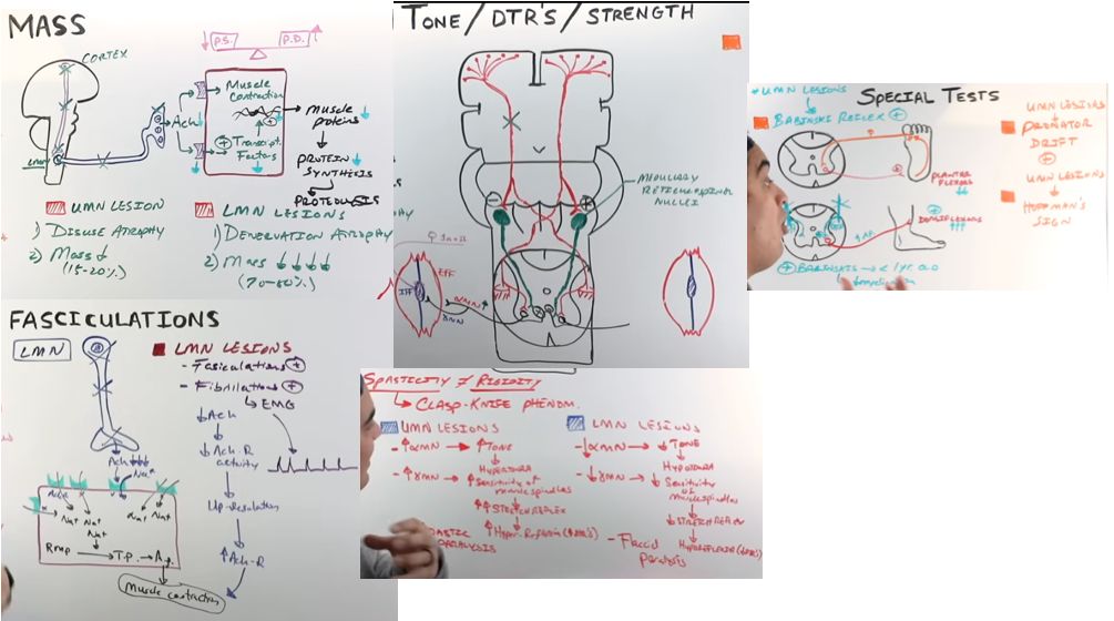
- Spastic paralysis[…]: loss of CST input to medullary reticulospinal β†’ less inhibition of Ξ± motor neurons β†’ hypertonia.
- Hypertonia[…] that is velocity-dependent (more resistance with faster movement), in one direction, often with clasp-knife[…] phenomenon. Contrast with PD rigidity which is velocity-INDEPENDENT and bidirectional.
- Hyperreflexia[…], clonus, and positive Babinski sign[…].
- NO atrophy or fasciculations.

LMN lesion signs (anterior horn cell or peripheral motor nerve):
- Flaccid paralysis[…].
- Hypotonia[…], hyporeflexia[…], and absent Babinski.
- Atrophy and fasciculations[…].

ALS[…] uniquely shows BOTH UMN and LMN signs simultaneously.
Spinal Cord :: Meninges and Lumbar Puncture

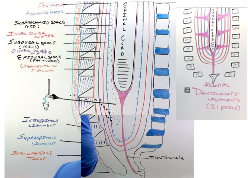
For LP: palpate the iliac crest[…] (corresponds to the L4 spinous process); insert the needle just above it at the L3/L4[…] interspace β€” below the conus medullaris (L1-L2) to avoid cord injury.

Layers (deep to superficial):
- Pia mater[…]: attached to the spinal cord surface; ends as the filum terminale[…] anchoring to the coccyx.
- Subarachnoid space[…]: contains CSF; extends to S2 (large lumbar cistern is the LP target).
- Arachnoid mater[…].
- Subdural space[…]: potential space.
- Dura mater[…].
- Epidural space[…]: contains fat and venous plexus.

The denticulate ligaments[…] are pia mater offshoots that suspend the cord laterally within the dural sac.
Spinal Cord :: Blood Supply and ASA Syndrome

Arterial supply: the vertebral arteries[…] and segmental radicular arteries feed one anterior spinal artery (ASA)[…] (supplies anterior 2/3 of cord) and two posterior spinal arteries (PSAs)[…] (supply posterior 1/3 β€” i.e., dorsal columns).

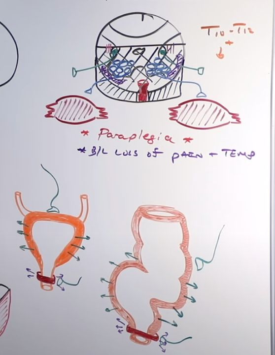
The artery of Adamkiewicz[…] is the largest radicular feeder, typically entering at T10-T12[…] on the left, supplying the lower thoracic and lumbar ASA territory.

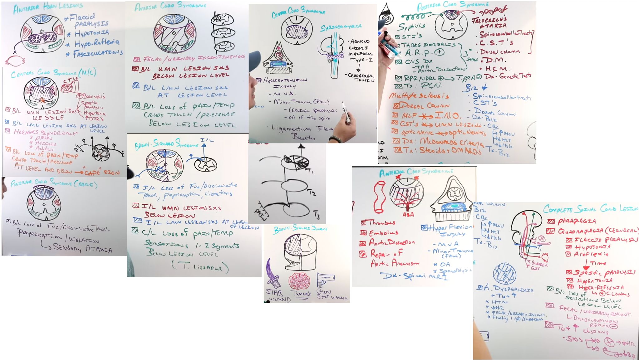
Anterior Spinal Artery (ASA) Syndrome[…]: knocks out the anterior 2/3 of the cord:
- Lateral CST β†’ BLE paraplegia[…] (or quadriplegia if higher).
- STT β†’ loss of bilateral pain and temperature[…] below the lesion.
- Anterior gray horn (at the level) β†’ LMN signs at that level.
- Lateral gray horn (if T1-L2) β†’ autonomic dysfunction.

DCML/dorsal columns are SPARED[…] (PSA territory) β€” so vibration and proprioception are preserved.
Spinal Cord Lesion Syndromes

Anterior gray horn lesion[…] (e.g., polio, West Nile, SMA): LMN signs at the level β€” flaccid paralysis, hypotonia, hyporeflexia, atrophy, fasciculations. ALS[…] shows additional UMN signs from CST involvement.

Central cord syndrome[…] (often from hyperextension injury in elderly or syringomyelia): hits the lateral CST and crossing anterior white commissure fibers. Features:
- Bilateral UMN signs[…] below the lesion, somatotopically affecting BUE > BLE (cervical/thoracic fibers are more medial).
- Bilateral cape-like loss of pain and temperature[…] at the level (crossing STT fibers).
- DDX: syringomyelia[…] β€” central canal dilation, often from Chiari I or cord injury.

Posterior cord syndrome (tabes dorsalis)[…]: hits dorsal columns β€” loss of vibration, proprioception, and fine touch[…] below the lesion, with sensory ataxia and a positive Romberg sign. Classic cause: neurosyphilis (tertiary)[…].

Brown-SΓ©quard syndrome (cord hemisection)[…]: ipsilateral UMN paralysis and DCML loss BELOW the level; contralateral STT loss (pain/T) starting 1-2 segments BELOW[…] the lesion; LMN signs and ipsilateral total sensory loss AT the level.
Subacute Combined Degeneration
  • Subacute combined degeneration (SCD)[…] is caused by (3) vitamin B12 or Copper deficiency OR NO overdose[…]
  • Pathology: demyelination of the dorsal columns[…] AND the lateral corticospinal tracts[…] β€” "combined" referring to ascending sensory + descending motor tracts.
  • Clinical features:
    • Loss of vibration and proprioception[…] β†’ sensory ataxia and positive Romberg.
    • UMN signs[…] (spasticity, hyperreflexia, Babinski) from CST involvement.
    • Often accompanied by peripheral neuropathy[…] and macrocytic anemia.
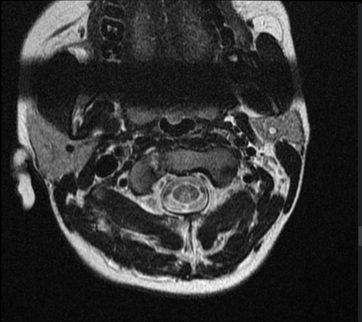
  • Diagnosis: low serum B12, elevated methylmalonic acid (MMA)[…] and homocysteine. MRI shows symmetric T2 hyperintensity in the dorsal columns (inverted V or rabbit-ears sign on axial).
  • Treatment: IM cyanocobalamin[…] replacement; deficits may be partially reversible if treated early.

pic credit: Batta N, Subacute combined degeneration of cord - vitamin B12 deficiency. Case study, Radiopaedia.org (Accessed on 14 Jul 2026) https://doi.org/10.53347/rID-29761