EEG

EEG

Radiology · 215 cards · 48 labeled figures

EEG - Basic Neurophysiology
  • we need >6[…]cm2 of synchronized cortical activity for anything to be detected on scalp EEG
  • the resting potential inside the cell is -70[…]mV established by Na/K ATPase[…] channel
  • depolarization leads to Na+ rushing into the cell making the resting potential less[…](-) (aka more[…]+) while the extracellular area becomes more (-)[…]
  • hyperpolarization leads to K- rushing out of the cell making the resting potential more[…](-) (aka less[…]+) while the extracellular area becomes more (+)[…]
  • Excitatory neurotransmitters such as glutamate[…] bind to postsynaptic receptors and cause an excitatory postsynaptic potential (EPSP)[…]
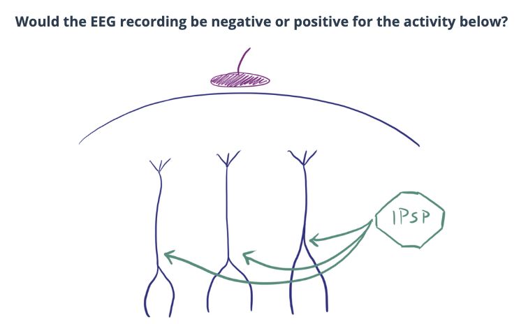
  • Inhibitory neurotransmitters, such as GABA[…], cause an inhibitory postsynaptic potential (IPSP[…])
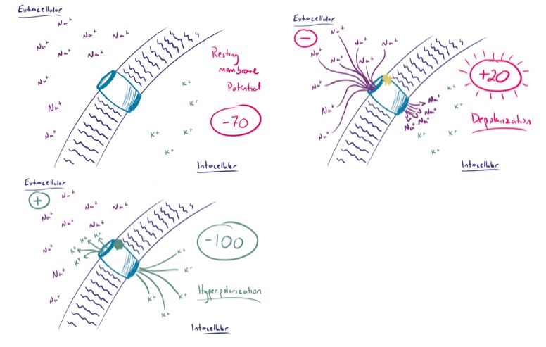
EEG - Basic Neurophysiology
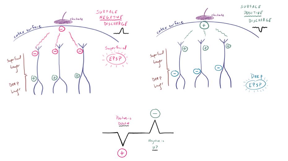
  • EEG electrodes on the scalp pick up the extra[…]cellular voltage which are a summation of voltages from many EPSP[…]s and IPSP[…]s over a broad area of cortex
    • superficial[superficial or deep?] depolarizations create (-)[…] EEG signals
    • deep[superficial or deep?] depolarizations lead to (+)[…] EEG signals
  • On EEG
    • positive potentials will show up as down[down or up]going waves 
    • negative potentials will show up as up[down or up]going waves 
EEG - Montages
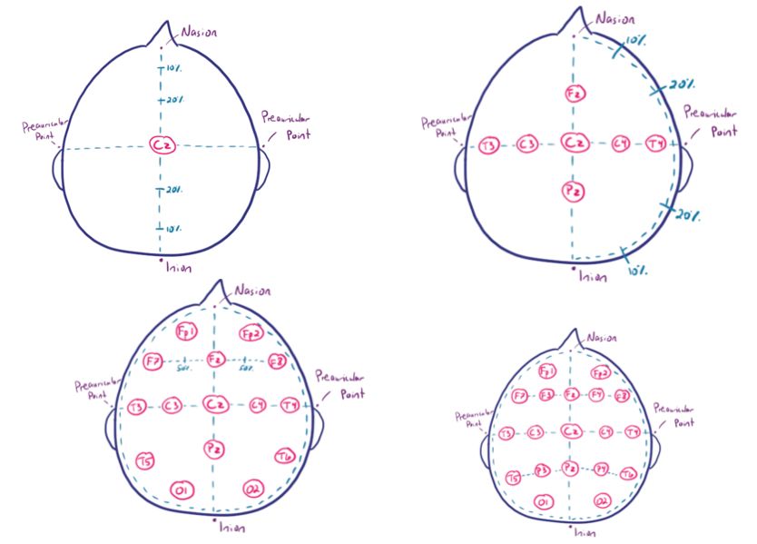
  • The most commonly used montage is the international, standardized 10-20[…] System which splits the skull into increments of 10[…]-20[…]% to place each electrode - Basic Principles:
    • odd[…] numbers on the left
    • even[…] numbers on the right
    • F[…] for frontal[…]
    • T[…] for temporal[…]
    • P[…] for parietal[…]
    • O[…] for occipital[…]
    • Fp[…] for frontopolar[…] which corresponds to the eye blink[…] artifact
    • Z[…] used as subscript after the location is used to denote midline[…]
EEG - Montages
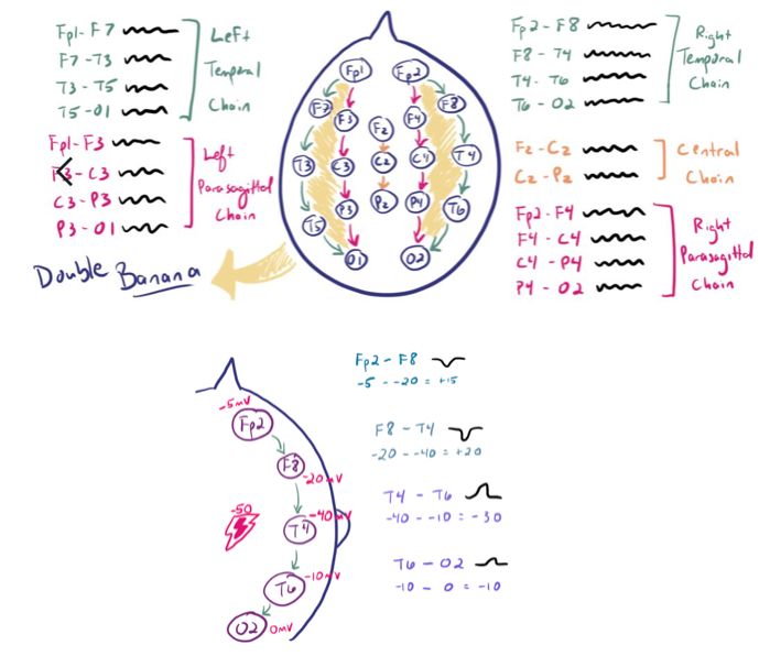
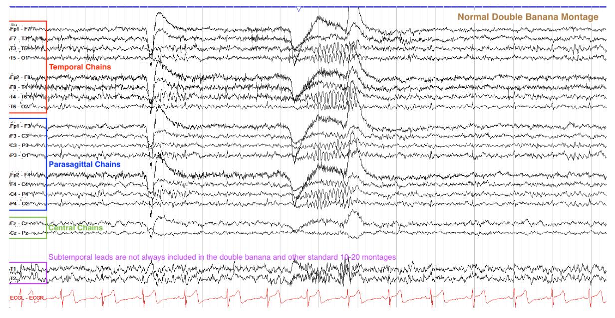
  • In a Bipolar[…] montage, aka the "Double Banana[…]," each electrode is linked to a contralateral chain[…] 
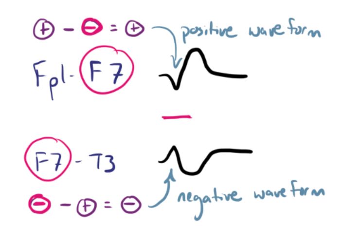
    • in this montage: if the 1st[…] electrode voltage in the chain is higher[…] (ex: Fp2 = -20) than the 2nd[…] electrode in the chain (ex: F8 = -50), you get a positive[…] deflection which shows up as a down[…]ward wave
    • if the 1st electrode voltage in the chain is lower[…] (ex: Fp2 = -50) than the 2nd electrode in the chain (ex: F8 = -20), you get a negative[…] deflection which shows up as a up[…]ward wave
    • In a phase reversal[…], the electrode of maximal voltage will affect the surrounding tracings
      • negative[…] phase reversal: generally seen with epileptiform[…] activity where the tracings will face toward[toward or away from?] each other
      • positive[…] phase reversal: generally seen with artifactual[…] activity where the tracings will face away from[toward or away from?] each other
EEG - Settings
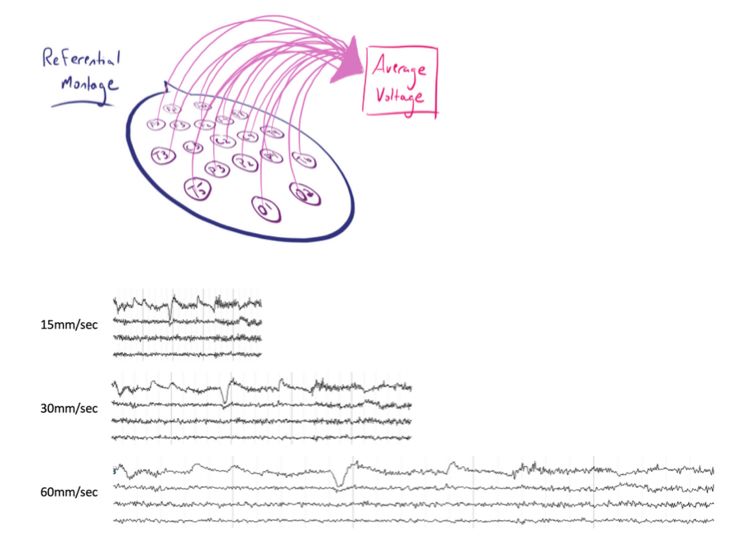
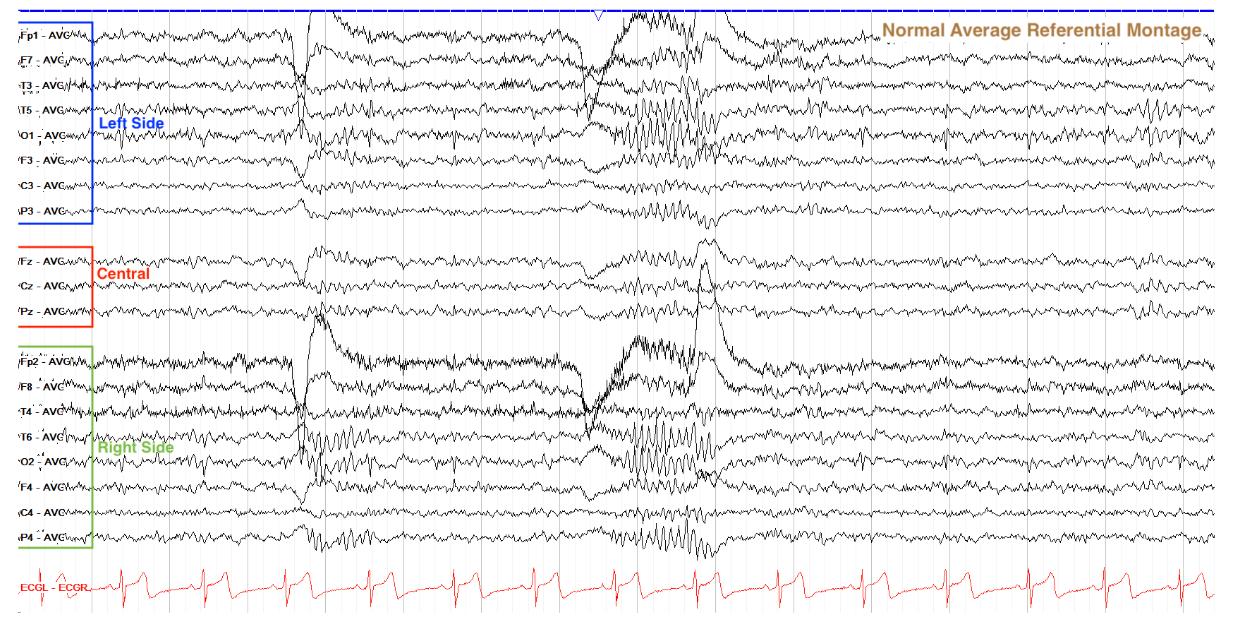
  • In a Referential[…] montage, electrodes are compared to a single refernce point
    • standard adult reading speed is 30[…]mm/sec; for neonates it's 15[…]mm/sc
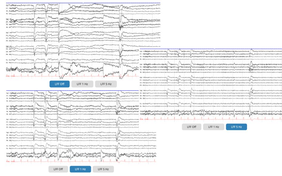
    • standard low frequency filter (LFF)[…] (which filters out frequencies below a certain threshold) is 1[…]Hz
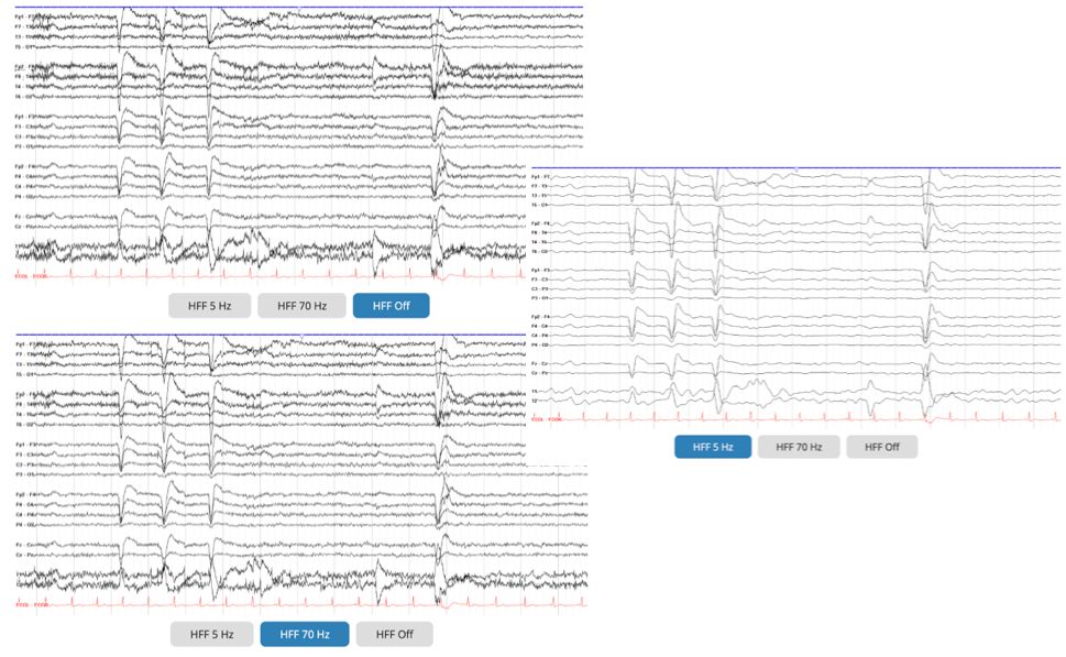
    • standard high frequency filter (HFF)[…] is 70[…]Hz
    • the notch filter[…] selectively removes 60[…]Hz activity that arises from electrical interference[…] such as wires and equipment
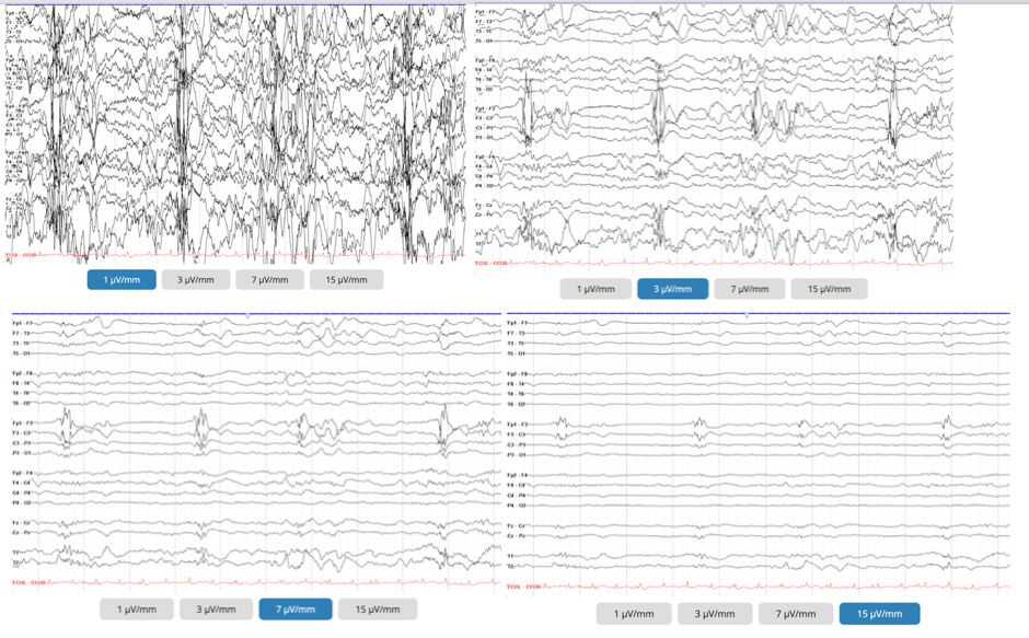
    • standard sensitivity[…] is 7[…]µV/mm; this value of the tracing is the height to change a waveform; a higher[…] number will smaller[…] the waveform appears (ex: instead of 5 µV to move a pen 1mm, it takes 10mm to move the pen 1mm)
    EEG - Frequencies
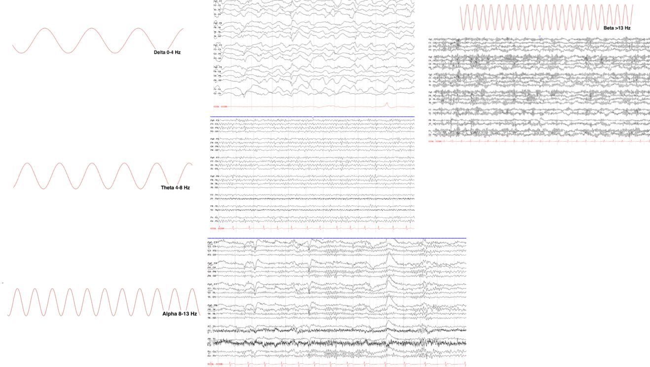
    • delta[…]0-4[…]Hz and the slowest
    • theta[…]4-8[…]Hz and often re-emerges in drowsy[…] periods
    • alpha[…]8-13[…]Hz and the halmark of the normal awake adult brain
    • beta[…]13-30[…]Hz and often found with benzodiazepene[medication?] use
    • higher[…] frequency is inversely related to lower[…] amplitude
    EEG - Morphology of Wave
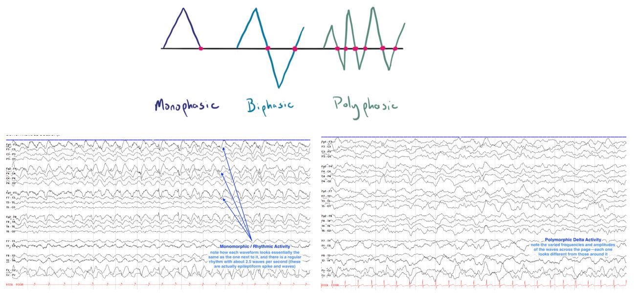
    • monophasic[…] wave begins on 1 side of baseline and crosses baseline only once
      • often concerning for epileptiform[…] activity such as in the case of rhythmic[…] activity
    • biphasic[…] wave begins on 1 side of baseline and crosses baseline twice
    • polyphasic[…] wave begins on 1 side of baseline and crosses baseline multiple times
    EEG - Organization 
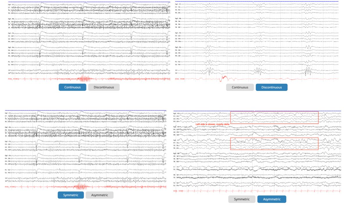
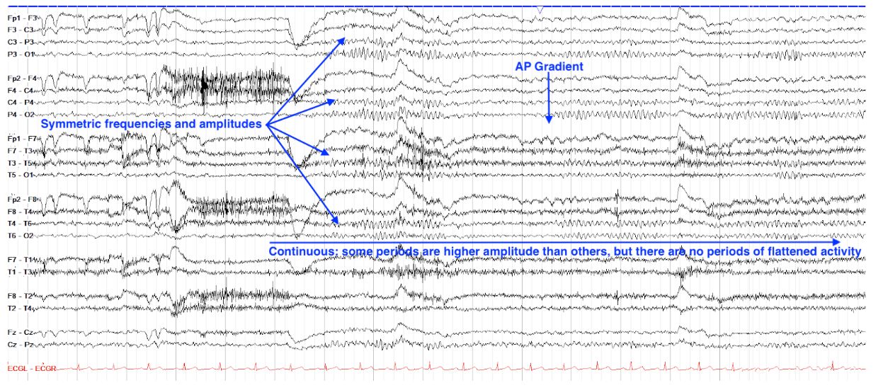
    • Continuity[…] - look if there are interruptions[…] (ex: flat[…] or attenuated[…] activity) which we call "discontinuous[…]" waveform
    • Symmetry[…] - look if the L and R sides appear similar in amplitude[…] and frequency[…]
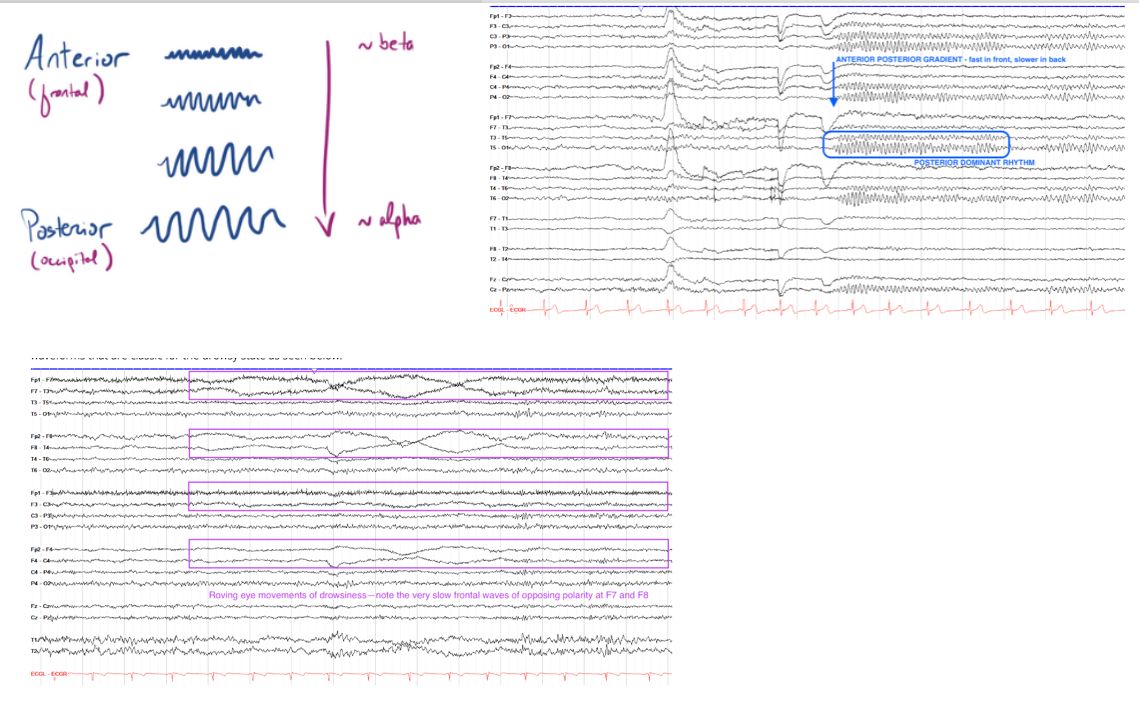
      • normal brains have an anterior-posterior gradient[…] where faster[…]lower[…] amplitude frequencies are near the front[front or back?] of brain while slower[…]higher[…] amplitude frequencies are near the back[front or back?] of brain
    • Posterior Dominant Rhythm (PDR)[…] - this is the frequency at rest[…] of the occipital[…] region when eyes are closed[…]
      • normal rhythm = alpha[…] range but specifically in adults it should be 8.5[…]-12[…]Hz
      • should be symmetric[…] from L to R; if there is >50[…]% amplitude difference or >1[…]Hz difference frequency between sides, this is abnormal
      • note: L[…] side of skull is slightly thicker in most ppl so the PDR may be a bit more attenuated there
    • Variability[…] - this is the presence of shifts[…] in the waveform across the tracing; normal brain should have these
    • Reactivity[…] - this is the presence of shifts[…] in the frequency[…] according to external stimuli[…]
    • State[…] - this is whether they are in awake[…]drowy[…], or asleep[…] states
      • Awake: symmetric[…] PDR w/predominant alpha[…] and beta[…] rhythm, with eye blink[…] artifact and movement[…] artifact; may see myogenic[…] artifact and chewing[…] artifact as well
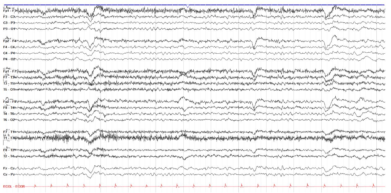
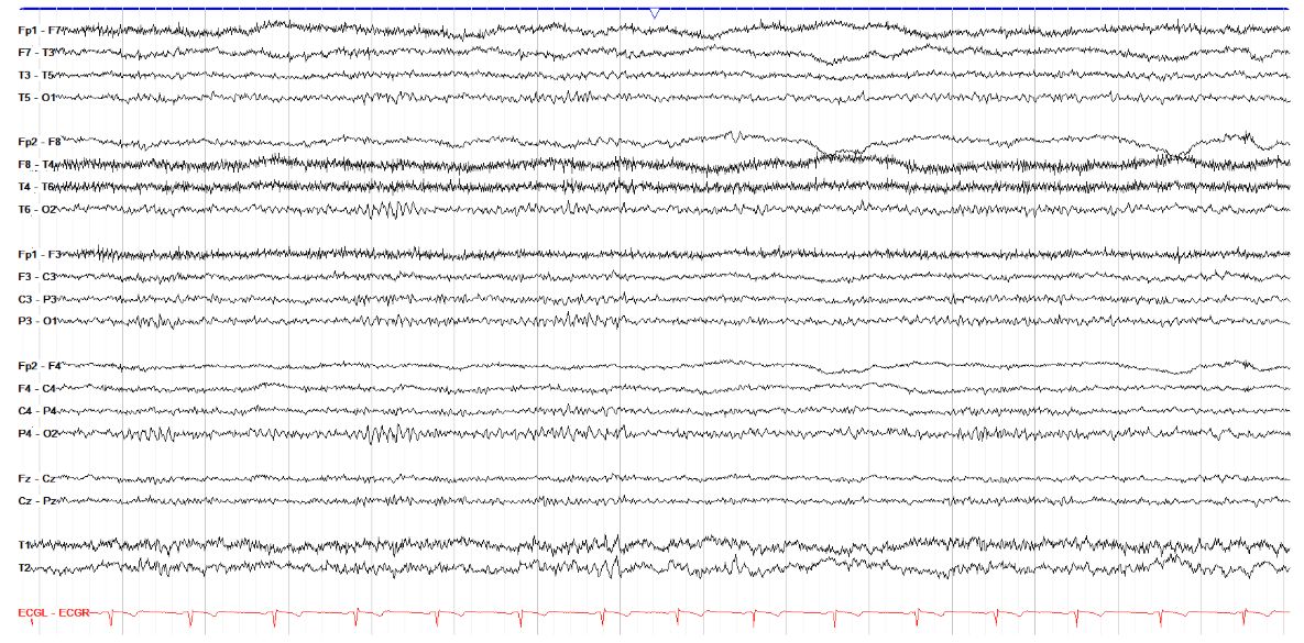
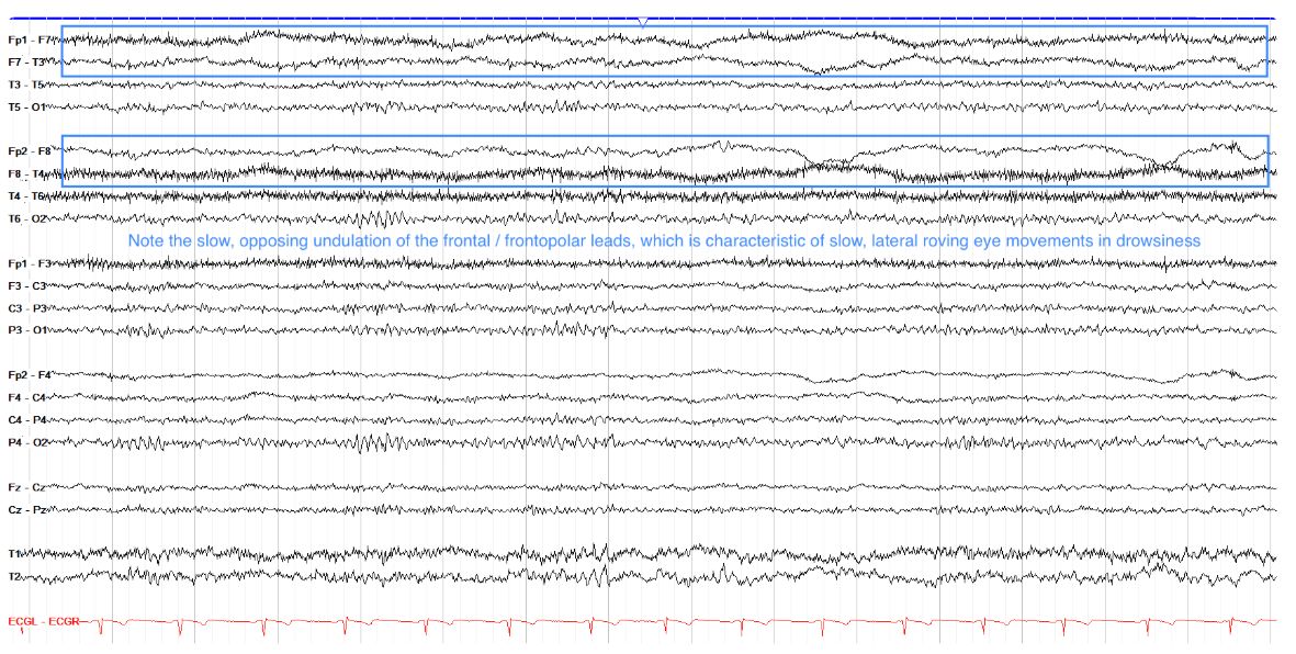
      • Drowsy: mild diffuse slowing[…] of rhythm w/decreased frequency[…] of eye blink and roving[…] eye movements
        • corneas are (+)[…] charged so when eyes look R then F8 sees the cornea (+) charge while F7 sees (-) charge
      • Sleep: discussed separately
    EEG - Provocation 
    • We use 2 types of provocation to assess underlying SZ risk
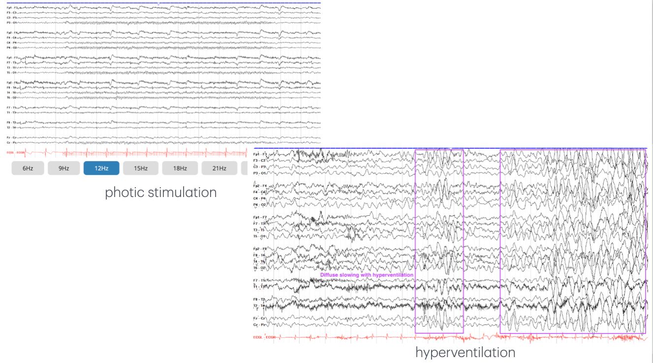
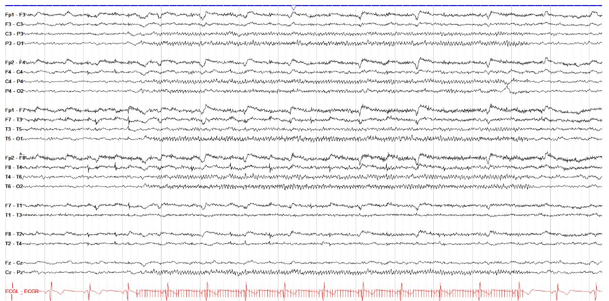
      • Photic Stimulation[…]: light is flashed in train of increasing frequencies to look for photic driving[…] where background is time-locked/sync'd to each light flash 
        • pts w/SZ may devlop a photo[…]paroxysmal response w/epileptiform activity
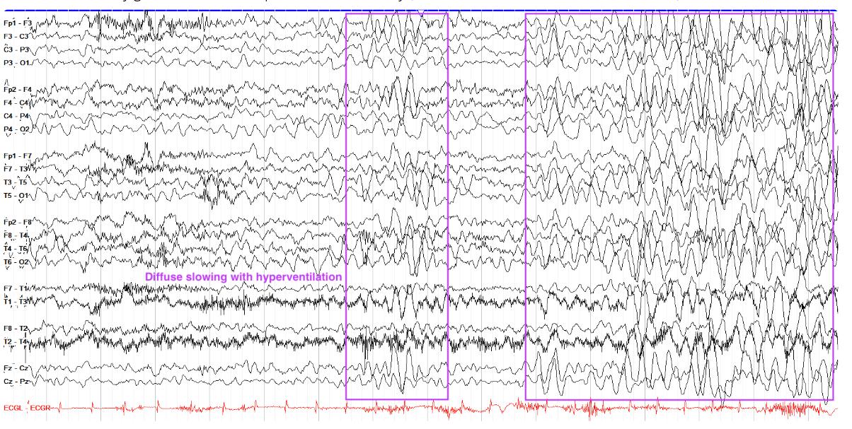
      • Hyperventilation[…]: this is when you ask patient to breathe large deep breaths very quickly and aggressively
        • avoid in pts w/cardiopulmonary[…] issues or recent stroke 
        • a classic response is diffuse slowing[…] in response
    EEG - Stage 1 Sleep
    • decreased eye blinks[…]
    • slow roving[…] eye movements
    • gradual loss of PDR[…] 
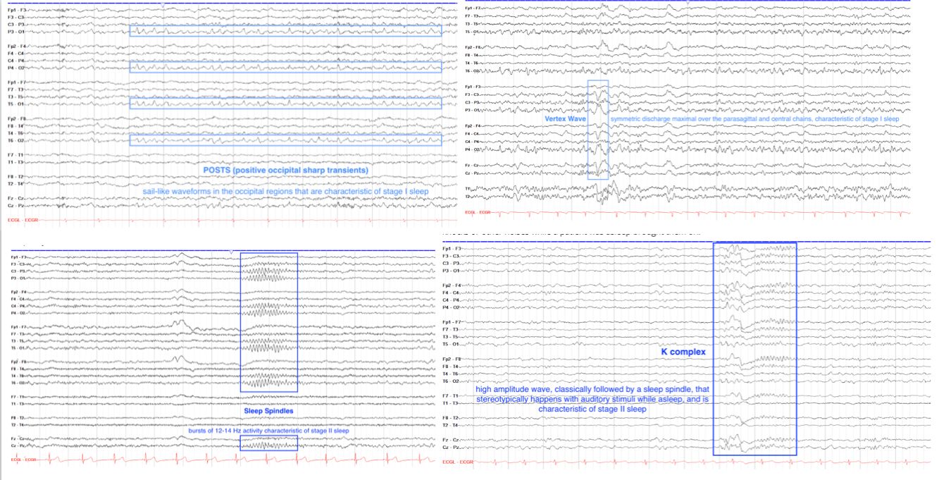
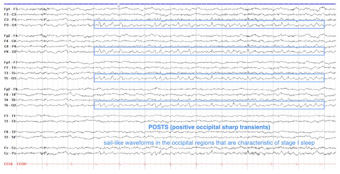
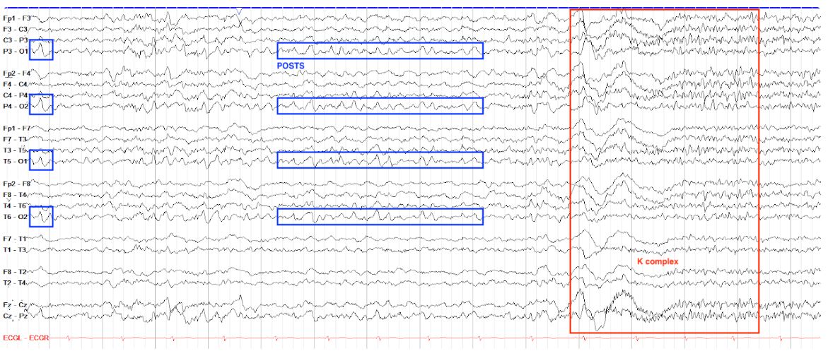
    • +positive occipital sharp transients[…] of sleep (aka POSTS[…])
      • these are positive deflections in the occipital[…] leads that look like sharp waves but non-epileptiform
      • classically described as "sail-like[…]" appearance
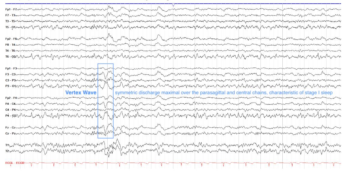
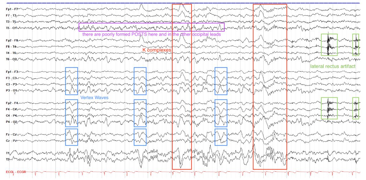
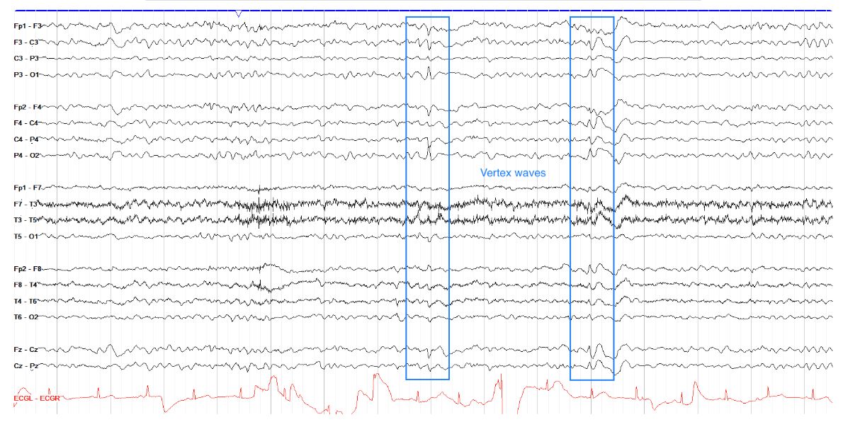
    • +vertex[…] waves
      • bilateral phase reversing[…] discharges over the central[…] regions
    EEG - Stage 2 Sleep
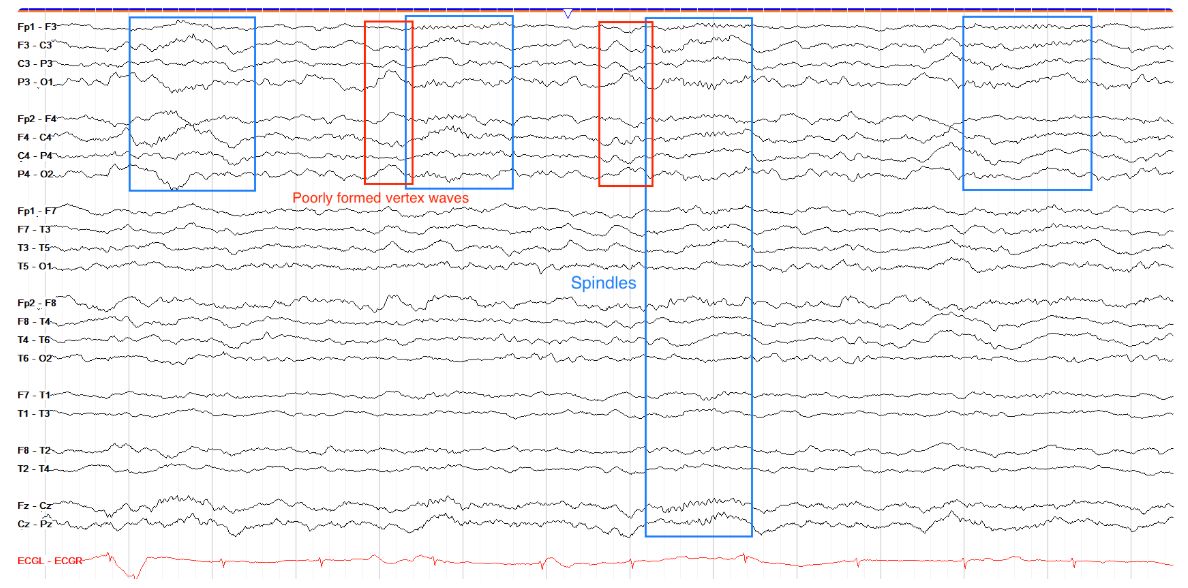
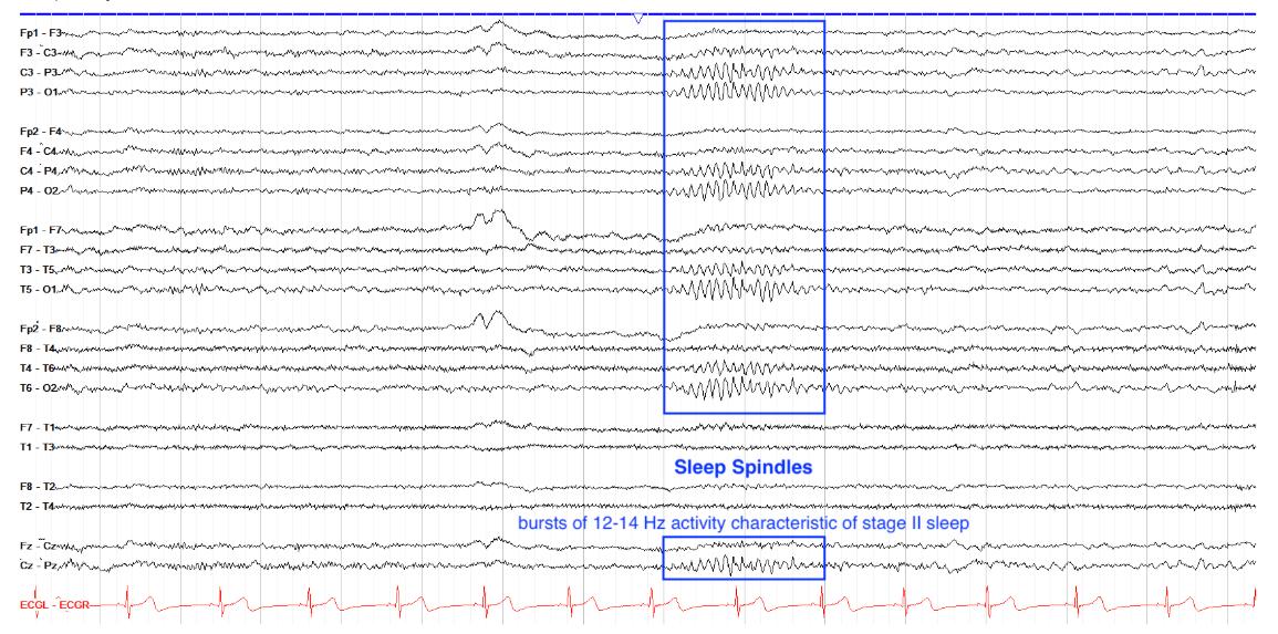
    • sleep spindles[…]
      • symmetric bursts of 12[…]-14[…]Hz activity arising from the reticular[…] nucleus of the thalamus
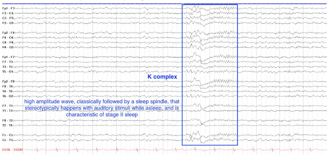
    • K complexes[…]
      • high amplitude[…], symmetric waveforms with initial negative[…] followed by a slow positive[…] phase
      • often followed by a sleep spindle[…]
    EEG - Stage 3 Sleep
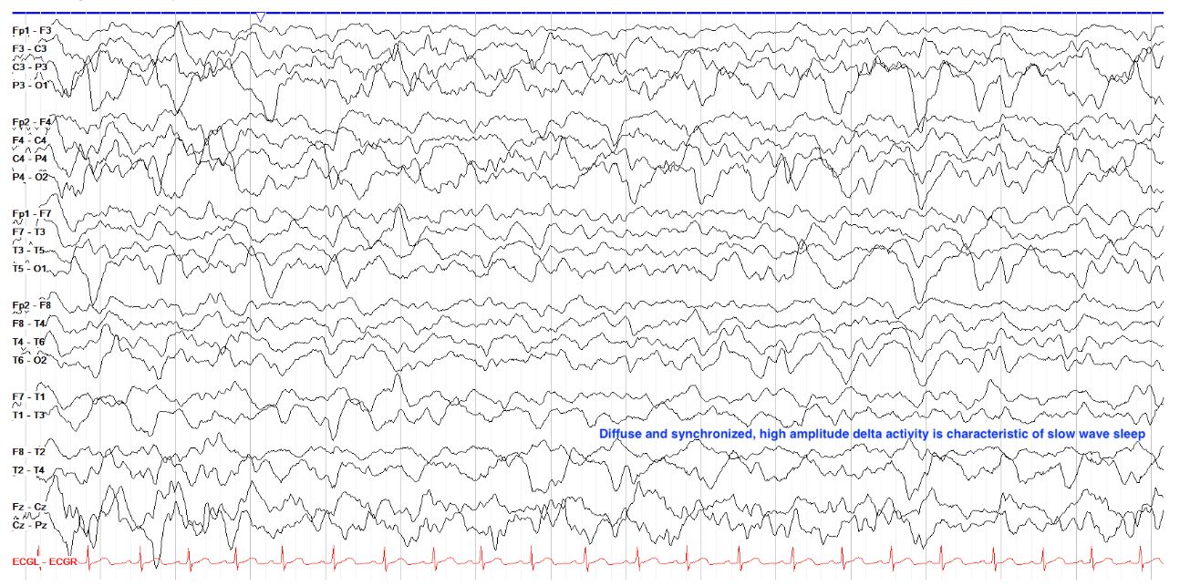
    • aka "Slow Wave[…]" Sleep
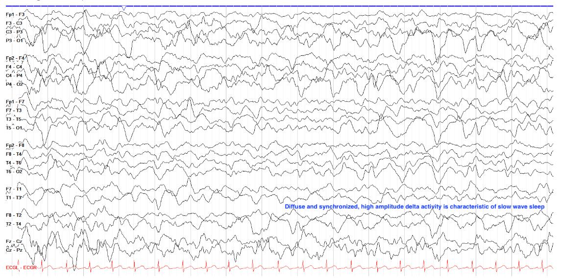
    • high[…] amplitude, synchronized delta[…] activity
    • meds such as benzos[…] reduce the amount of this stage
    EEG - REM Sleep
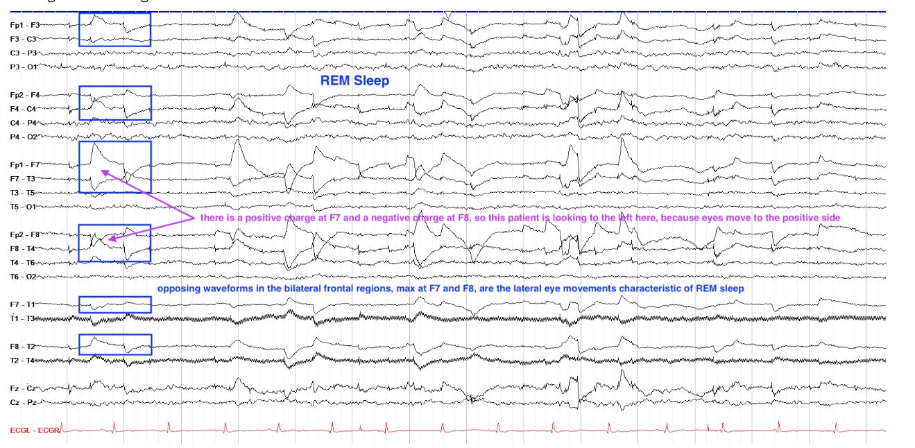
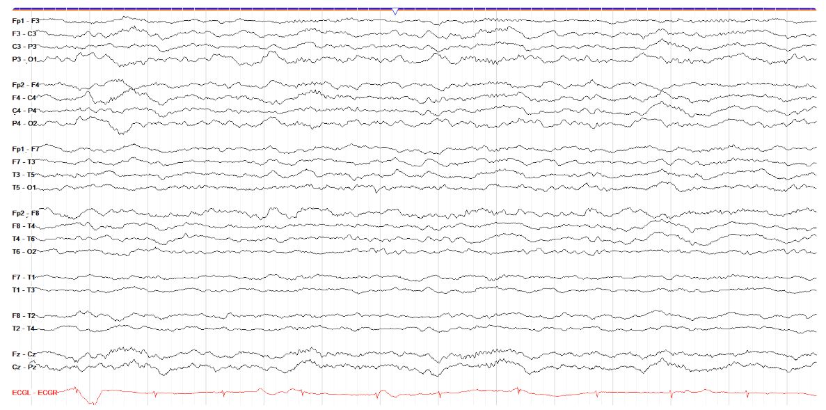
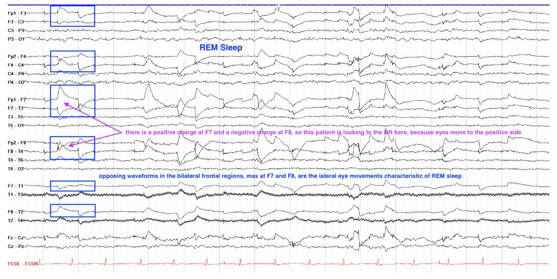
    • marked by diffuse attenuation[…] of amplitudes w/range of frequencies
    • the rapid eye movements are seen as sharply contoured opposing waveforms in the left[…] and right[…] frontal[…] regions
      • these arise bc the cornea is (+)[…] charged > pt looks R > R eye cornea moves closer to the F8[…] electrode (causing positive[…] charge) but the L eye looks away from the F7[…] electrode (causing negative[…] charge) > leads to +[…] charge the direction you're looking and -[…] charge the opposite side
    EEG - Artifact
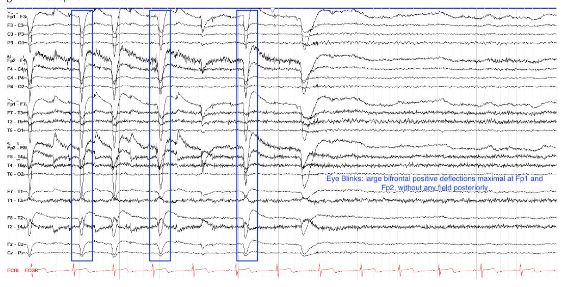
    • Eye Blink[…] Artifact
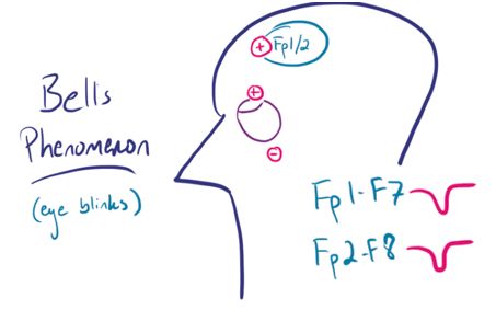
      • arise d/t Bell's[…] Phenomenon
        • cornea is +[…] charged, retina is -[…] charged
        • when blinking, eyes slightly roll up[…], causing cornea to move closer to frontal[…] electrodes Fp1[…]Fp2[…]
        • on EEG you'll see a positive[…] wave
    EEG - Artifact
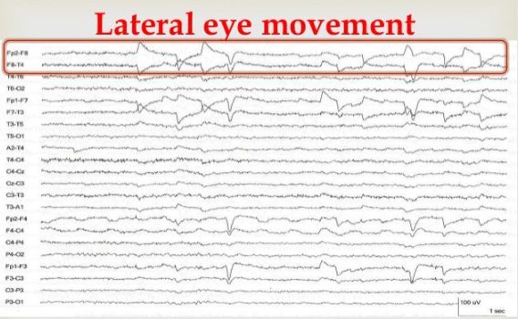
    • Lateral Eye Movement[…] Artifact
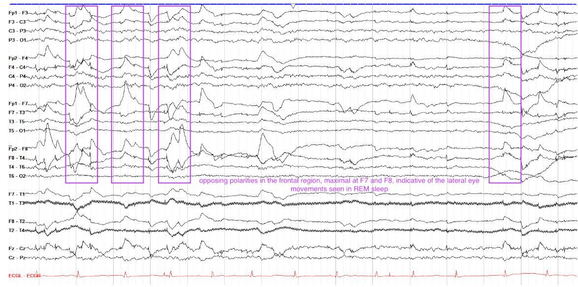
      • you'll see opposing polarity waves in F7[…]F8[…] leads
      • recall: cornea = +[…] charge, retina = -[…] charge
      • when you look R, the R eye cornea gets closer to F8 > +[…] charge while the L eye > -[…] charge 
    EEG - Artifact
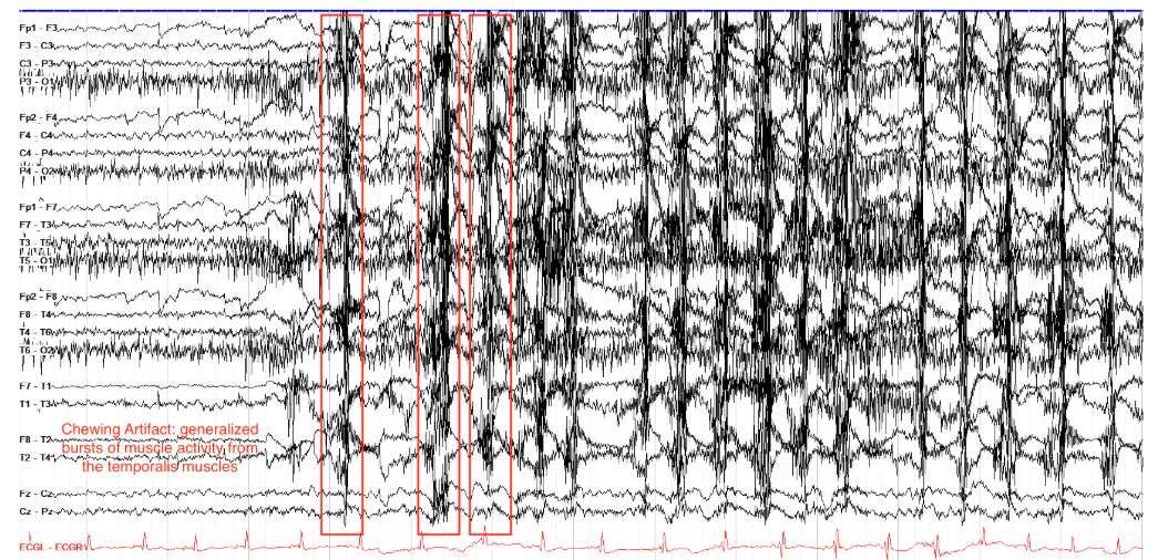
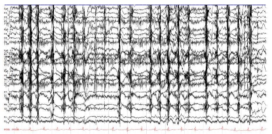
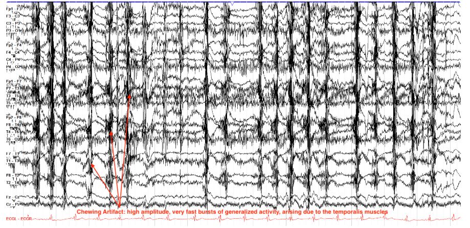
    • Chewing[…] Artifact is muscle artifact from the temporalis[…] muscle marked by sudden onset, intermittent bursts of generalized fast activity
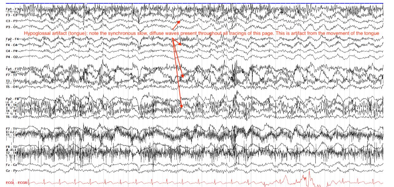
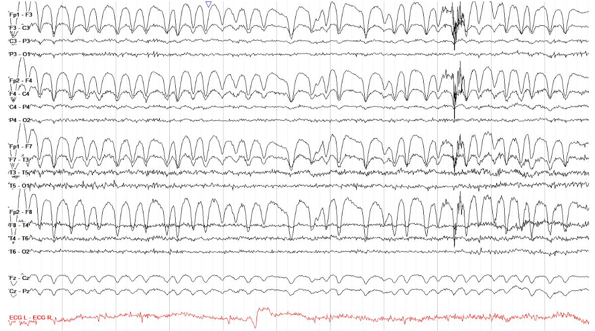
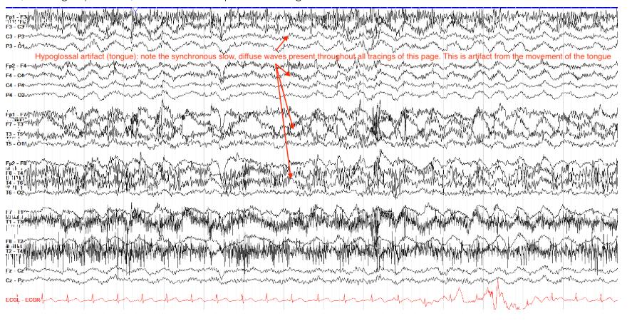
    • Tongue Movement[…] Artifact also known as Hypoglossal[…] Artifact arises due to movement of the tongue[…]
      • one notable point is that it is reproducible[…] for example when you ask the patient to say "la la la"
    EEG - Artifact
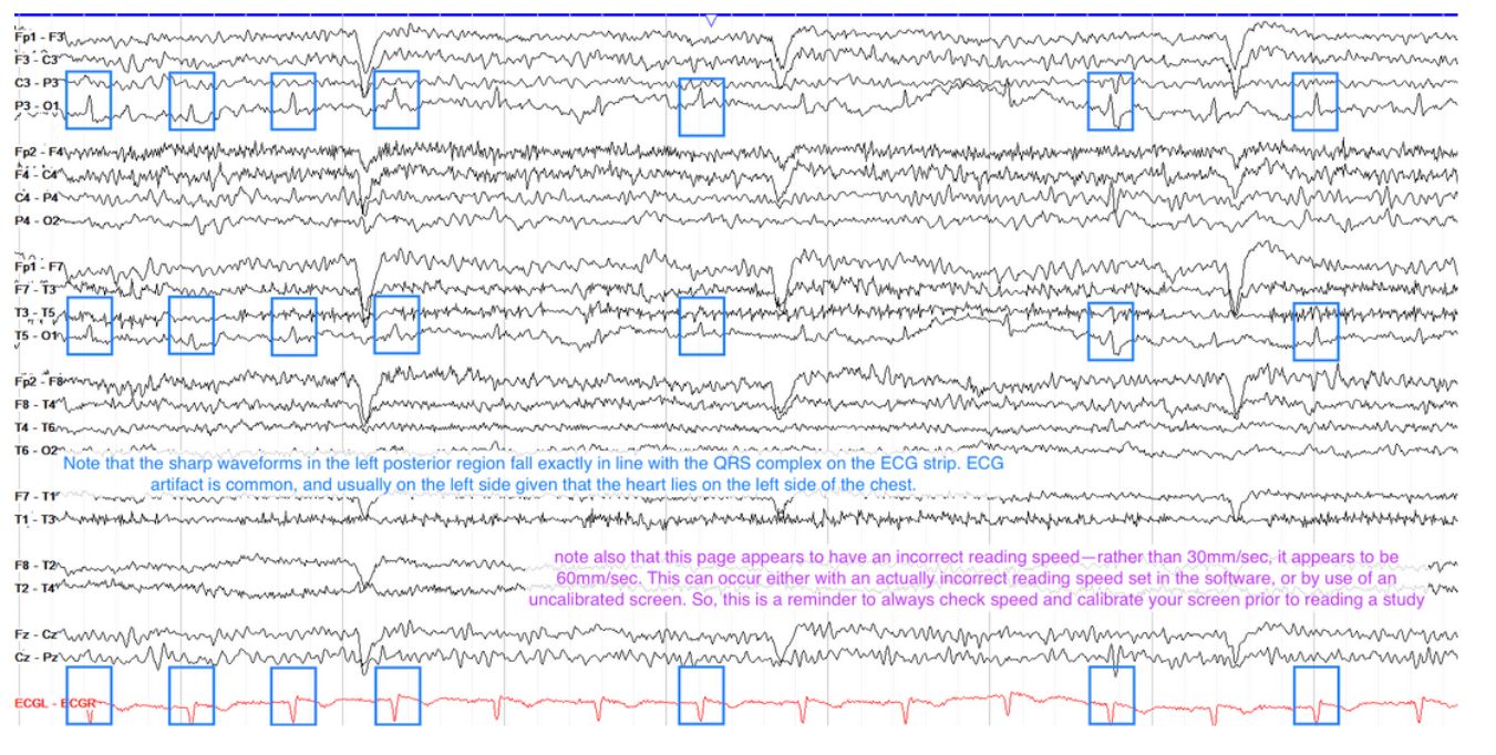
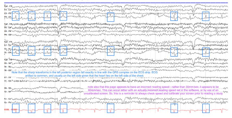
    • ECG[…] artifact has waveforms time locked to the QRS[…] complex
      • more often seen on the L[…] side because the heart[…] is located there
    EEG - Artifacts
    • Electrical[…] Artifact - d/t wires (~60[…]Hz in US and 50Hz in Europe)
      • can use the notch[…] filter to help remove this
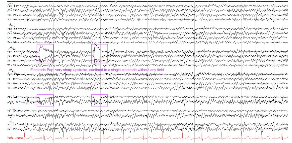
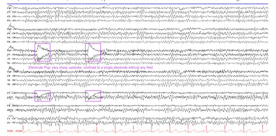
    • Electrode[…] artifact - a good example is electrode "pop[…]" often caused by a loose[…] electrode 
      • this will look like a sudden, deep up[up or down?]slope with a slow down[up or down?]slope
    EEG - Artifacts
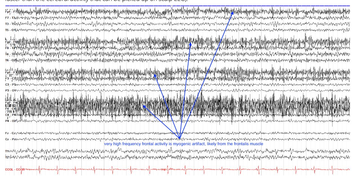
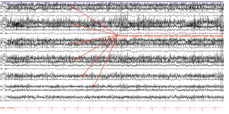
    • Myogenic[…] Artifact - often found in the frontal[…] region d/t frontalis muscle[…]temporal[…] region d/t temporalis[…] muscle
    EEG - Artifacts
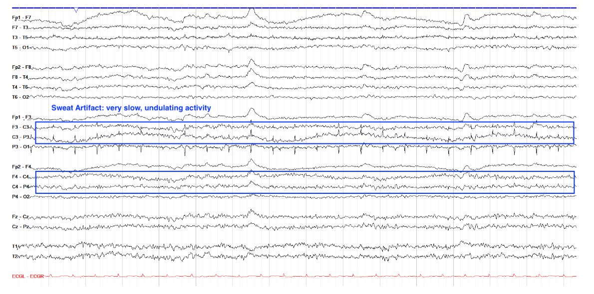
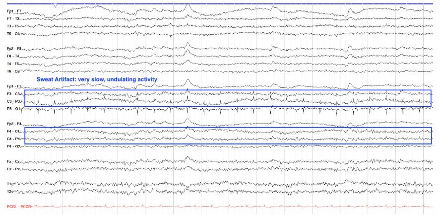
    • Sweat[…] Artifact - very low amplitude, slow[…] frequency d/t the NaCl-[…] charge picked up by electrodes

    EEG - Artifacts
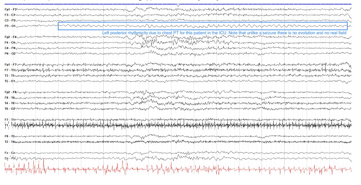
    • Chest PT[…] Artifact - esp seen in intubated ICU patient -- will look like a monomorphic seizure but there will be no evolution[…] and no real field
    EEG - Artifacts
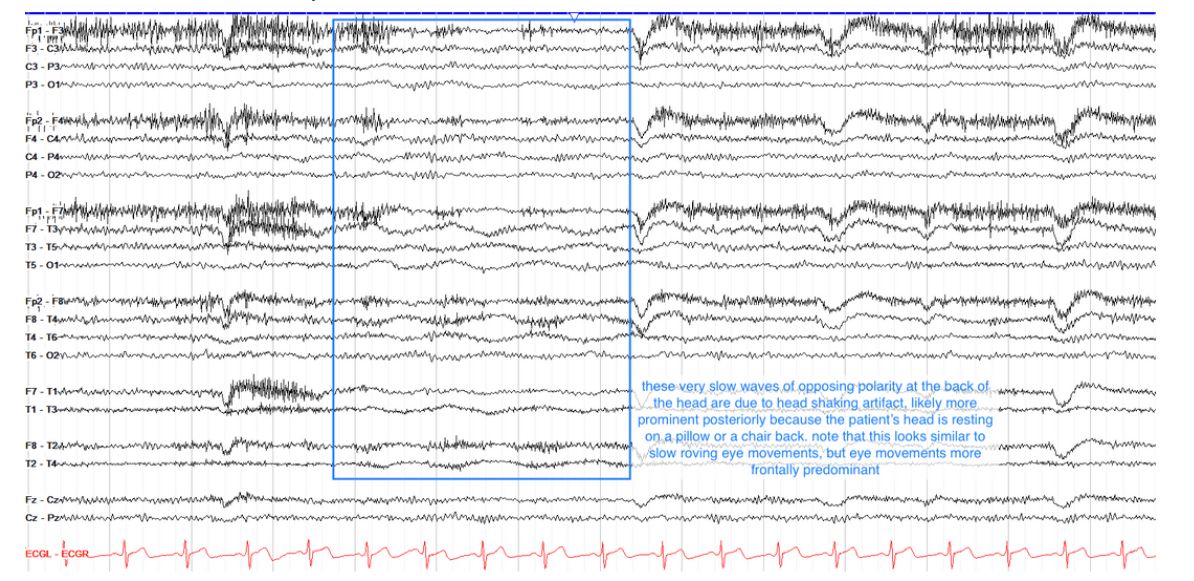
    • Head Shaking[…] Artifact - slow, low amplitude activity that will look similar to eye movement[…] artifact but unlike that artifact, this is not localized to the anterior[…] leads alone
    EEG - Artifacts
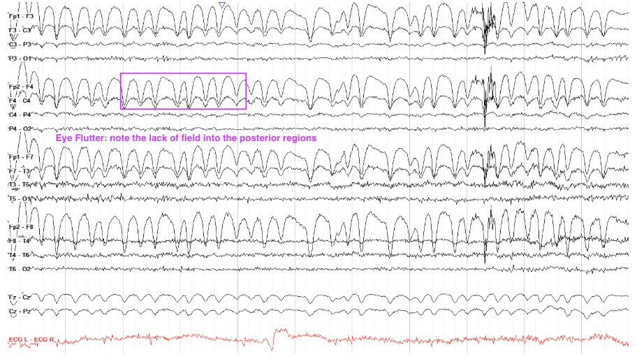
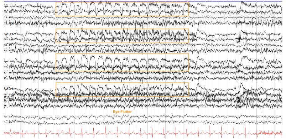
    • Eye Flutter[…] Artifact - similar to eye blinks but repetitive and note that they will NOT appear in the posterior[anterior or posterior?] fields
    EEG - Normal Variants
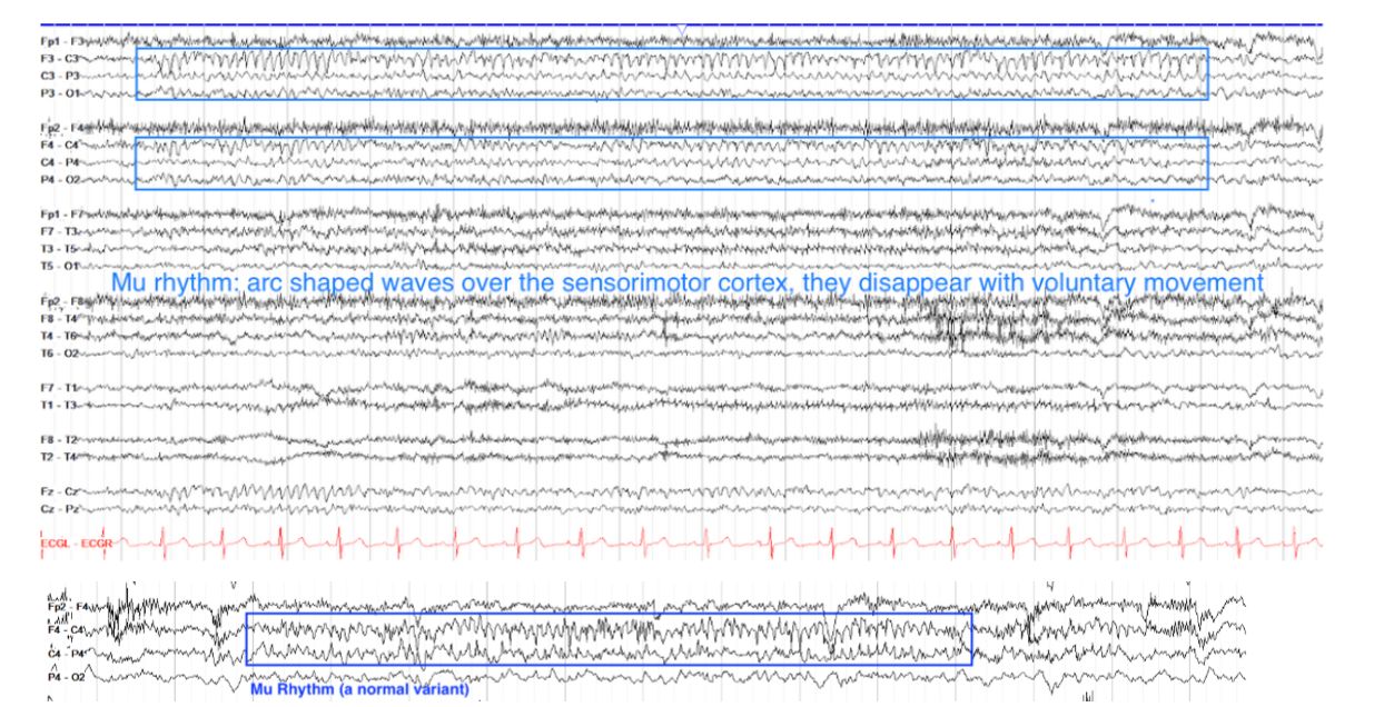
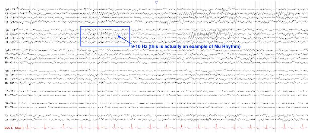
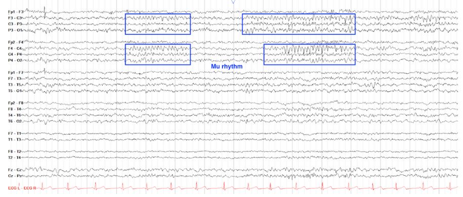
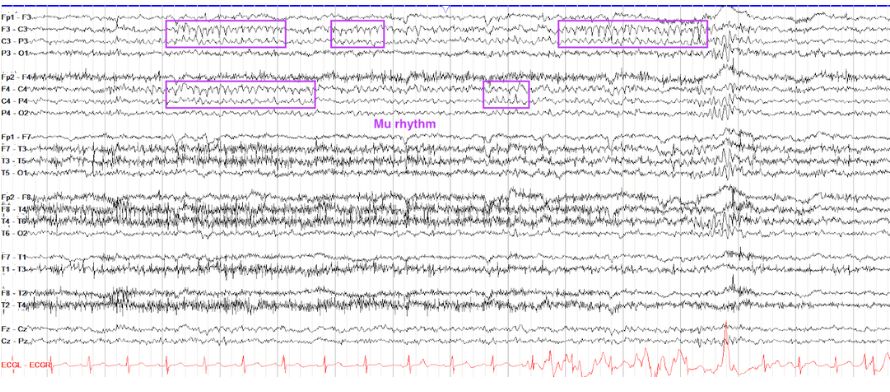
    • Mu[…] Rhythm 
      • similar to PDR[…] for occipital[…] region, this is the idling activity of the sensorimotor[…] region
      • goes away with thoughts of and actual motor[…] activity
      • appears as alpha[…] activity (usually 7-11Hz) over the parasagittal[…] regions (e.g., around the "C[…]" leads)
    EEG - Normal Variants
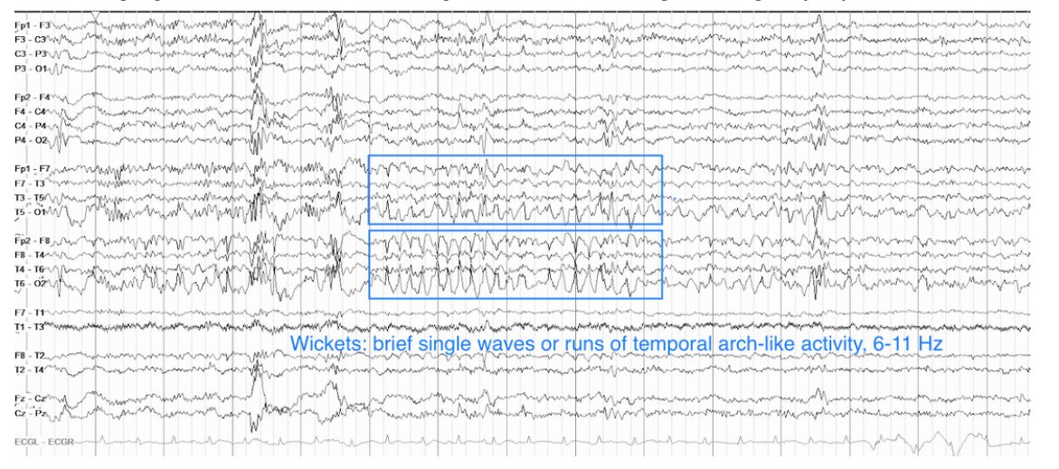
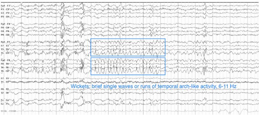
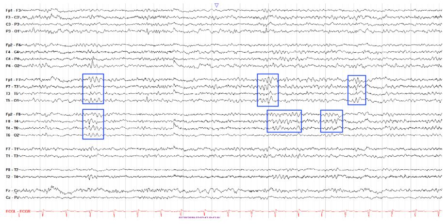
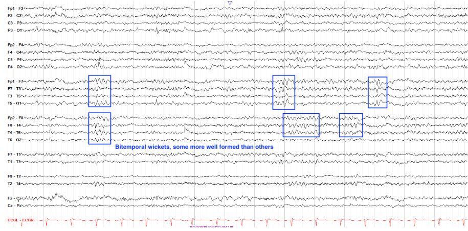
    • Wicket[…] Waves - similar in appearance to wickets[…] used in cricket
      • arch-like, alpha[…]-range rhythm (7[…]-11[…]Hz) 
      • seen in the temporal[…] region
      • unlike SZ: they will NOT have an aftergoing slow[…] wave, will NOT change the background[…], will NOT propogate into the neighboring part of the electrode chain[…], and will NOT evolve[…] over time
    EEG - Normal Variants
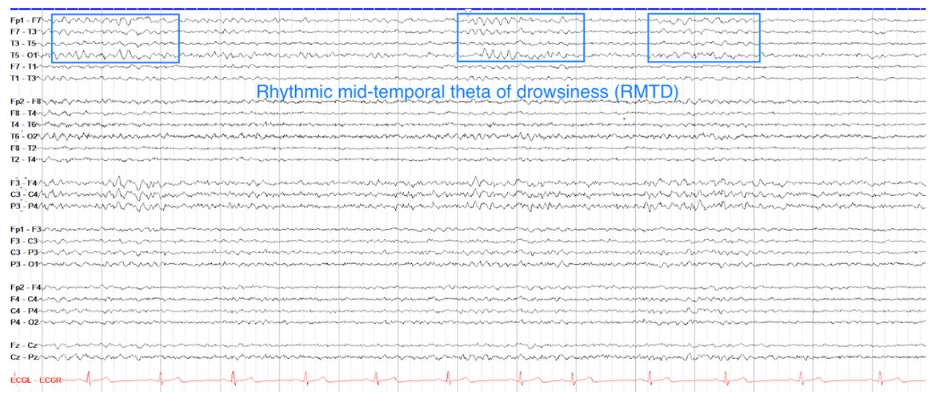
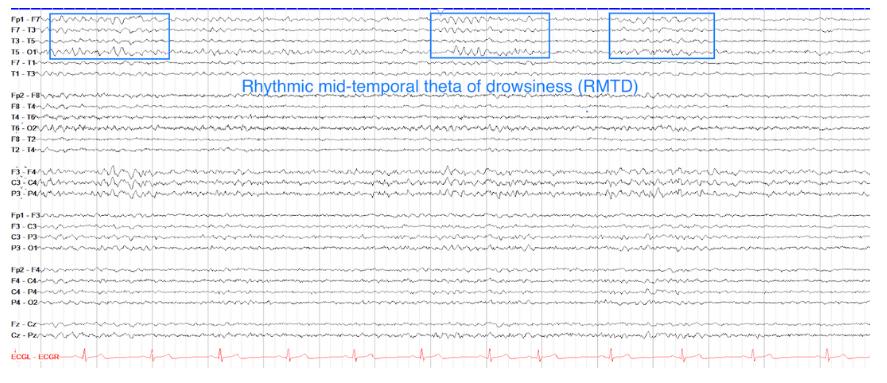
    • Rhythmic Mid-Temporal Theta of Drowsiness (RMTD)[…]
      • sharply contoured, rhythmic theta[…] activity
      • restricted to temporal[…] regions
      • it does not evolve[…] like a seizure would
    EEG - Normal Variants
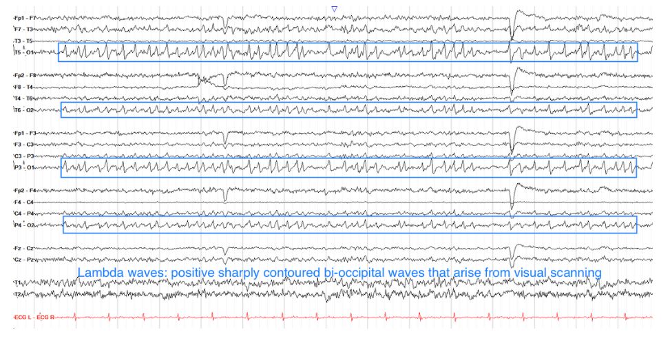
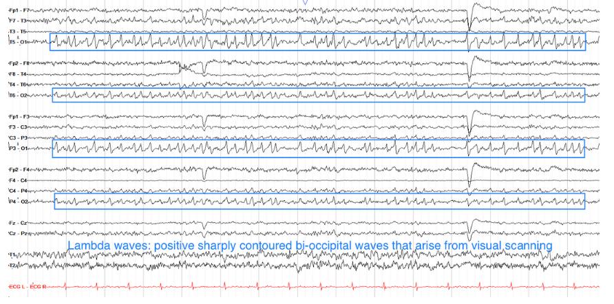
    • Lambda[…] Waves - usually arise in awake[…] state when patient is visually scanning something as with reading[activity?]
      • these are BL, symmetric, sharply contoured positive waves in occipital[…] leads with a "sail-like[…]" appearance
      • similar to POSTS[other variant] but these come w/ other e/o wakefulness like eye blink[…] artifact, myogenic[…] artifact, etc rather than in early sleep[…] stage
    EEG - Normal Variants
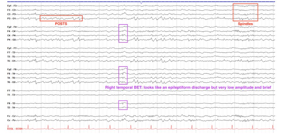
    • Benign Epileptiform Transients of Sleep (BETS)[…] - aka "Small Sharp Spikes (SSS)[…]"
      • this is a normal[normal or abnormal?] finding in drowsy[…] and asleep[…] states
      • have a low amplitude[…] and duration[…] 
      • mostly seen in temporal[…] leads
    EEG - Normal Variants
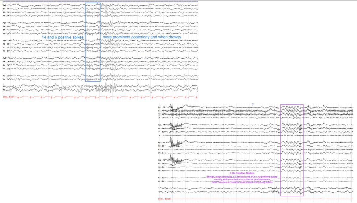
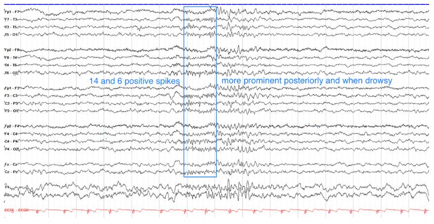
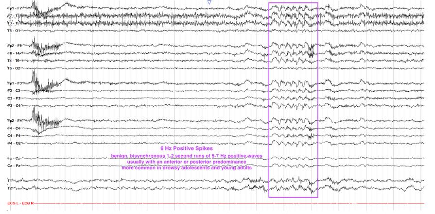
    • 14 and 6 Positive Spikes[…] 
      • these are 1[…]-2[…] second bursts of sharply contoured positive waveforms in frequencies of either 14[…]Hz or 6[…]Hz; often bilateral/sync'd
    EEG - Nonepileptiform Abnormalities
    • slowing[…] 
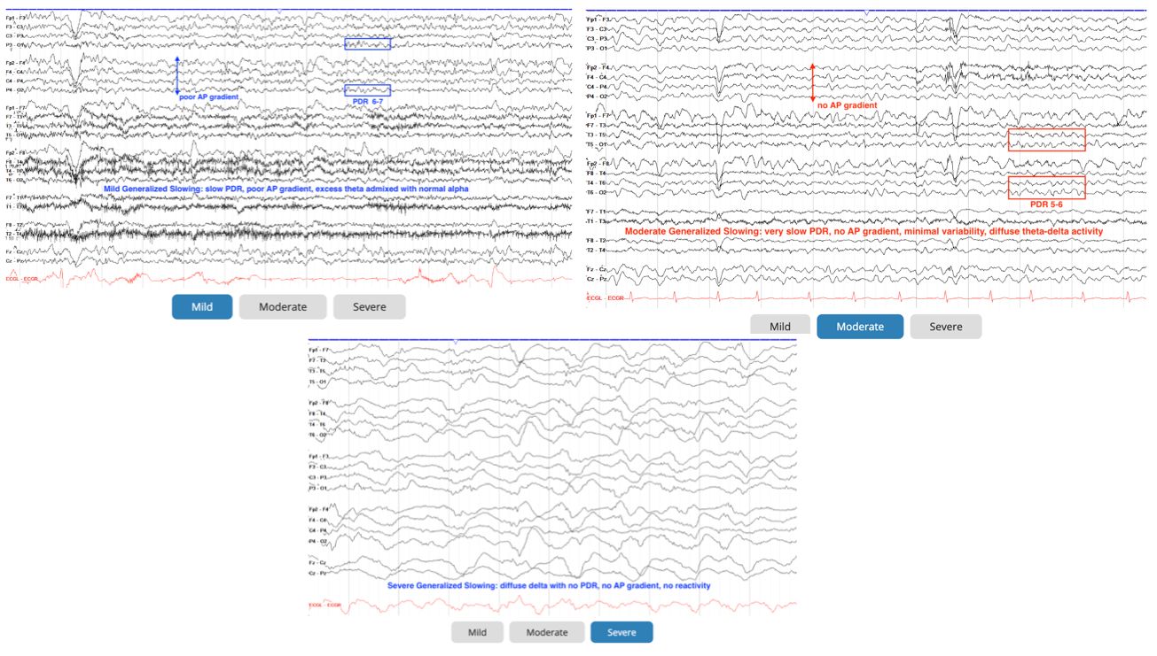
      • generalized slowing[…] 
        • mild[…]: mostly alpha[…]-theta[…] rhythm
        • moderate[…]PDR[…] is messed up or gone, mostly theta[…]-delta[…] rhythm
        • severe[…]: mostly delta[…]
      • focal slowing[…]
        • continuous[…] - c/f structural lesion like tumor/bleed/stroke/demyelinating
        • intermittent[…] - c/f smaller structural lesion such as FCD or small tumor
        • polymorphic[…] 
        • monomorphic[…] - more c/f epileptiform activity
    EEG - Nonepileptiform Abnormalities
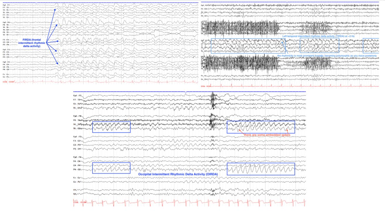
    • "IRDA[…]s"
      • Frontal (FIRDA)[…]: does not travel back to the posterior[…] regions
      • Generalized (GIRDA)[…]: travels back to the posterior[…] regions
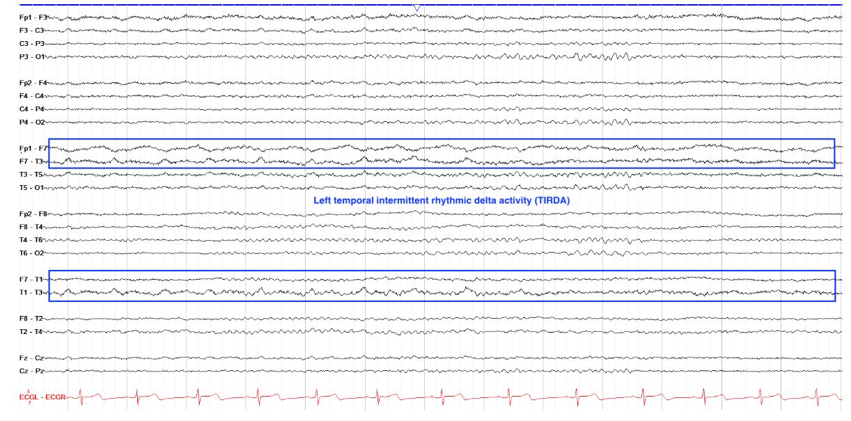
      • Temporal (TIRDA)[…]: this is an epileptiform discharge w/high risk seizures 
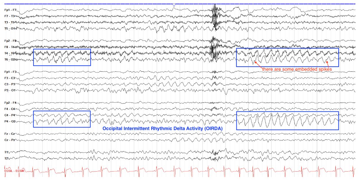
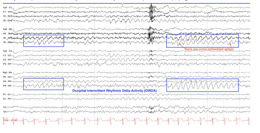
      • Occipital (OIRDA)[…]: this is an epileptiform discharge
    intermittent rhythmic delta activity (IRDA)
    EEG - Nonepileptiform Abnormalities
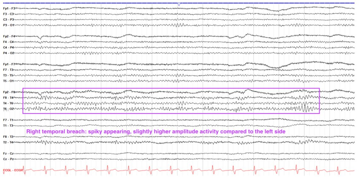
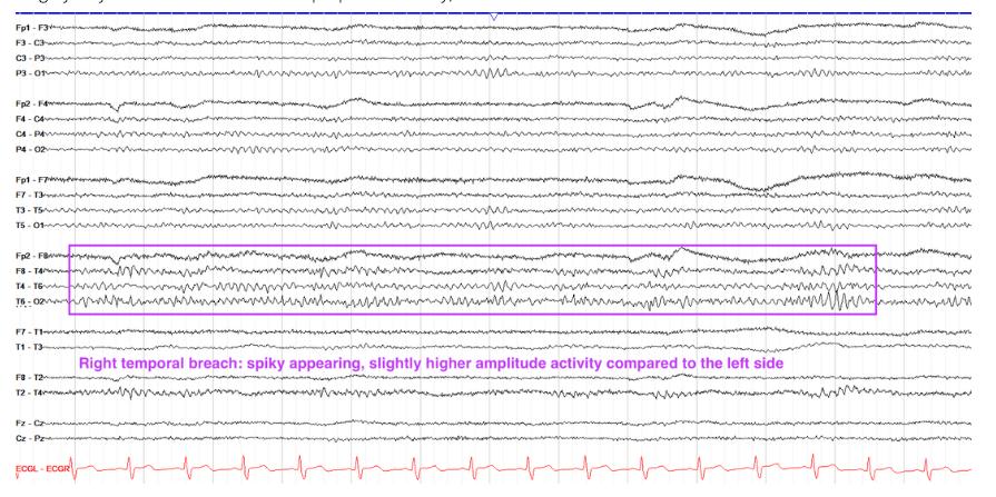
    • Breach[…] Activity: higher amplitude, spiky/irregular activity over an area of skull that has been previously opened[…] d/t surgery
    EEG - Nonepileptiform Abnormalities
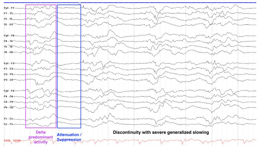
    • attenuation[…] - decrease in amplitude[…]
      • often d/t fluid (blood/CSF)[…] between skull/brain or stroke or tonic seizures
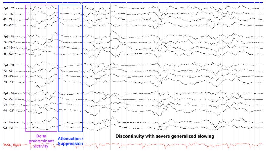
    • discontinuity[…] - intermittent periods of attenuation[…]
      • extreme sedation leading to intentional discontinuity is called burst suppression[…] (requires >50[…]% of the record to be suppressed)
    EEG - Nonepileptiform Abnormalities
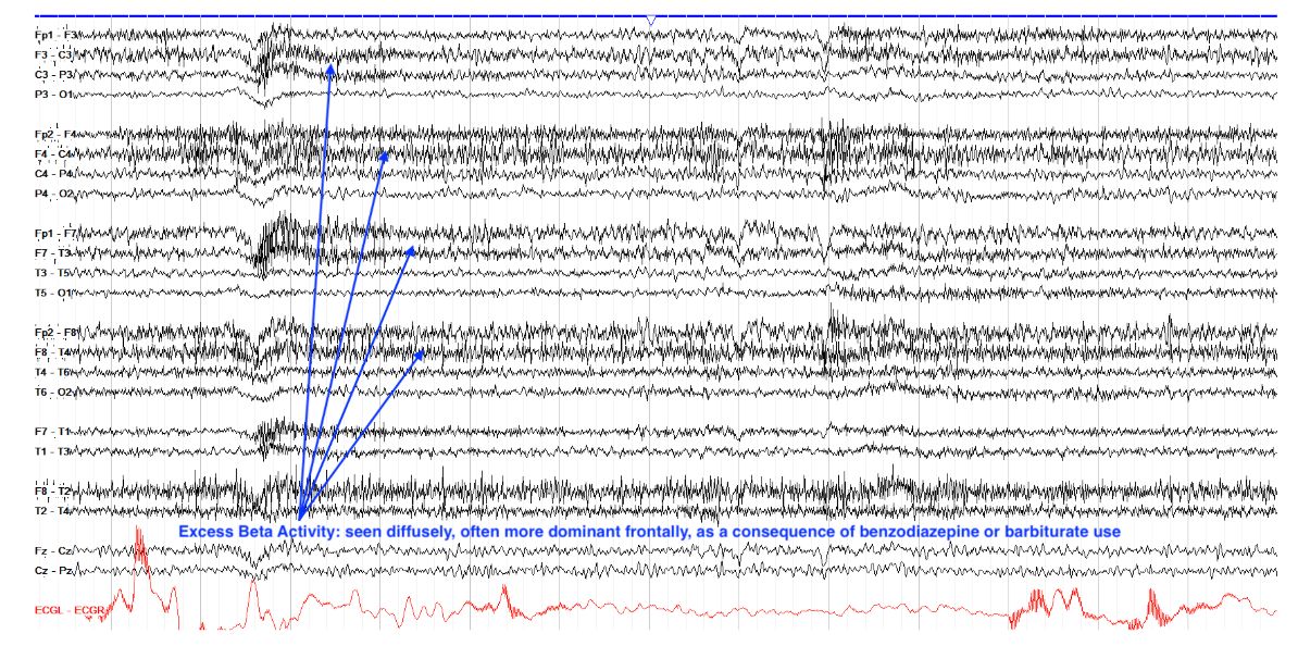
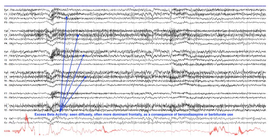
    • excess beta[…] activity is often 2/2 benzo[med class?] or barbiturate[med class?] use
      • typically a benign[benign or worrisome?] finding
    EEG - Nonepileptiform Abnormalities
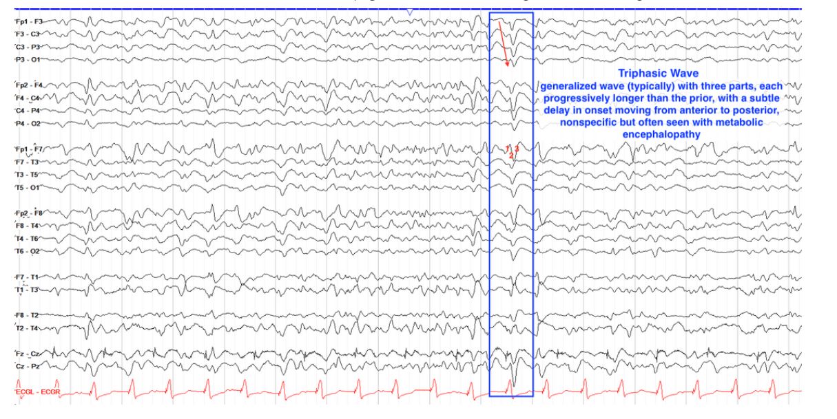
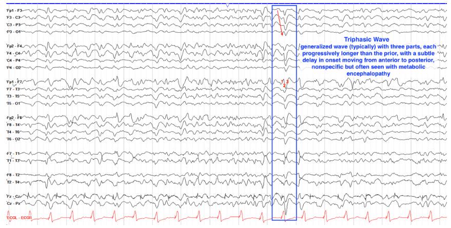
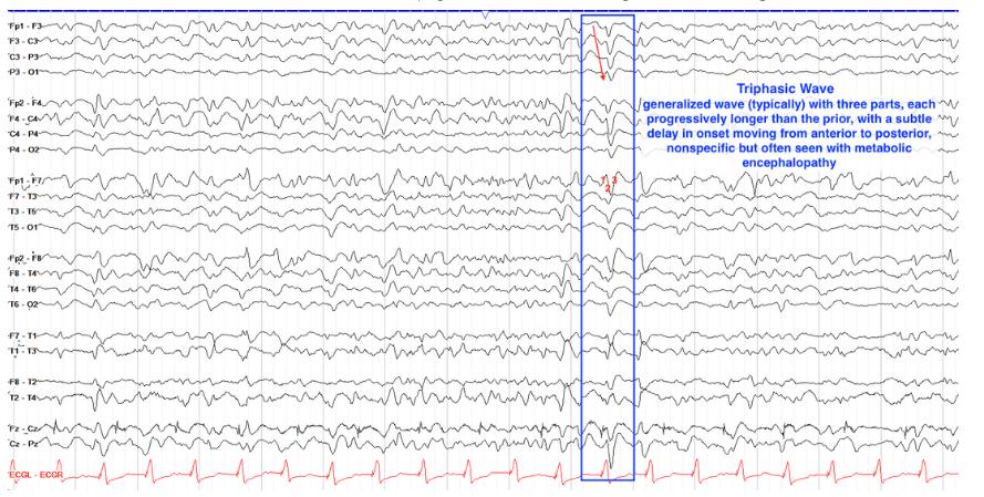
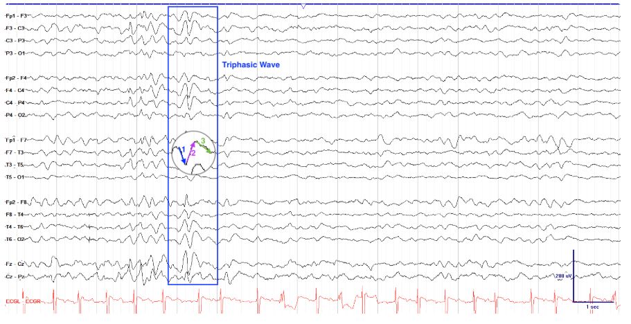
    • Triphasic[…] Waves
      • classically associated with metabolic[…] encephalopathy
      • often generalized[focal or generalized?]
      • comprised of 3[…] parts, each slightly longer[…] than the last part
      EEG - Epileptiform Activity
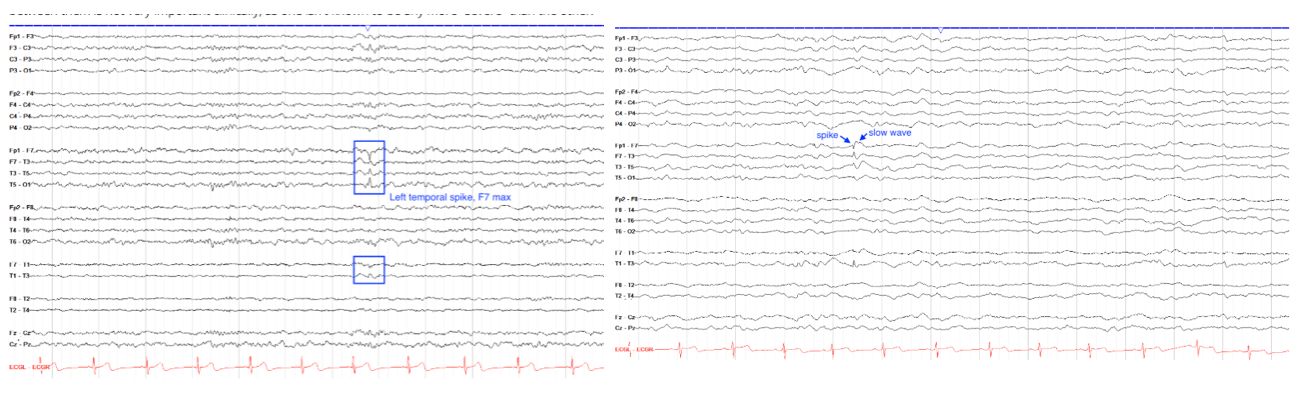
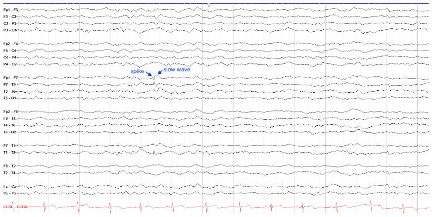
      • Sharp[…]: single epileptiform discharge w/disruption of background[…]
      • Spike[…]: faster version of above 
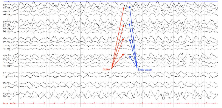
      • After a sharp or spike, there is typically a slow wave[…] which is the refractory period of the affected neuron population, often higher[…] amplitude than precedent spikes/sharps
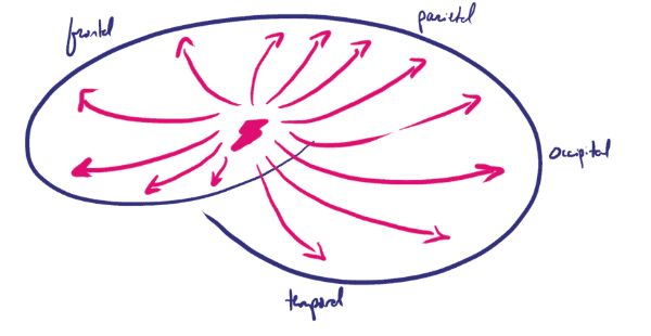
      • Interictal should always disrupt the background[…] and have a field[…] (aka "ripples[…]" of a discharge in the surrounding EEG electrodes)
      EEG - Epileptiform Activity
      • generalized[…] discharges cannot be localized as they involve the entire cortex simultaneously
        EEG - Epileptiform Activity
        • Periodic Patterns
          • discharges must occur for at least 6[…] cycles total
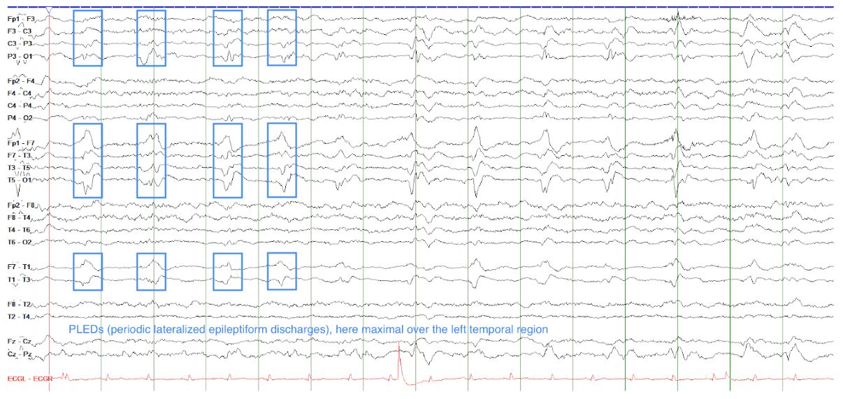
          • ex) LPD[…]s aka PLED[…]s
          • these can be part of a seizure if they reach 2.5[…]Hz for at least 10[…] seconds

        LPDs = lateralized periodic discharges
        PLEDs = periodic lateral epileptiform discharges
        EEG - Epileptiform Activity
        • Rhythmic Patterns
          • FIRDA[…] and GIRDA[…] are not necessarily epileptogenic
          • when you see consistently lateralized rhythmic pattern such as theta[…] or delta[…] this should raise c/f focal cortical issue
            • TIRDA[…] is often associated w/underlying focal temporal lesion (FCD, tumor, etc)
            • OIRDA[…] is more common in kids and usually epileptogenic but not as much as temporal
        FIRDA = frontal intermittent rhythmic delta activity
        GIRDA = generalized intermittent rhythmic delta activity
        TIRDA = temporal intermittent rhythmic delta activity
        OIRDA = occipital intermittent rhythmic delta activity
        EEG - Epileptiform Activity
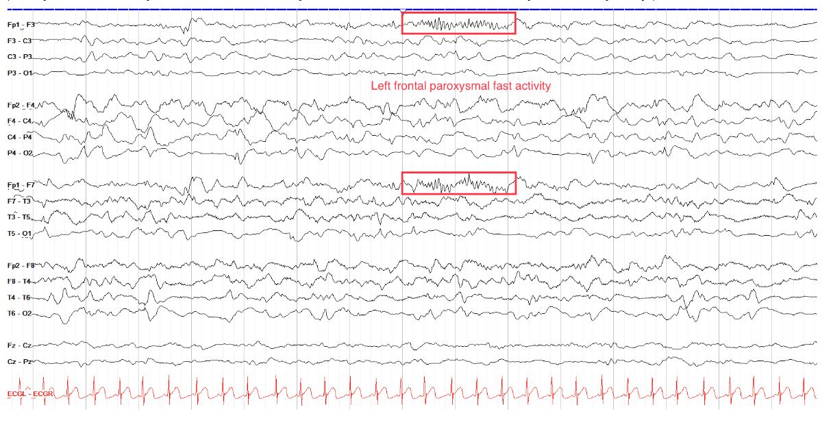
        • Paroxysmal Fast[…] Activity
          • usually benzo[medication?] effect
          • often benign[benign or problematic?] and usualy seen in frontal[location?] regions
        EEG - Epileptiform Activity
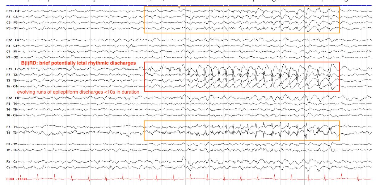
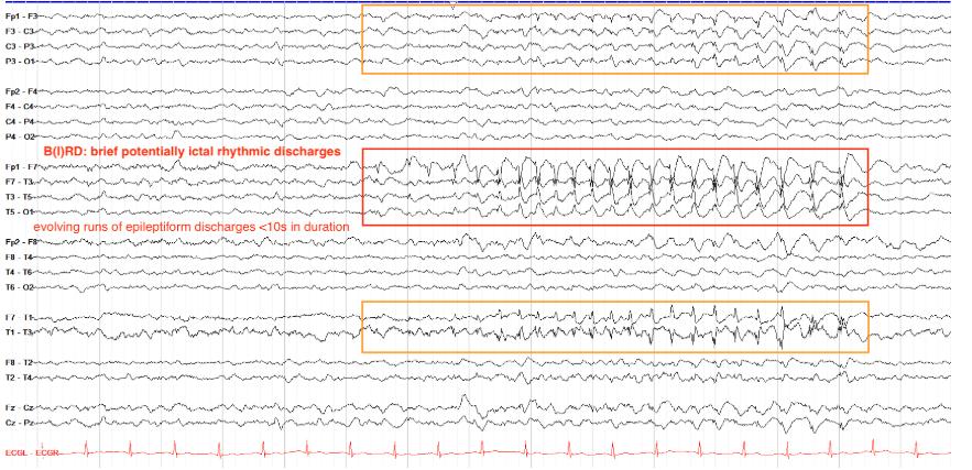
        • Brief Potentially Ictal Rhythmic Discharges (BIRDS)[…]
          • these are rhythms that evolve but do not meet the minimum 10[…] second criteria to constitute a SZ
          • often seen in pts with NMDA[…]r encephalitis or other critically ill patients
          • over 90% will go on to have SZ[…] eventually
        EEG - Epileptiform Activity
        • Photoparoxysmal[…] Response
          • often seen as emergence of interictal activity as a result of photic stimulation[…]
        EEG - Seizures
        • Electrographic SZ Patterns
          • must evolve[…] temporally and/or spatially
          • can be either generalized[…] or focal[…]
          • must last greater than 10[…] seconds (less than this are considered BIRDs[…]
        • Generalized[…] SZ
          • nonlocalizable[…] onset 
          • must evolve[…] in frequency (ex: becoming faster until offset)[…] or distribution (ex: spread from one region to another)[…]
          • all of these lead to LOC because consciousness requires activity of the RAS[…] + at least 1[…] cerebral hemisphere
            • this explains why pts can have generalized sublinical[…] SZ (no outward sx apart from LOC)
          • Subtypes
            • GTC[…]
            • Tonic[…]
            • Atonic[…]
            • Absence[…]
            • Myoclonic[…]
        • Focal[…] SZ
          • Subtypes
            • Focal Aware[…]: pt retains consciousness + memory[…] of SZ, may even be able to talk during SZ
            • Focal Impaired Aware[…]: pt has +LOC
            • Focal to BL Tonic Clonic[…]: occurs when foci spreads to contralateral hemisphere[…]
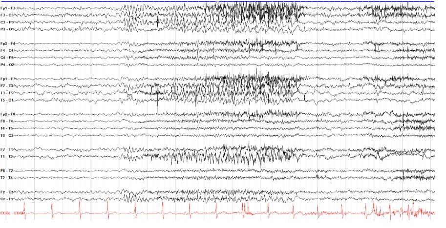
        • Tonic Clonic[…] SZ: inital period of tonic[…] motor activity (stiffening) and on EEG myogenic[…] artifact > then will evolve into intermittent spike[…]/polyspike[…] bursts corresponding to the jerking[…] muscle activity then slow wave[…] with postictal attenuation[…]
          • GTC[…]
          • Focal[…] evolving to BL Tonic Clonic
        • Tonic[…] SZ: muscle stiffening without evolution to clonic
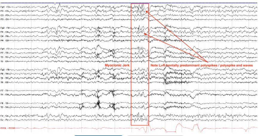
        • Myoclonic[…] SZ: brief involuntary jerks except the discharges run for longer and often lead to significant myogenic[…] artifact with postictal attenuation[…]
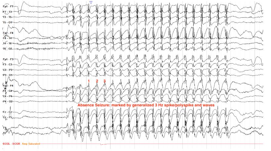
        • Absence[…] SZ: abrupt/brief cessation of awareness[…], often w/eye flutter[…] or blinking[…], w/rapid return to normal[…] afterward
          • EEG: characteristic generalized[…] 3[…]Hz spike and wave[…]
        BIRDS = brief intermittent rhythmic discharges
        LOC = loss of consciousness
        RAS = reticular activating system
        EEG
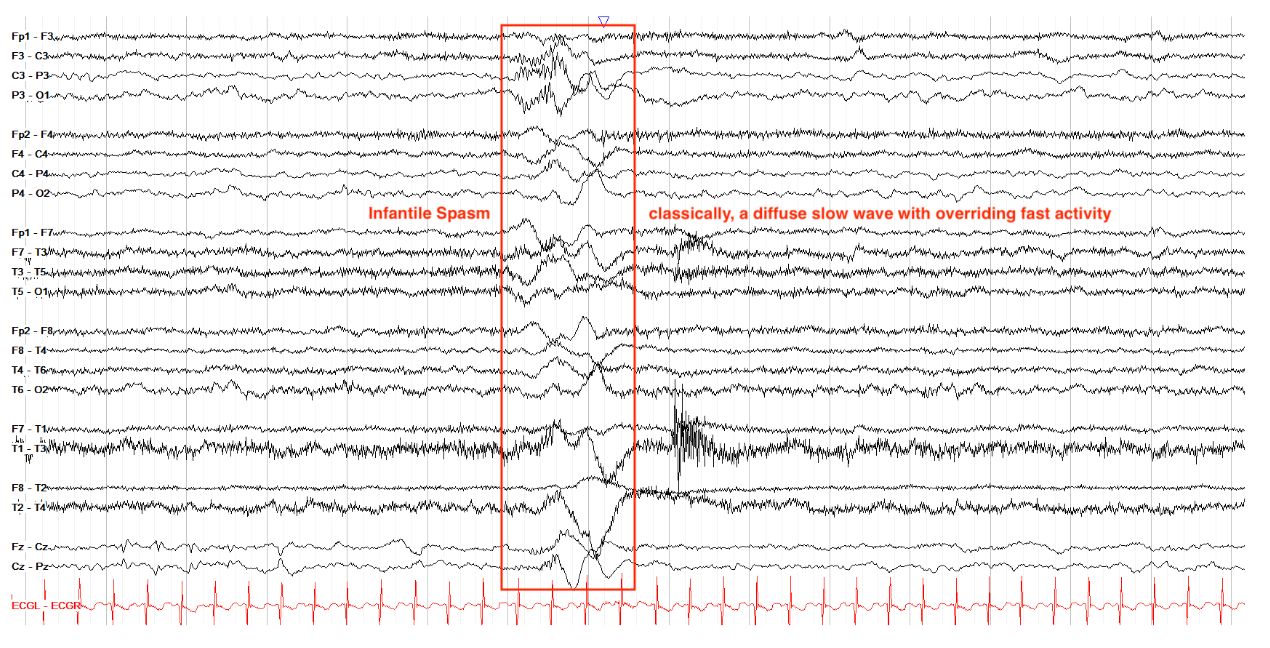
        • Spasms
          • most commonly seen iso infantile[…] spasms aka West[…] Syndrome
          • can also be seen in other epilepsy syndromes esp in kids such as LGS[…]
          • EEG: background of hypsarrhythmia[…] w/high amplitude[…], very disorganized[…] activity, and multifocal spikes[…]
        EEG
        Would a neuron changing from a potential of -70mV to -30mV make it more or less likely to propagate a signal?
        More likely[…]

        resting membrane potential of neurons is -70mV, and depolarization leads to propagation of a signal through a neuron while hyperpolarization leads to inhibition of signal propagation. Any positive shift in the neuron's intracellular voltage (ex from -70 to -10mV) pushes it closer to depolarization and firing, while any negative shift in the neuron's intracellular voltage (ex from -70 to -90mV) pushes it away from depolarization and towards a continued resting state.
        EEG

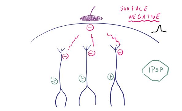
        negative[…]

        Recall that IPSPs promote hyperpolarization, in which K+ moves out of the cell and causes the interior of the cell to become more negative than usual. Thus, the extracellular space becomes relatively positive.In this case, the IPSP targets the deeper portions of the neuron column, so the deep extracellular area becomes positive while the adjacent superficial area becomes relatively negative. It is this superficial negative charge that the scalp EEG electrode is able to see, and thus you'd see an upward deflection for this electrode on the tracing.
        EEG
        In an EEG report, you read that epileptiform discharges were seen in F7 and T1. If so, which region(s) of the brain are involved?
        Left anterior temporal region[…]

        Recall that despite being "F" electrodes, F7 and F8 actually overlie the left and right anterior regions, respectively. T1 and T2 are left and right subtemporal, T3 and T4 are over the left and right mid-temporal regions, and T5 and T6 are over the left and right posterior temporal regions. Thus, the discharges in question most likely come from the left temporal lobe. Note, however, that in documenting and interpreting scalp EEGs, it is better to say "region" than "lobe," because recall that to detect cerebral activity scalp EEG requires a relatively broad area--at least 6 square centimeters--of cortex to be involved. With such a broad area, you can't say for certain that the entirety of the discharges in question were from the temporal lobe itself and not, say, from an adjacent sulcus of the frontal or parietal lobes.

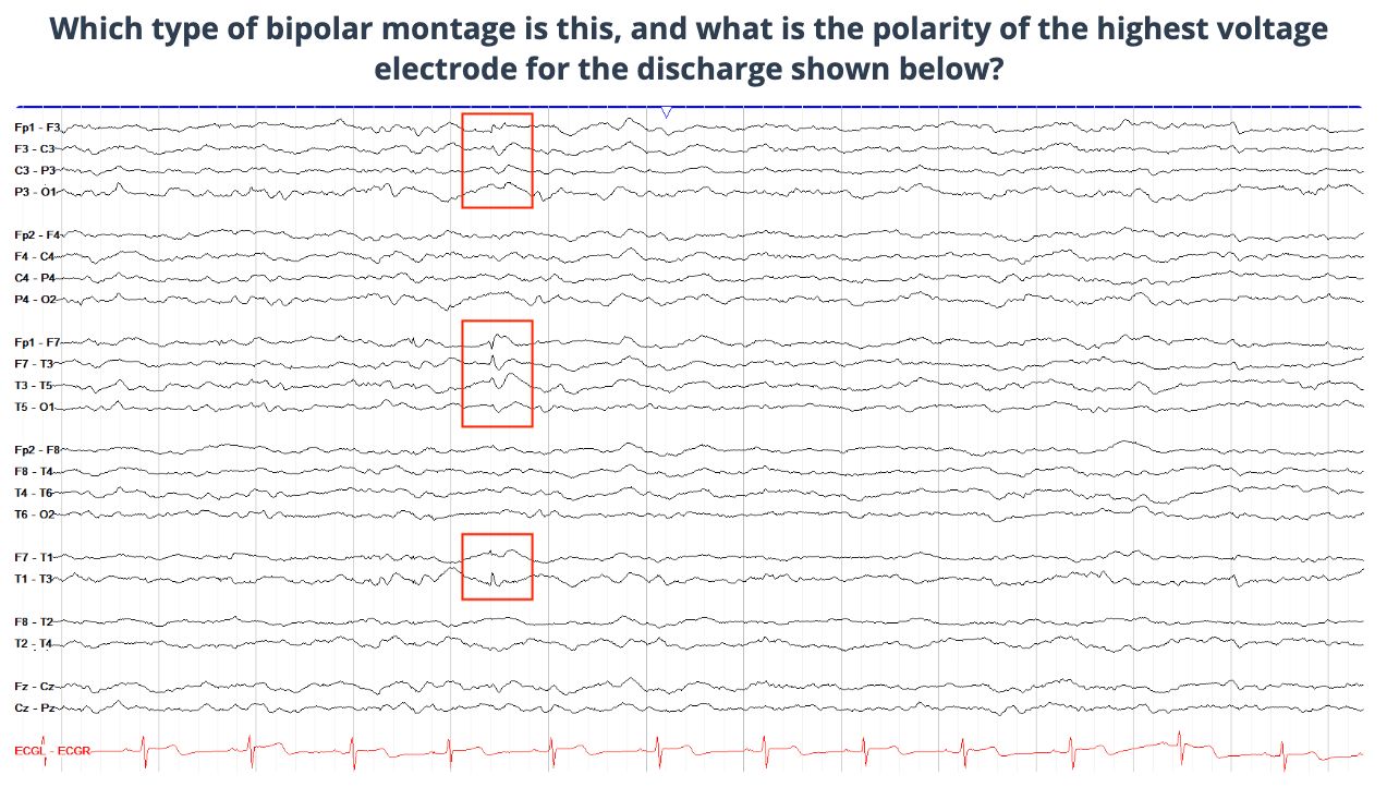
        EEG

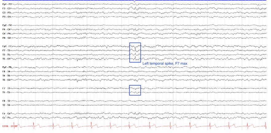
        Bipolar Montage, negative polarity, F7 phase reversal[…]

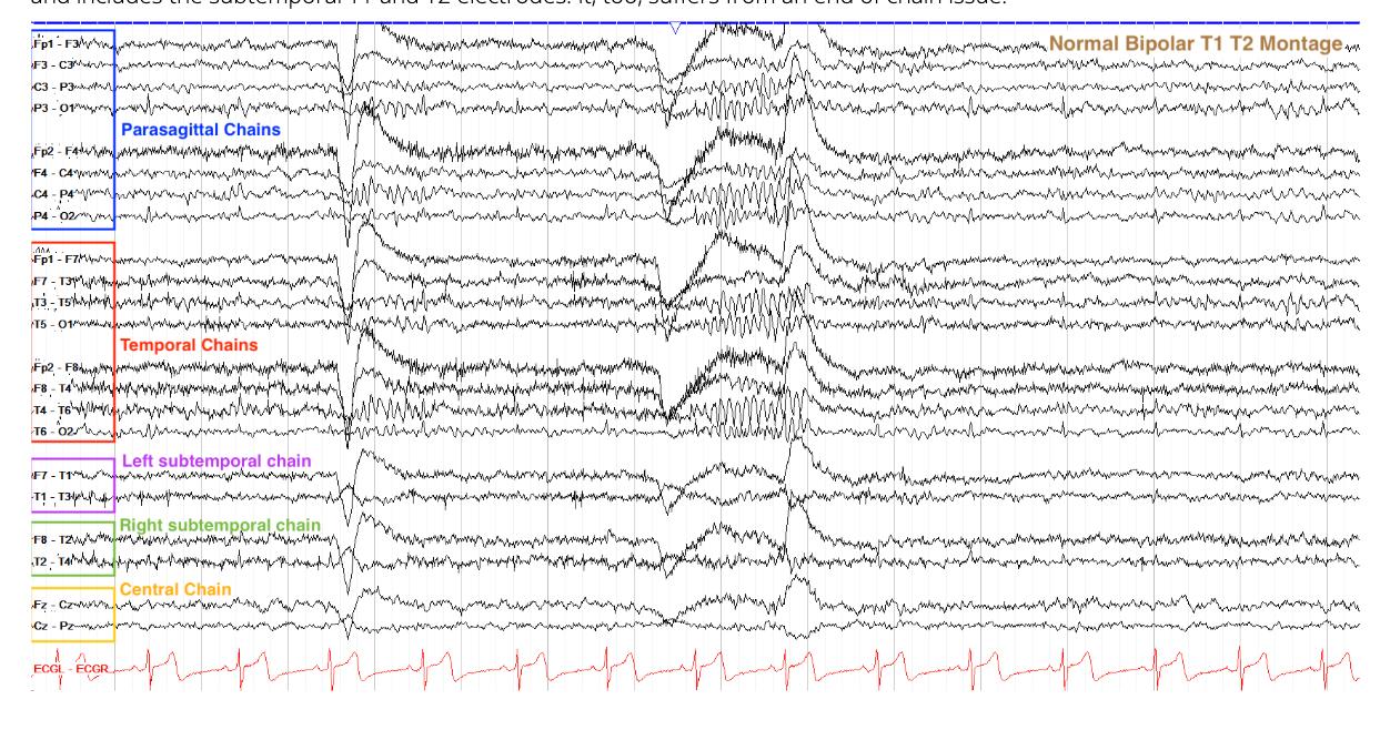
        This tracing shows pairs of electrodes in chains going from anterior to posterior, with the parasagittal chains on top of the temporal chains; the subtemporal and central chains are also present. So, this is a bipolar T1-T2 montage (if the temporal chain was on top of the parasagittal, it would be a double banana).

        Regarding the discharge, we clearly see a phase reversal between Fp1-F7 and F7-T3, meaning that F7, the middle electrode of this set of pairs, has the maximal voltage of this discharge. There is also a field farther out (field being the presence of a disruption of the background that mimics the maximal point of phase reversal, but "tapers" off as you get farther away from the point of maximal voltage). The phase reversal points inward, so we know it is a negative phase reversal.

        EEG
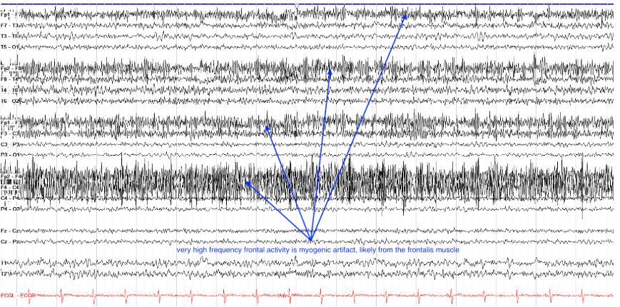
        How might you optimize the low or high frequency filters to better view any underlying activity in the frontal regions on this tracing?


        Lower the high frequency filter[…]

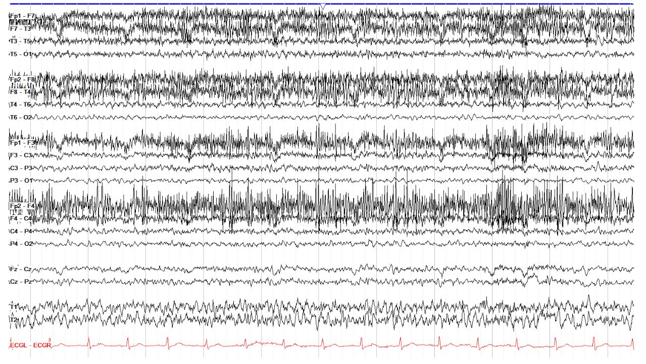
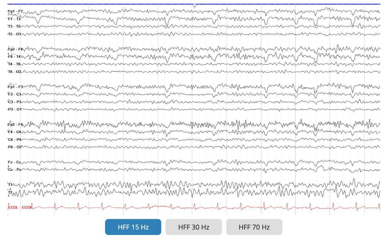
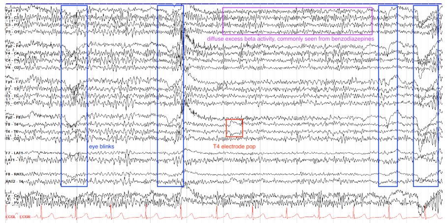
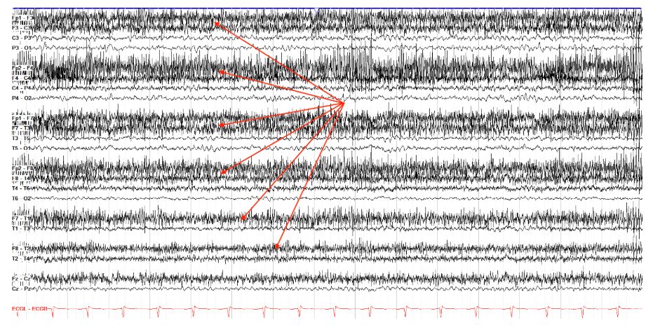
        This tracing shows a lot of myogenic activity, seen as very high frequency activity most dominant over the frontal regions. This is common, but can obscure underlying slower frequencies. If this is a problem, you can lower the high frequency filter from 70 Hz to somewhere between 15-30 Hz to see if you can remove some of the artifact. However, in doing this be conscious that you may also be removing some physiologic beta activity. In this tracing, in fact, there appears to be diffuse excess beta in addition to the myogenic artifact; excess beta is most commonly found in the setting of benzodiazepine use.


        How can you change the sensitivity to make the marked waveforms easier to see, without overlap into the adjacent chains?


        Decrease the sensitivity[…]


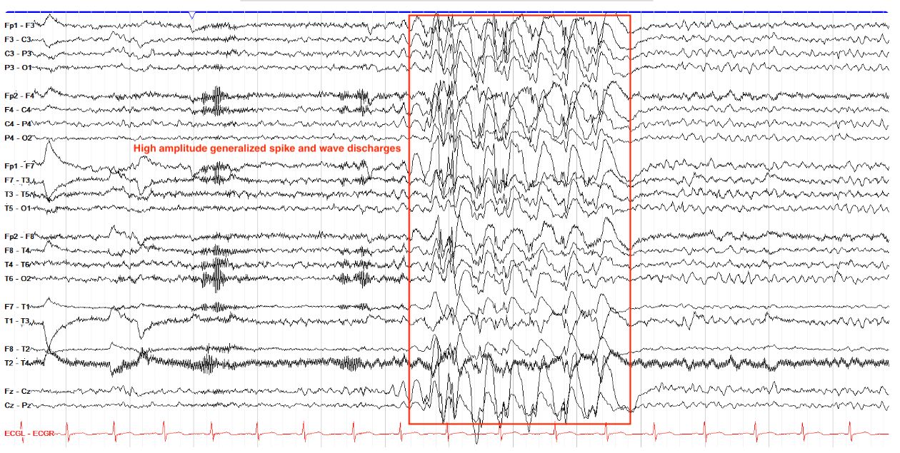
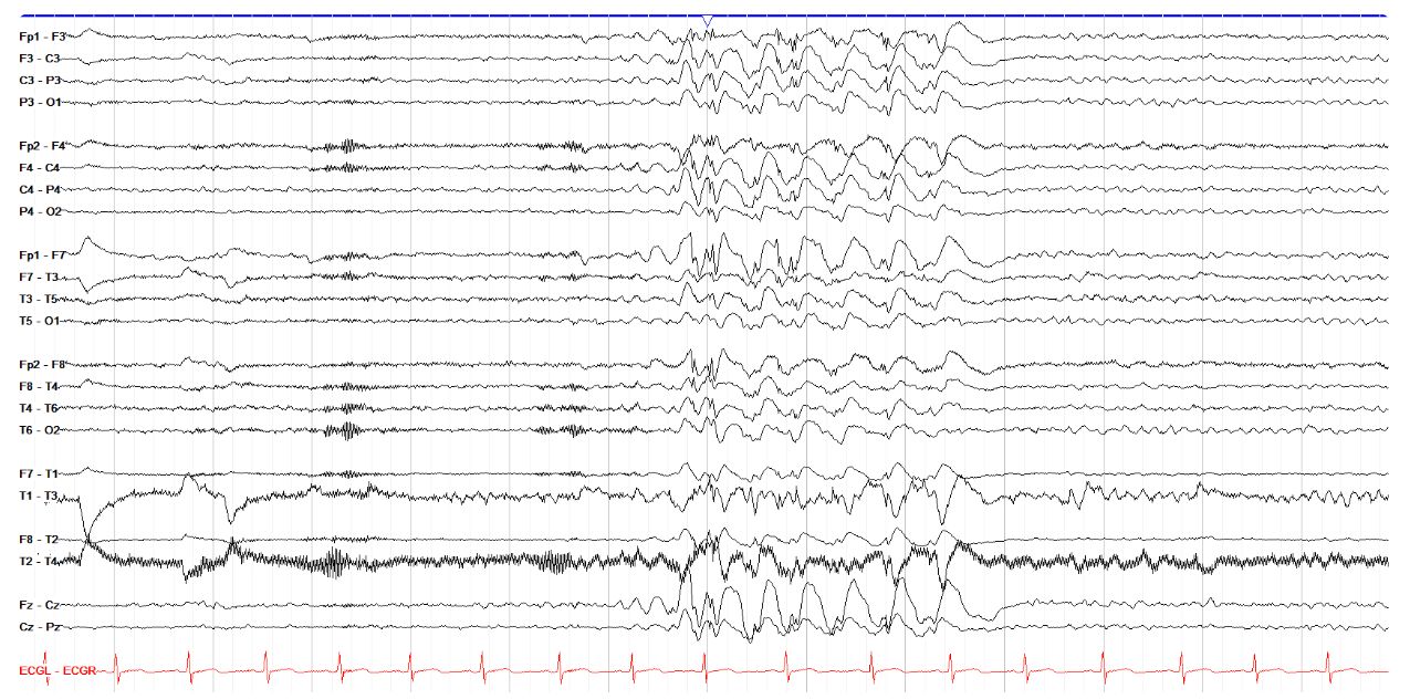
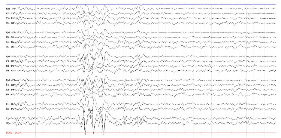
        Recall that sensitivity is a measurement of how much voltage is required to cause a certain amount of deflection of a wave. Lower sensitivities require more voltage to make a wave of a certain height compared to higher sensitivities. So, in this example of very high amplitude generalized spike and waves, with some overlap into adjacent chains making it hard to clearly see the details of the waves, you can decrease the sensitivity (going from, say 7 μV/mm to 15 μV/mm) to make the waveforms appear smaller and more easily seen. The image below is the same set of waveforms but at a lower sensitivity of 15μV/mm. Note that this also makes all the surrounding waves smaller and harder to read.

        EEG
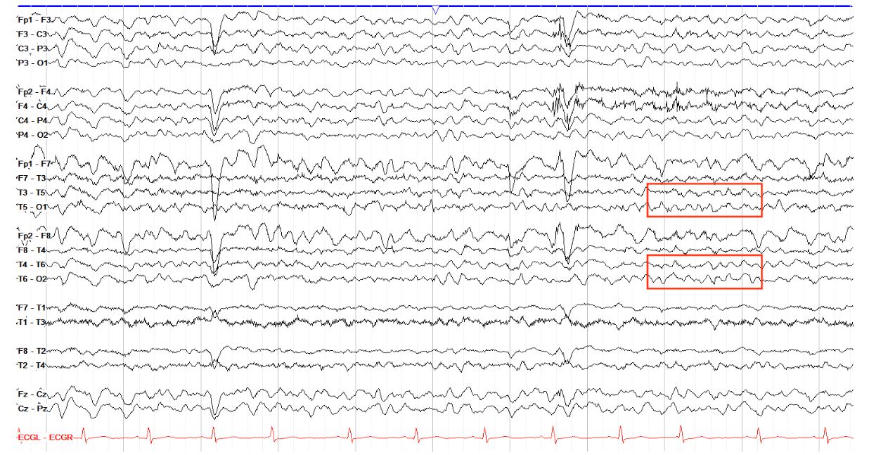
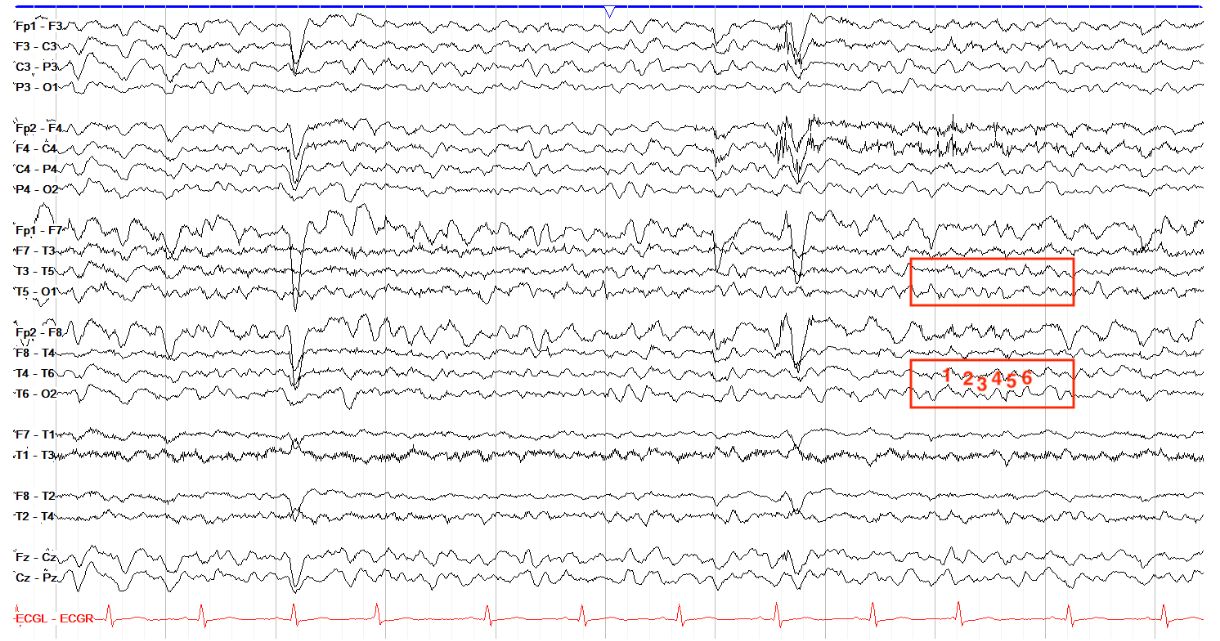
        How would you categorize the frequency of the tracing area marked below?

        theta (6Hz)[…]

        The selected area shows approximately 6 waveforms per second, making it theta as it falls within the theta range of 4-7 Hz. Recall that delta goes from 0-4 Hz, alpha from 7.5-12 Hz, and beta is 12 Hz and above. The dominant frequency of the awake adult brain should be alpha, while theta tends to emerge more in children and in periods of drowsiness.

        EEG
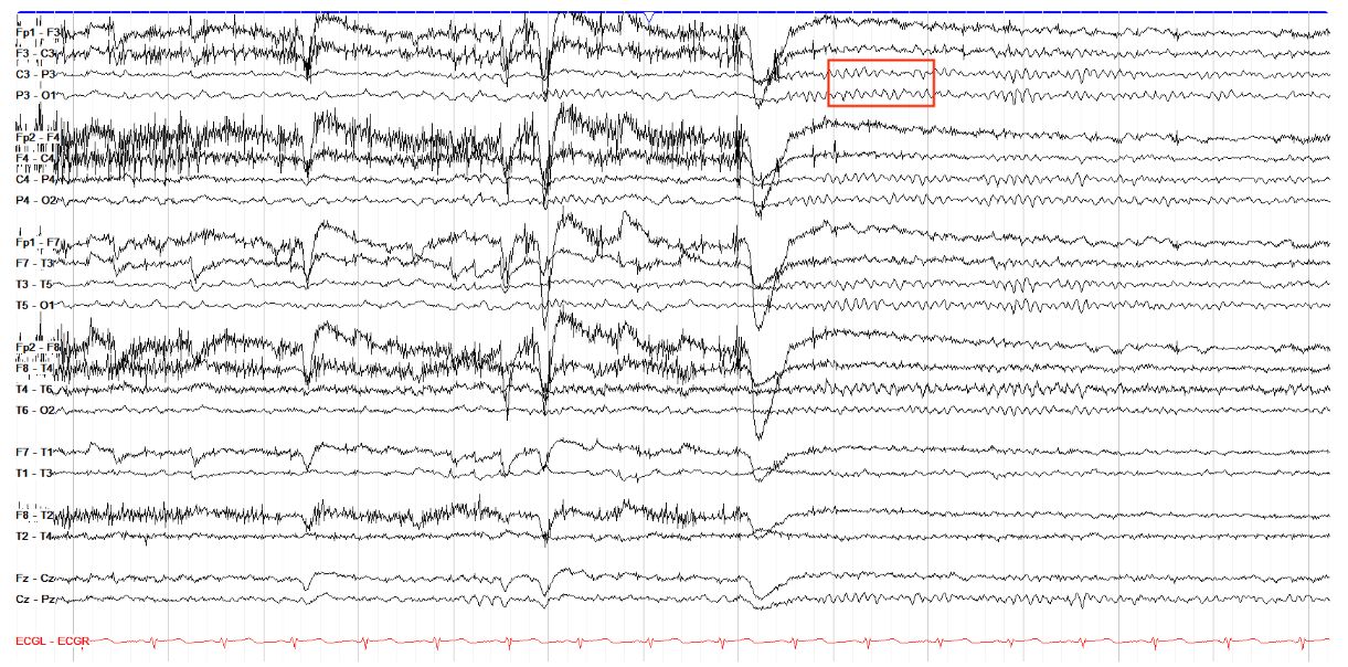
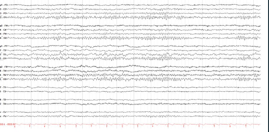
        What is the frequency (the category and numbered hertz) of the area marked below?


        Alpha, 10 Hz[…]

        In counting the number of waves in this one second increment, there are 10, making this an example of an alpha frequency. This is actually an example of what is called the posterior dominant rhythm, which is discussed in the normal awake section and is a defining hallmark of the normal awake EEG.

        EEG
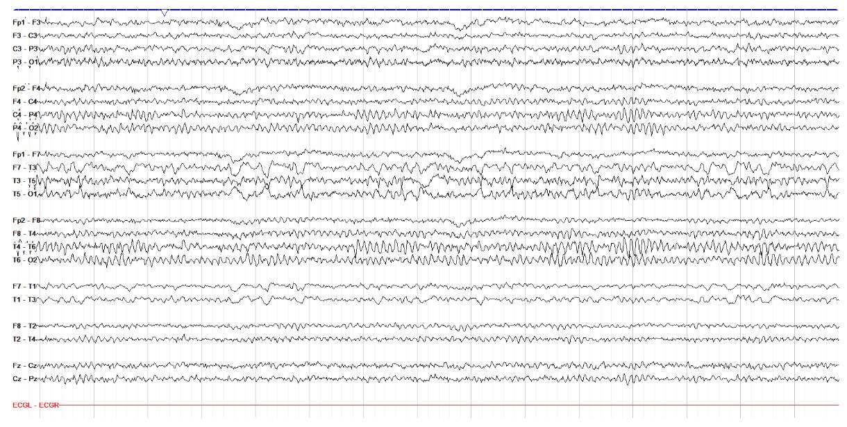
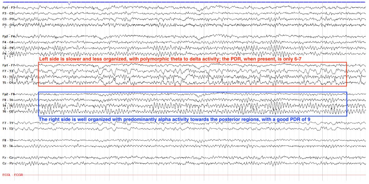
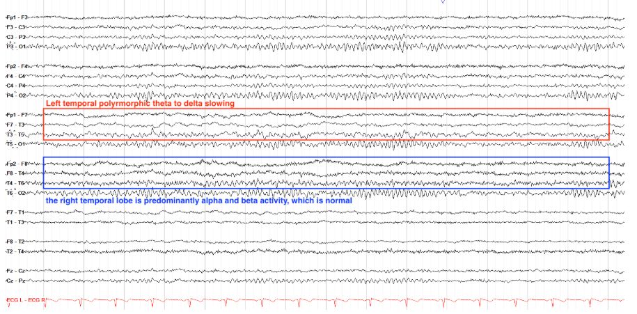
        How would you describe the left hemispheric findings below in terms of frequency and morphology?

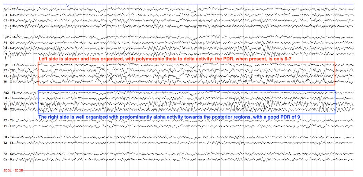
        Polymorphic Theta to Delta Slowing[…]


        This tracing shows a persistent slowing over the left hemisphere. Note how the right side has predominantly alpha activity in the posterior regions--this is the posterior dominant rhythm, discussed in the Normal Awake section. On the left side, however, this alpha activity is absent, replaced by a "messier" and slower theta to delta activity that varies in appearance (including both frequency and amplitude), making it polymorphic.

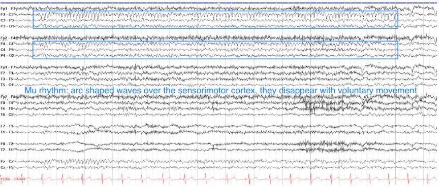
        EEG
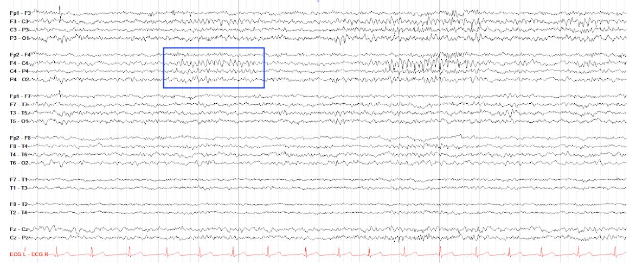
        What is the frequency of F4-C4 in the the area marked below?

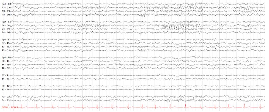
        Alpha, 9-10 Hz[…]


        In counting the waveforms, there 9-10 per second in the area marked, making this an alpha rhythm. This is a normal tracing page, and the area marked is actually an example of Mu rhythm, which is an alpha rhythm representative of the resting basal state of the sensorimotor cortex. It typically emerges in periods of calm and subsides with movement or thoughts of movement.


        EEG
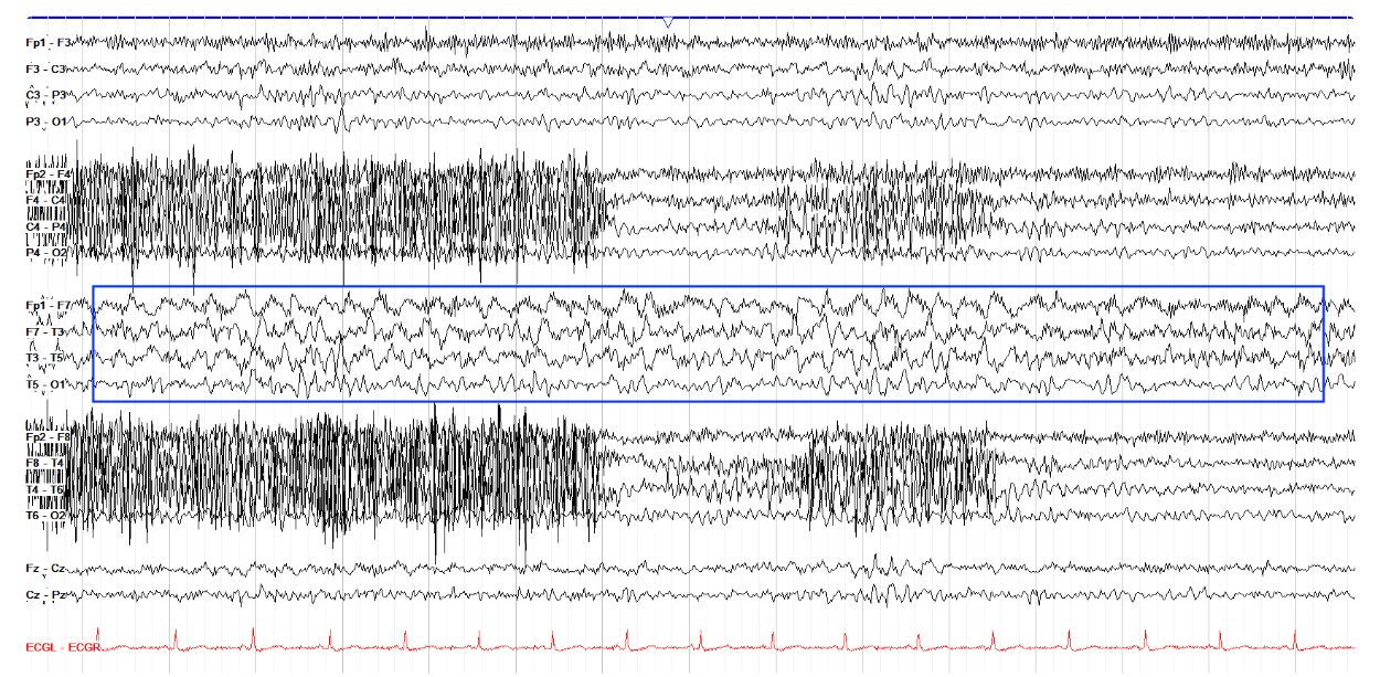
        How would you describe the dominant morphology in the area below?

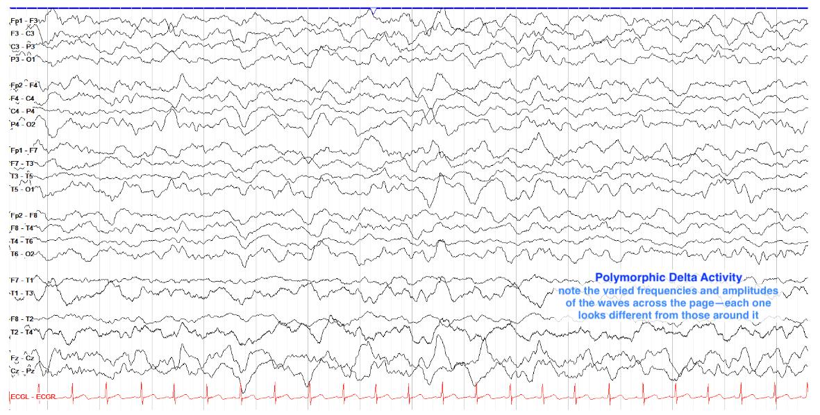
        Left temporal polymorphic delta[…]

        This tracing shows persistent delta activity in the left temporal chain. The delta activity varies from second to second and each wave looks different than those around it, making it polymorphic. Note that there is admixed beta, theta and alpha frequencies amongst the delta, as a real EEG almost never has just a single frequency (the brain is a very active place!).

        Note also that while much of the right hemisphere is obscured by high frequency myogenic artifact (the very fast activity seen in first half of the page in the right temporal and parasagittal chains--see the artifact section for more details), there is not a similar delta slowing in the right temporal chain. This sort of lateralized slowing suggests underlying cerebral dysfunction of the slowed region; when present in the temporal lobe, it can also suggest increased seizure potential.

        EEG
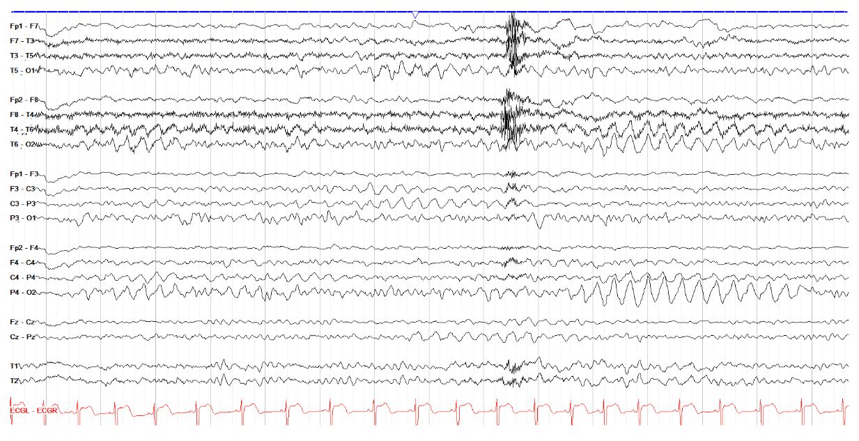
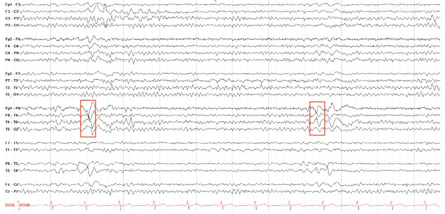
        Describe the activity seen in the occipital region below.

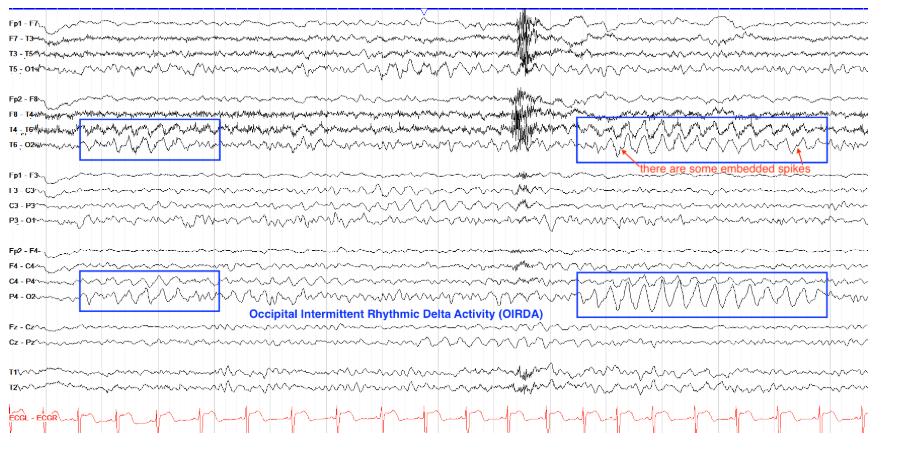
        Occipital intermittent rhythmic delta activity (OIRDA)[…]


        This page shows periods of fairly monomorphic or rhythmic delta frequency activity in the occipital regions, more dominant on the left as marked below but also present near the middle of the page on the right side. This pattern is called occipital intermittent rhythmic delta activity (OIRDA). It is more commonly seen in children and can sometimes be associated with increased epileptogenic potential. In this case, there are subtle spikes seen prior to some of the waves; two are marked below.


        EEG
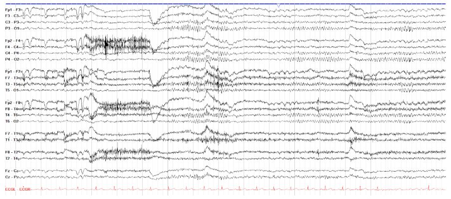
        Is this tracing both continuous and symmetric?

        Yes[…]

        EEG
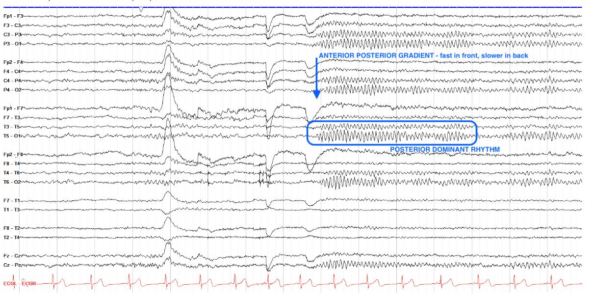
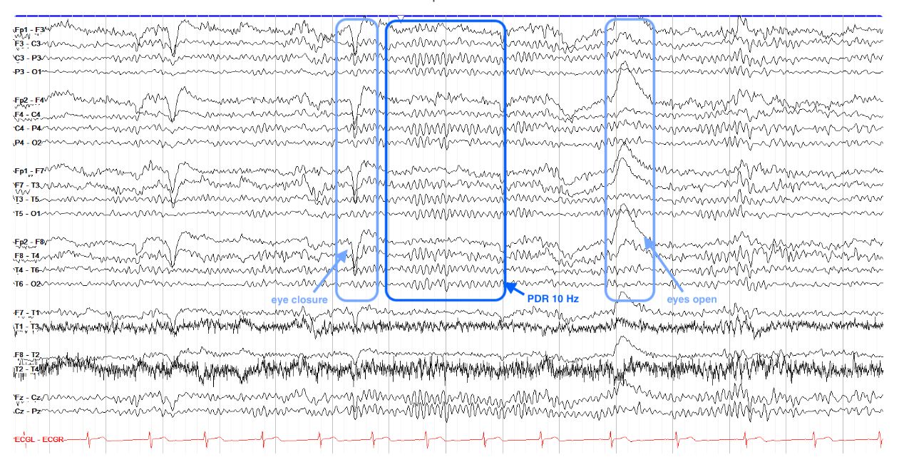
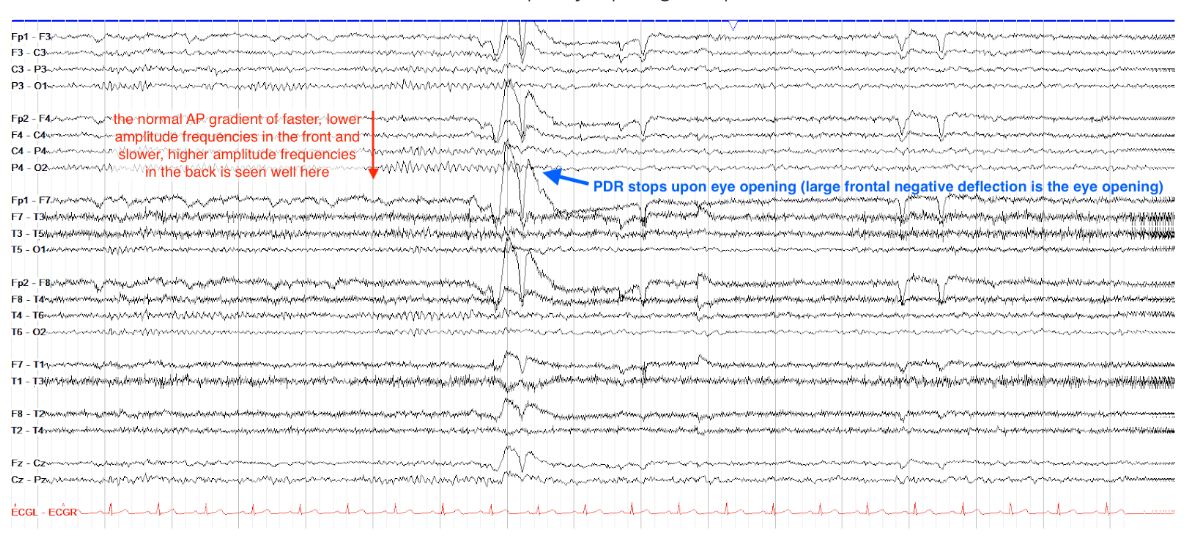
        What is the PDR on the tracing below?

        10[…]

        In this normal EEG tracing, note first that the anterior-posterior gradient is intact and normal, with faster, lower amplitude frequencies seen over the frontal regions and slower, higher amplitude frequencies seen over the posterior regions. Furthermore, the PDR, best seen after the eye closure (the large frontal positive wave right before the blue box, due to Bell's Phenomenon), is a crisp and symmetric 10 Hz. Note that the PDR recedes upon eye opening (the large frontal negative wave) several seconds later, as expected.


        EEG
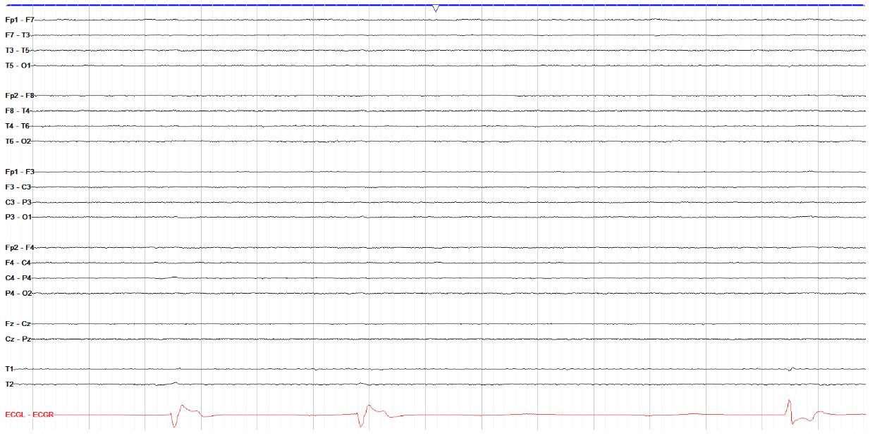
        Does the tracing below show variability?

        No[…]


        This is not a trick question, just an extreme example. This tracing of an unfortunately brain dead patient has no background activity or variability. Note the cardiac tracing does still have activity, but shows multiple abnormalities including profound bradycardia. Compare this flat EEG tracing to the other examples on this page to get a sense of how the EEG tracing should change second to second.

        EEG
        Interpret the background of the tracing below
        (AP gradient, PDR, variability/reactivity, state of consciousness)

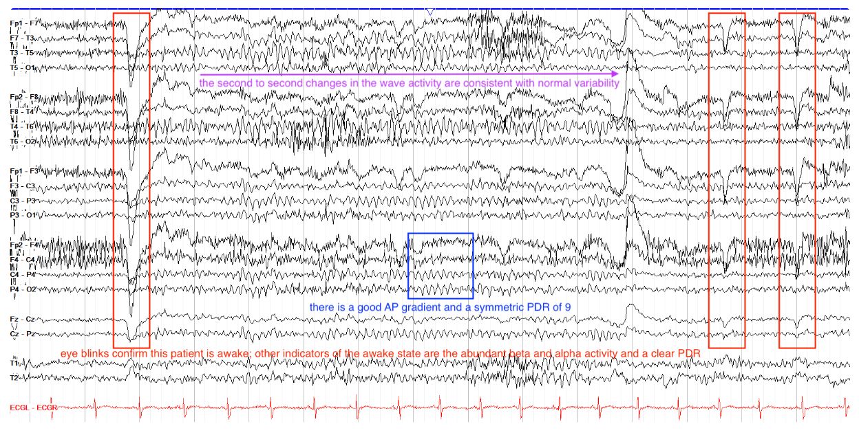
        normal awake EEG, PDR 9 Hz[…]

        The key to any EEG interpretation is a consistent approach. Look for a good anterior-posterior gradient, which this tracing shows with faster, lower amplitude beta activity in the frontal regions and slower, higher amplitude alpha activity in the occipital regions. Next, find the PDR and ensure it is symmetric in both frequency and amplitude, which this one is. If the PDR is present, the patient is awake, but eye blinks can help to confirm this. Variability and reactivity are almost always present if all of the above factors are present and normal.

        EEG
        Patient underwent photic stimulation. What does EEG show?

        photoparoxysmal response[…]
        EEG
        What is the PDR on the tracing below?

        7.5 - 8 Hz[…]


        This tracing has a mildly slow PDR--recall that the normal range for the PDR is 8.5 - 12 Hz even though the alpha range, which the PDR was historically named after, is from 7 - 12 Hz. Other notable findings here include a normal AP gradient and eye blinks suggesting the patient is awake. There is also good variability in waveforms across the page.
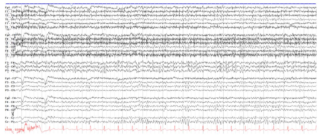
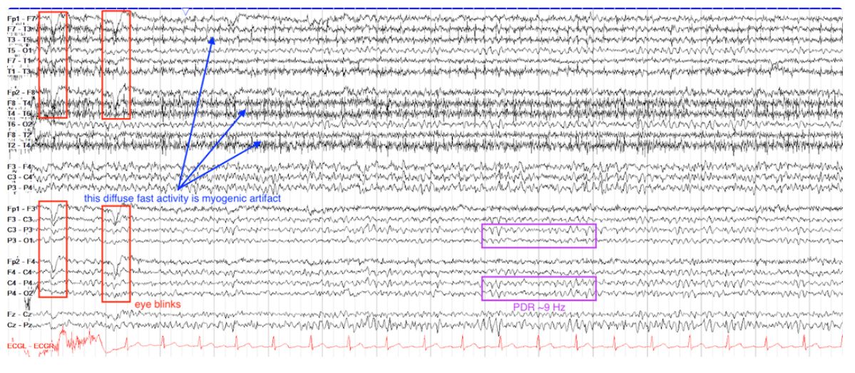
        EEG
        In which state is the patient on the tracing below?

        Awake[…]

        This tracing shows significant myogenic artifact with a normal approximately 9 Hz PDR and multiple eye blinks, all consistent with the patient being awake. Of note, this is a less commonly seen montage that focuses on the temporal chains, by placing the lateral temporal leads (T1, T3) together with the usual temporal chains on top. The middle chain is a transverse central chain comparing left to right. The bottom two chains are the usual parasagittal chains.

        EEG
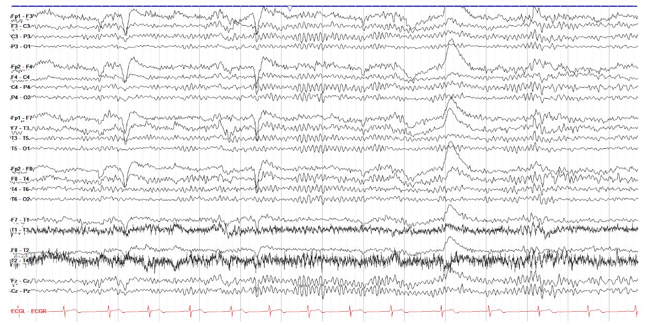
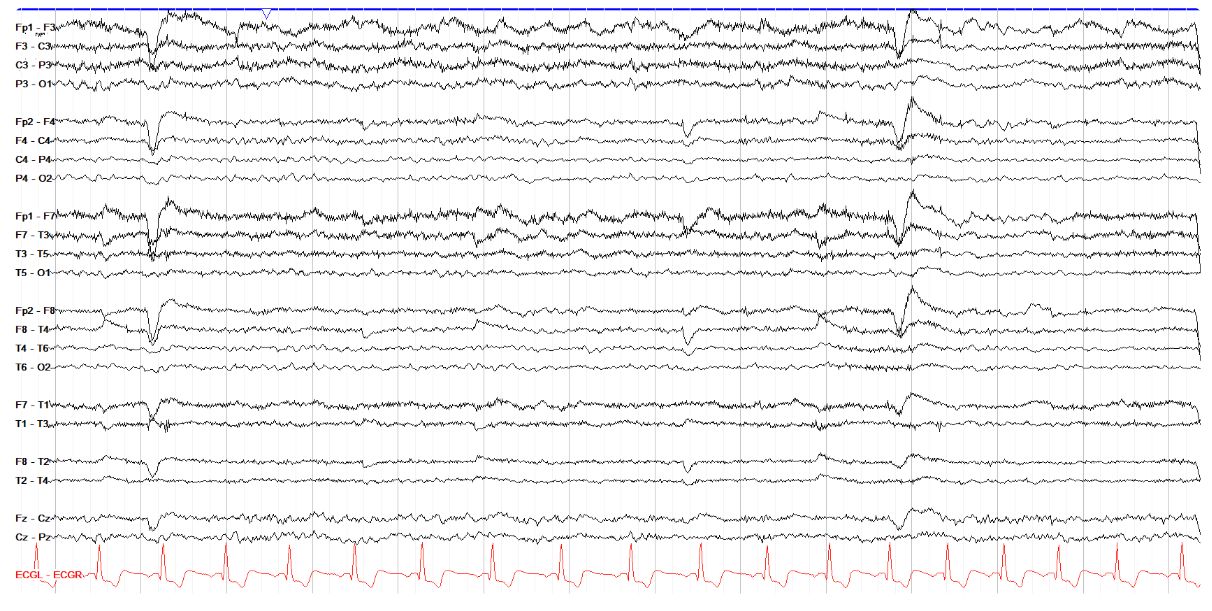
        Is there a normal AP gradient on this tracing?

        Yes[…]
        EEG
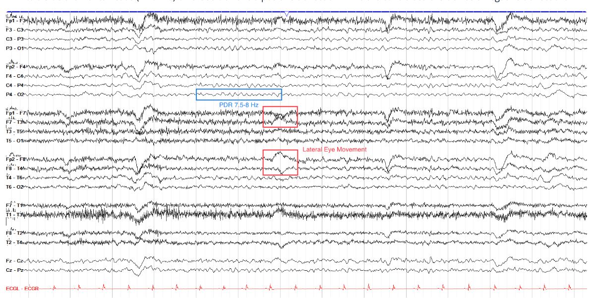
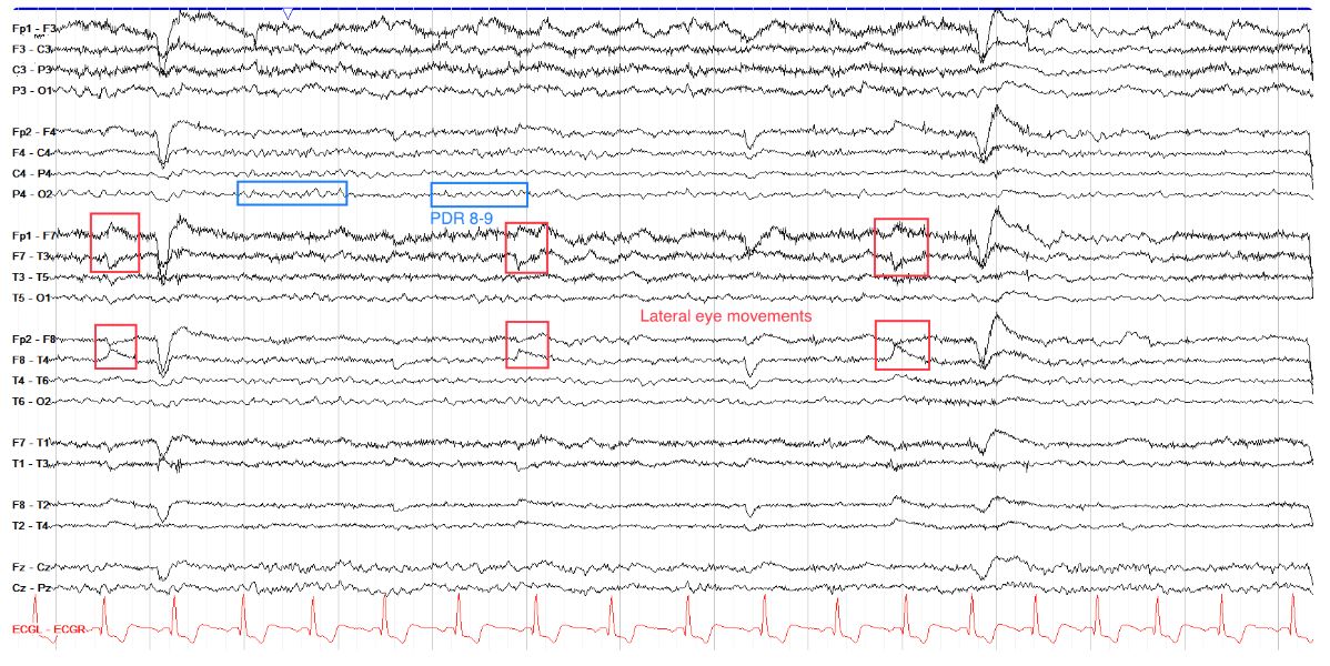
        What is the PDR on the tracing below?

        8-9 Hz[…]

        This normal awake tracing does not have the most clear PDR, but there are segments where a lower amplitude, somewhat sloppily formed PDR of approximately 8 Hz is seen, perhaps best shown in the blue box below. Note also on this page several lateral eye movement artifacts, seen as opposing polarities in the frontal electodes F7 and F8, due to the cornea's positive charge. We also see here several eye blinks (the large frontal positive deflections in the 2nd, 7th and 9th seconds of the page) and some overlying myogenic artifact, seen as very fast low amplitude activity more prominent on the left than right side (see artifacts section).

        EEG
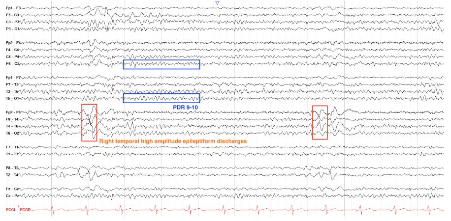
        What is the PDR on the tracing below?

        10 Hz[…]


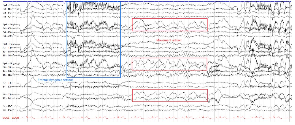
        This normal tracing has a PDR of 10, best seen in the middle portion the page. The PDR is symmetric in both frequency and amplitude (note, the amplitude is not exactly the same side to side but it is within the normal variation of no more than 50% difference). Noted on this page are several types of artifact, including frontal myogenic artifact (seen as high frequency activity in the frontal leads, often arising from the forehead) and movement artifact, seen as high amplitude, nonrhythmic activity over the right frontal region (it is possible the patient is lying with their right side of the head on a pillow, for example).

        EEG
        Does the tracing below show photic driving?

        No[…]

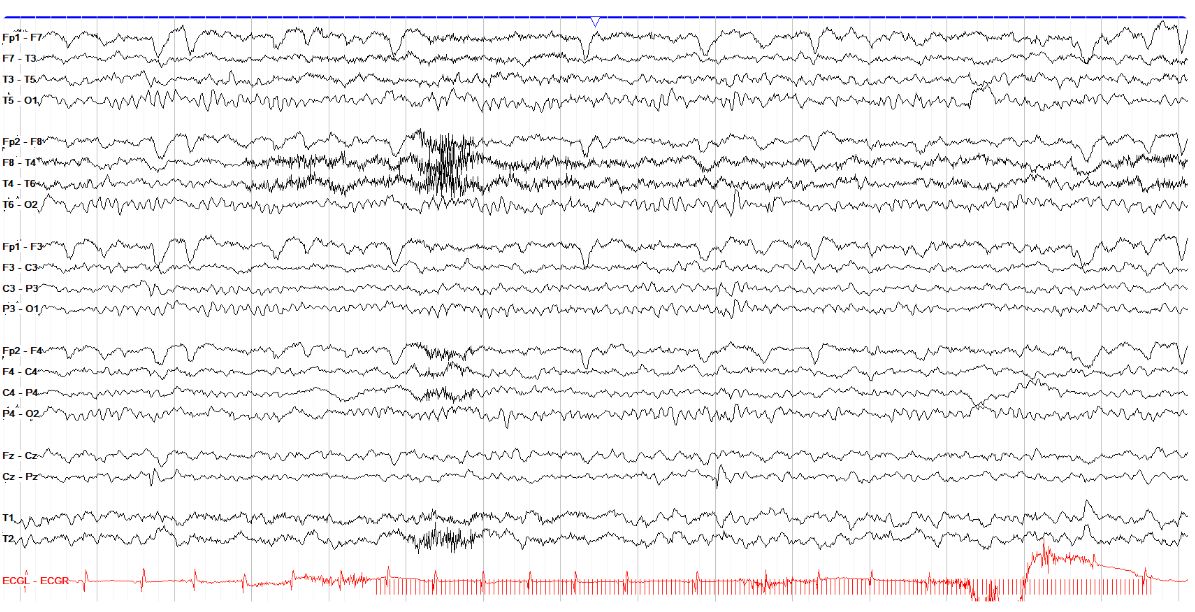
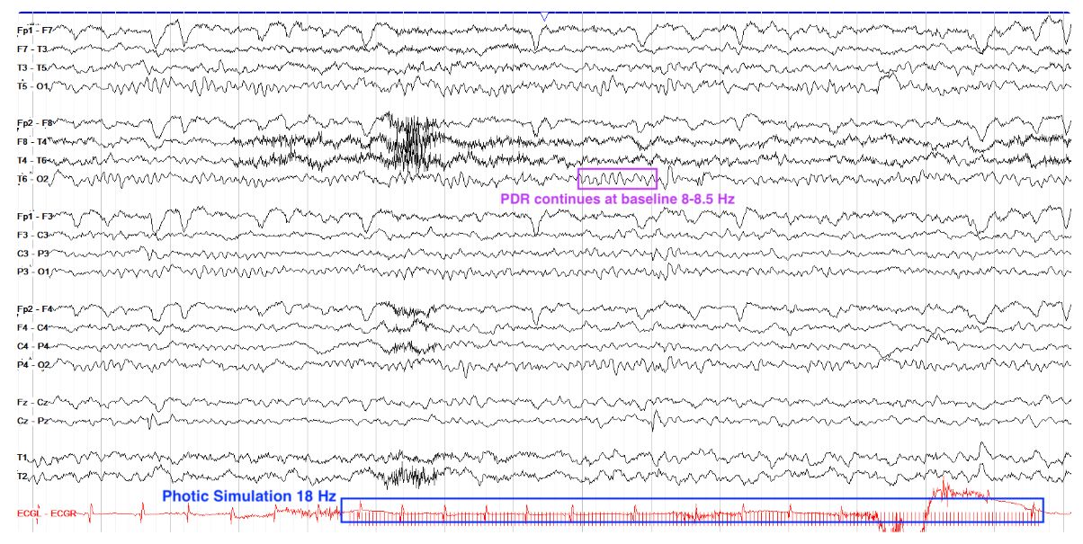
        This page does not show photic driving. There is a clear 8-8.5 Hz PDR prior to the onset of an 18 Hz train of photic stimulation, and that same PDR continues unchanged throughout the course of the stimulation.

        EEG
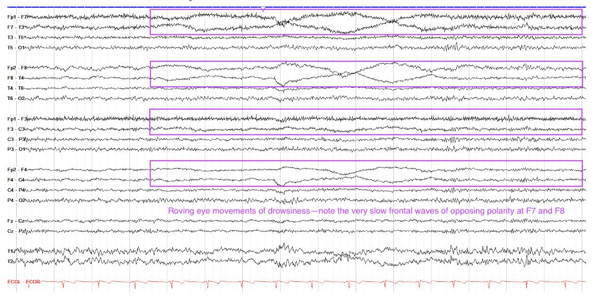
        What state is the patient in on the tracing below?

        Drowsy[…]


        This patient is awake but very drowsy. Recall that drowsiness is marked by diffuse attenuation and possibly mild slowing of the background, but you can still see a clear PDR in the posterior leads here. In the frontal and frontopolar regions, opposing slow undulations are seen in polarity, indicative of lateral roving eye movements. This occurs because the cornea is positively charged, and thus when you look to the right, the right eye's cornea gets closer to F8 and it sees a positive charge; at the same time, the left cornea moves away from F7 and thus it sees a negative charge. So, lateral eye movements lead to a frontal positive charge on the side to which you're looking, and a negative charge on the opposite side.

        EEG
        Which stage of sleep is seen below?

        Stage II[…]

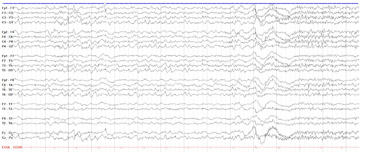
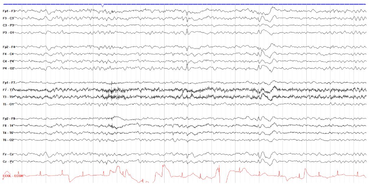
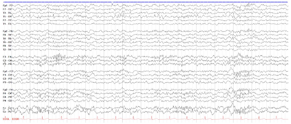
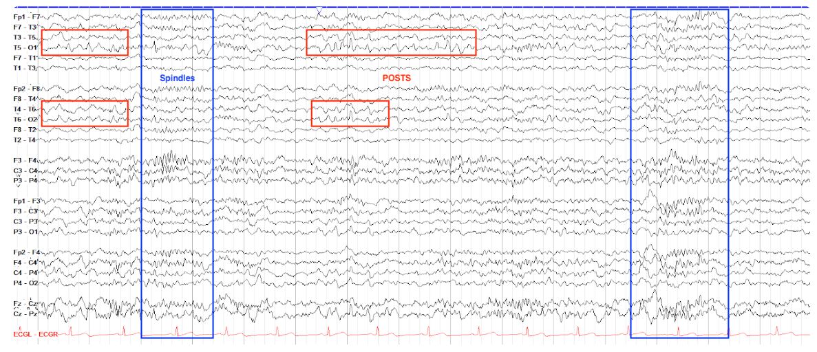
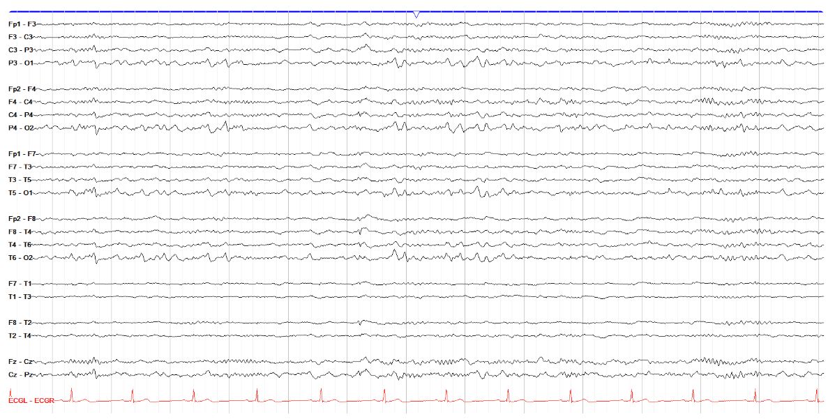
        Stage II sleep is characterized by all of the architecture from stage I -- including vertex waves and POSTS -- along with the arrival of sleep spindles and K complexes. Note the prevalence of POSTS on the first half of this page, then the high amplitude, diffuse and biphasic K complex followed quickly by a 12-14 Hz diffuse sleep spindle. This is an excellent example of a normal sleep tracing. There may also be a poorly formed vertex wave seen in the parasagittal leads in the fourth second of the page.

        EEG
        Which stage of sleep is seen below?

        Stage II[…]


        The presence of spindles--albeit subtle compared to prior examples--is the most obvious characteristic on this page, with some more diffuse than others. Thus this is--surprise!--stage II sleep. Again, there are still traces of stage I architecture via vertex waves, albeit very poorly formed (you know they are vertexes because, even though their formation is poor, there are subtle symmetric phase reversals over the parasagittal and central chains).

        EEG
        Which stage of sleep is seen below?

        Stage II[…]


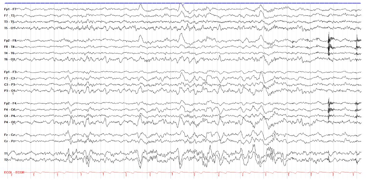
        This page shows several clear vertex waves, marked by high amplitude and symmetric discharges in the parasagittal regions and centrally, with a few less well formed ones in between the ones marked in blue boxes. There are also poorly formed POSTS in the occipital regions bilaterally. This tracing appears to be in the initial transition from stage I to stage II sleep, as some of the higher amplitude background activity in the middle and towards the end of the page are K complexes. Of note, the frontal bursts of very high frequency activity marked in green are lateral rectus muscle artifact (which are more classically seen in REM sleep); these are discussed in the artifact section.

        EEG
        In which stage of sleep is the patient below?

        REM[…]

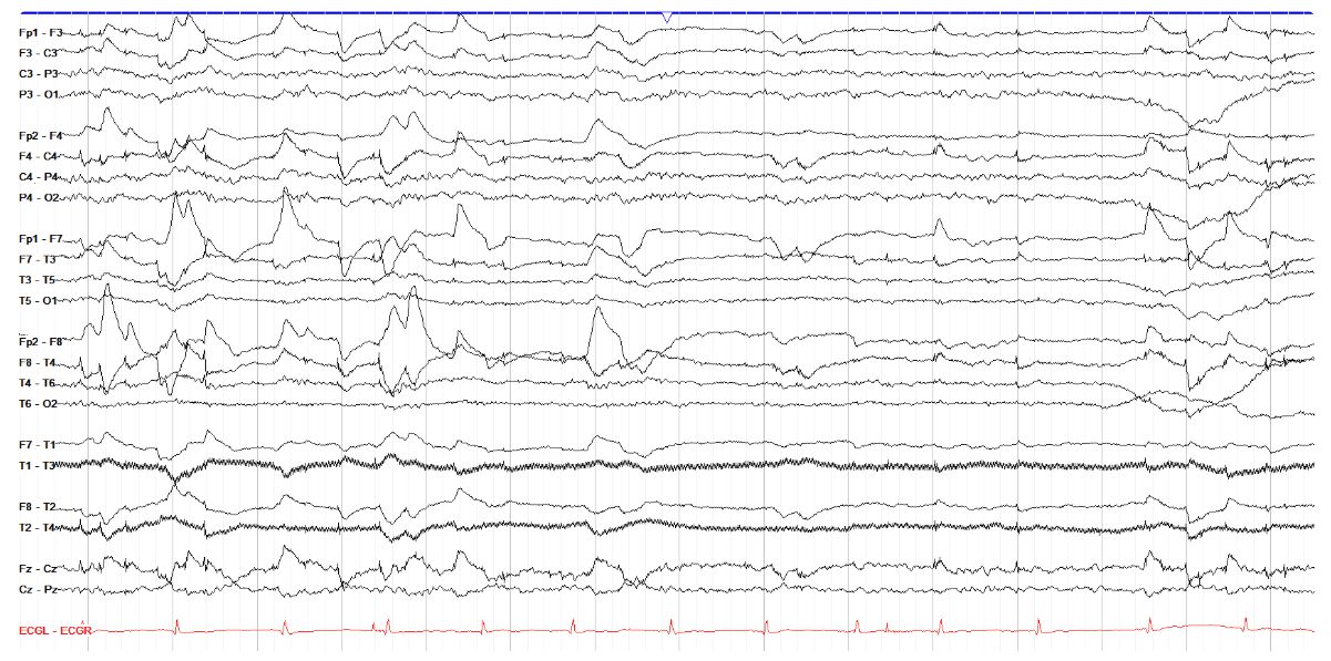
        This tracing shows multiple eye movements, mostly lateral eye movements, and very little other activity. This diffuse attenuation of activity with the present of eye movements is classic for rapid eye movement (REM) sleep. Recall the cornea is positively charged and the retina is negative, so when the eyes look up it causes the retina to move down and the frontal electrodes see a positive charge (and thus a negative charge when the eyes look down). Similarly, when the eyes look to the right, the cornea moves laterally and closer to the right frontal electrode (F8), which sees a positive charge, while the left side sees a negative charge as that cornea moves medially and away from the left frontal electrode (F7, in this example).

        EEG
        In which sleep state is the patient on the tracing below?

        Slow Wave Sleep (N3)[…]

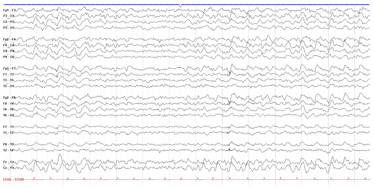
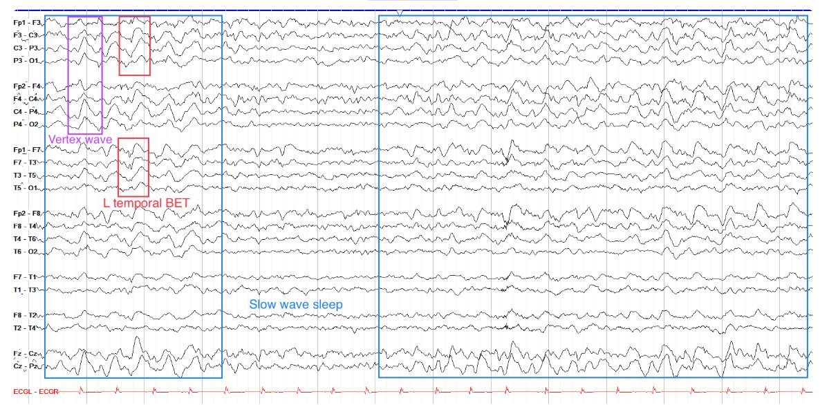
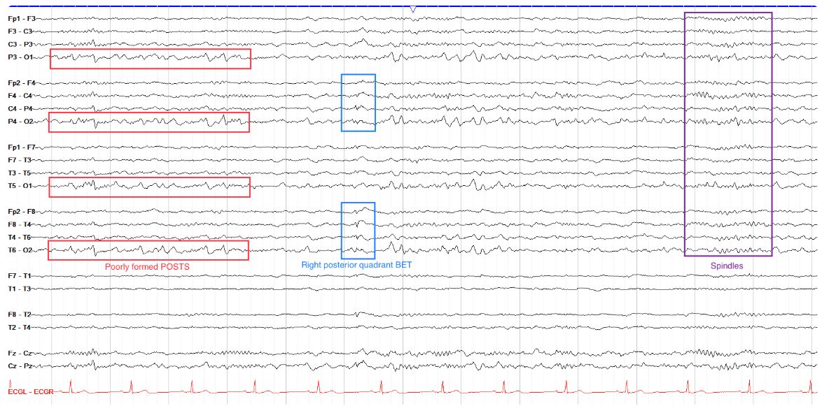
        This tracing shows diffuse, high amplitude delta activity with very little other sleep architecture, consistent with slow wave sleep. Note that poorly formed vertex waves are seen in the beginning of the page, which is normal given that sleep architecture from prior stages usually persists into slow wave sleep, but becomes less apparent and less organized. Of note, this tracing also shows a BET -- a benign epileptiform transient of sleep. Despite the name, this is a normal finding in sleep and is discussed more in the normal variants section. In brief, though, BETs differ from actual epileptiform discharges in that they are very small (

        EEG
        In which stage of sleep is this patient?

        Stage I[…]


        This tracing shows several vertex waves -- symmetric discharges maximal in the parasagittal and central regions. These are a hallmark of stage I sleep. You may also see POSTS (posterior occipital sharp transients of sleep) in stage I sleep. There are not yet any clearly seen K complexes or spindles on this page, which are not seen until stage II sleep.

        EEG
        In which stage of sleep is the patient below?

        Stage II[…]

        This tracing is somewhat different than those you've seen already, mostly due to its use an unusual montage. This is a specialized montage with a focus on the temporal regions (the two chains on top), a transverse section that compares the central region left to right (the middle chain), and the parasagittal chains on the bottom. In this montage, its important to note that the most posterior (occipital) leads in the temporal chains are not at the bottom of those chains, but rather in the middle; the bottom two lines in the temporal chains are actually the lateral temporal leads. Understanding this, the occipital leads show some POSTS, while spindles are seen elsewhere, making this consistent with stage II sleep.

        EEG
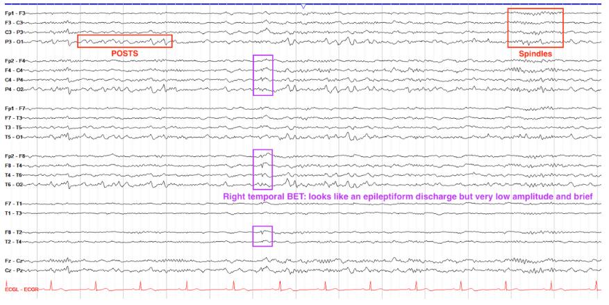
        In which stage of sleep is the patient below?

        Stage II[…]

        This tracing shows POSTS and sleep spindles, characteristic of stage II sleep. Again we see a BET, benign epileptiform transient of sleep (also called small sharp spikes, SSS). Note that BETs, like true epileptiform discharges, do have a clear field, but differ in that they are very small and do not disturb the background.

        EEG
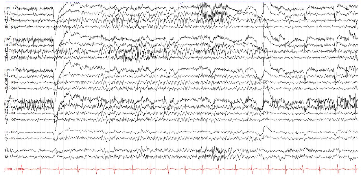
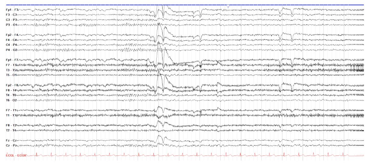
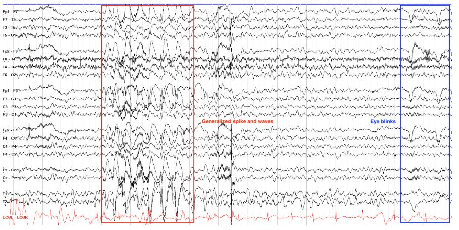
        Which of the waveforms below is/are eye blink(s)?

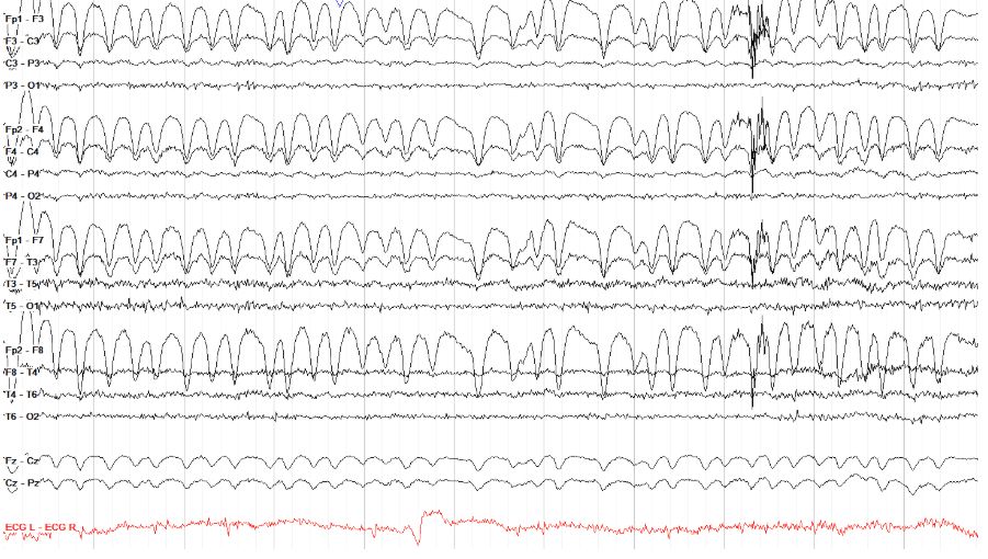
        R most; no posterior field[…]
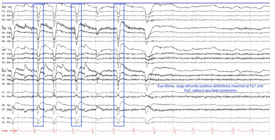
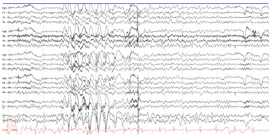
        This tracing shows multiple high amplitude generalized spike and waves with an anterior predominance, which are notable for a broad field that goes all the back to the occipital regions. However, toward the end of the page we also see a few eye blinks. Note how different they look to the generalized discharges--first, they have no field posteriorly; second, they have no preceding spike before the larger amplitude wave, and third, they have no disruption of the background.


        EEG
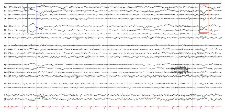
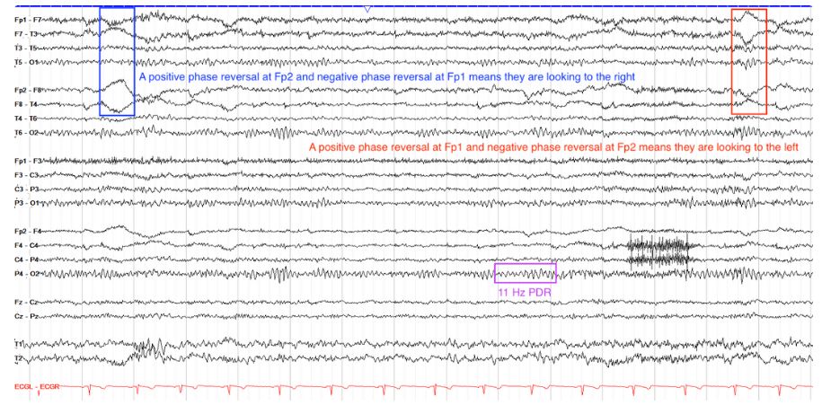
        To which side is the patient looking in each marked eye movement below?

        To the right in blue, and to the left in red[…]


        Recall that EEG helps you keep a positive attitude, so you always look to the positive side on EEG. As such, when considering F7 and F8--the electrodes maximally affected by lateral eye movements--when you see a positive phase reversal (the leads move away from one another) in the F8 leads, the patient is looking to the right, and should have a complementary, simultaneous negative phase reversal (the leads move toward one another) at F7. Other notable things on this tracing are a good PDR of around 11 Hz, a normal AP gradient, some myogenic frontal muscle artifact, and likely a drowsy state given the slow somewhat undulating frontal eye movement artifact in the temporal chains.

        EEG
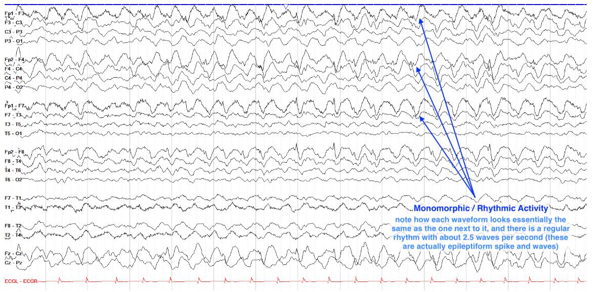
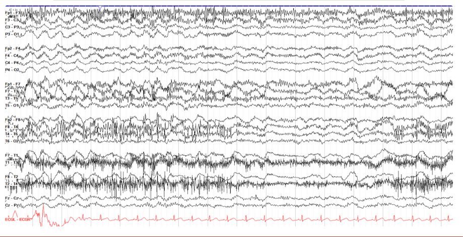
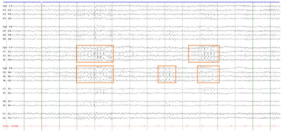
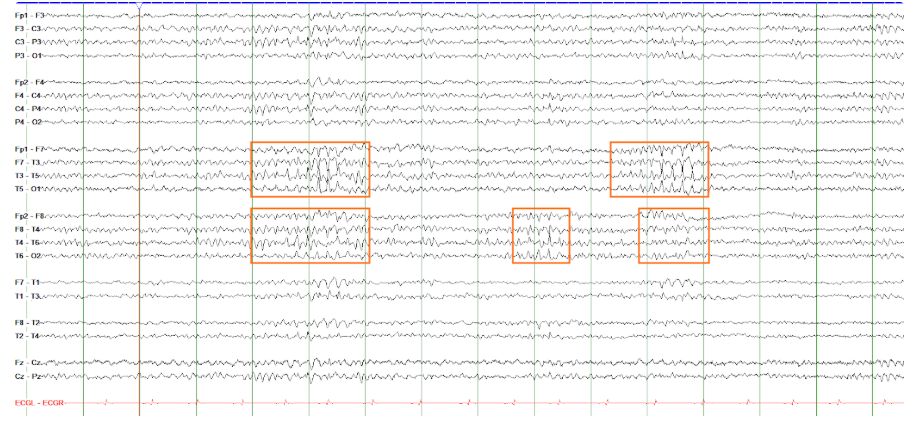
        Is the tracing below consistent with hypoglossal artifact?


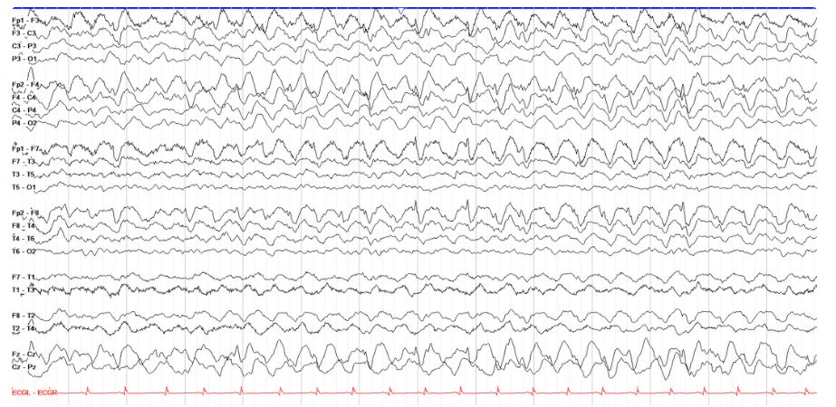
        No, these are bifrontal rhythmic spike and waves[…]

        Don't mistake this tracing for hypoglossal artifact! This page shows rhythmic 2.5 Hz bifrontal spike and wave activity. Notice first that the waves are not quite generalized, as they taper off into the occipital region in the temporal chains (hypoglossal artifact tends to be more generalized). Second, and even more importantly, notice the morphology--each waveform is composed of a small spike and an after going slow wave, which is not typical for hypoglossal artifact either.

        EEG
        How many kinds of artifact can you see in the tracing below?


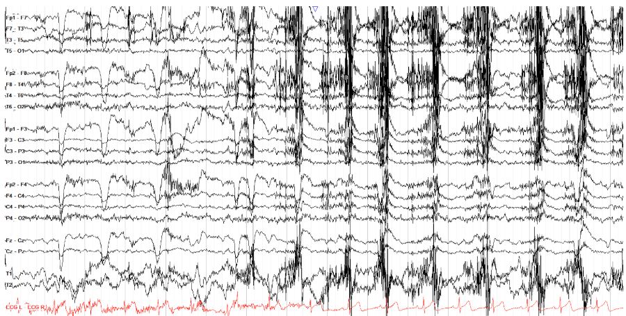
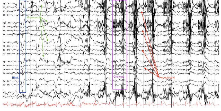
        Blink, Chewing, Hypoglossal, Myogenic, Movement[…]


        This page from a normal tracing shows many artifact types typical for the awake state. Most prominent are the early eye blinks and the aggressive chewing in the back half of the page with some hypoglossal movement seen in between the bursts of myogenic artifact from the chewing. Of note, there is also myogenic artifact elsewhere, most prominent frontally, and there are higher amplitude, disorganized movement artifacts also seen best frontally. Not marked on the page are a few lateral eye movement artifacts interspersed among everything else.

        EEG
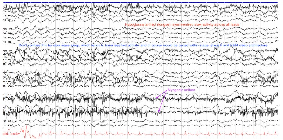
        Which two kinds of artifact are most prominent on the tracing below?

        Hypoglossal and Myogenic[…]


        This tracing shows prominent hypoglossal artifact, marked by diffuse slow activity. There is also a lot of myogenic artifact from the lateral temporal leads, likely from the temporals muscle. Towards the end of the page (the last three seconds), there is some movement artifact seen as "sloppy" appearing and disorganized slow activity more prominent frontally the temporal chains.

        EEG
        How many eye blinks are on the page below?

        4[…]
        EEG
        What activity might cause the artifact seen below?

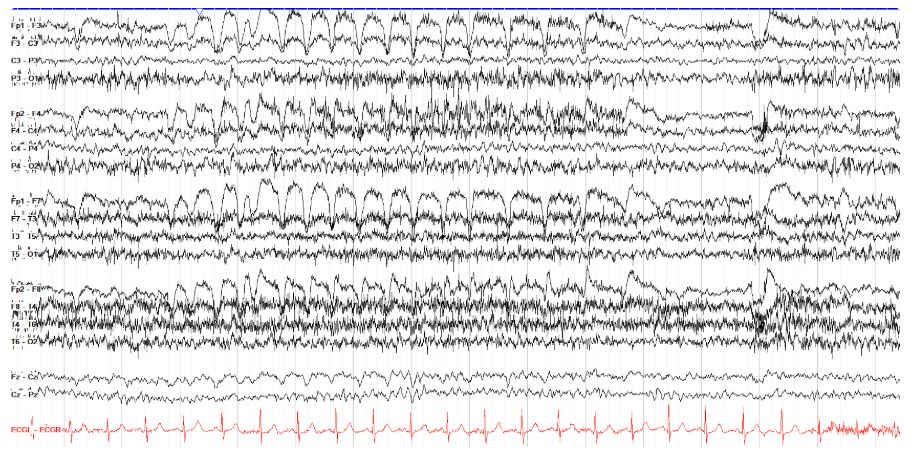
        Eye Flutter[…]

        Similar to eye blinks, note that the prominent positive deflections across this page do not extend posteriorly beyond the frontal leads. Given this constricted frontal area involved and the lack of any underlying spike waves to suggest epileptiform activity, these waveforms are most consistent with eye flutter. Perhaps the patient needs some eye drops.

        EEG
        Is the marking below cerebral or an artifact (and if so, what kind)?

        Myogenic[…]

        Here we see marked frontally predominant but also somewhat diffuse very fast activity. Remember that scalp electrodes really can't pick up activity much faster than beta from the brain, and so this marked activity is much too fast to be cerebral. The fact that it is bifrontally predominant and admixed with an otherwise normal background supports that its just myogenic artifact, likely from the forehead or temporalis muscles.

        EEG
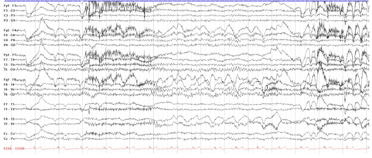
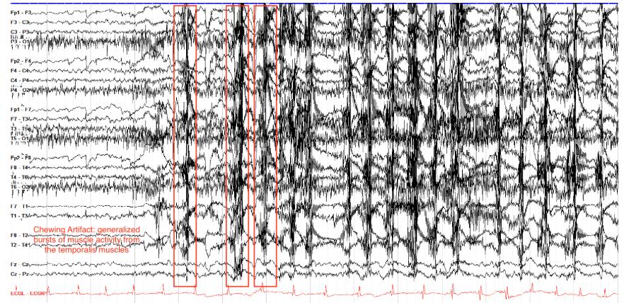
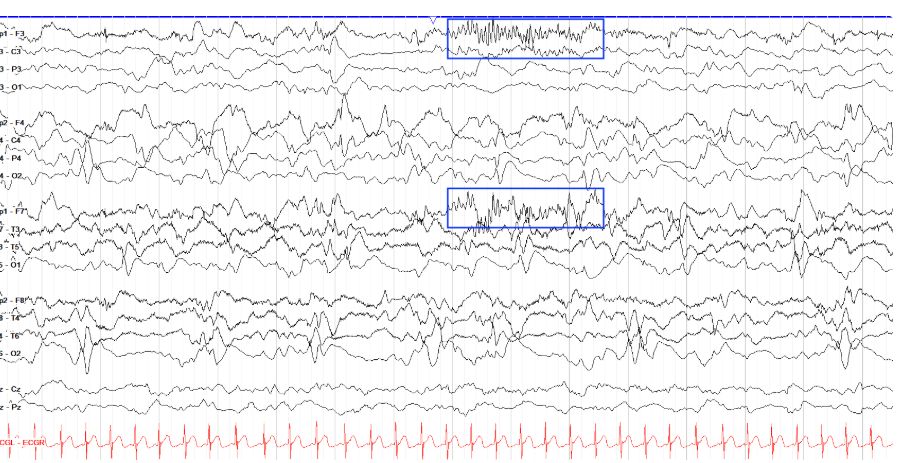
        Is the tracing below cerebral or an artifact (and if so, what kind)?

        Chewing[…] Artifact


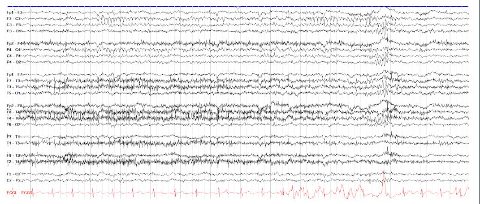
        Here we have intermittent, somewhat sporadically timed bursts of high amplitude, very very fast generalized activity that is consistent with myogenic activity. Underneath that you can see slower waves, but there's nothing evolving here. This is just chewing artifact, with those underlying slow waves being hypoglossal artifact (this doesn't have to be there, but often the two come together). Chewing artifact comes from the activity of the temporalis muscles used to chew.

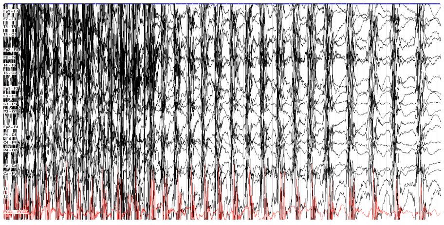
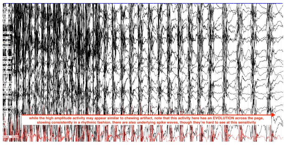
        Is the tracing below cerebral or an artifact (and if so, what kind)?

        Cerebral, this is a portion of a bilateral tonic clonic seizure[…]


        This is kind of a not fair question, because you can only see a single page, but this is not chewing artifact--its the back half of a focal to bilateral tonic clonic seizure. While chewing artifact can appear kind of similar, note here that there is a steady slowing in the frequency of the high amplitude activity, consistent with an evolution in seizure as it slows. You can also see, more in the last few seconds, that each high amplitude burst contains spikes, followed by the slow wave portions that make up the relatively less active portions of the page. Realistically, when reading a tracing you'll be able to see such a seizure from beginning to end and better follow its evolution.

        EEG
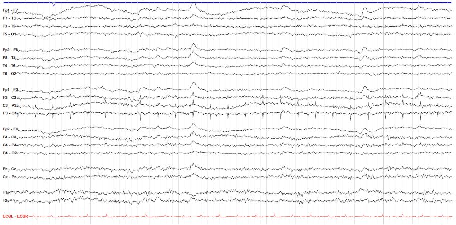
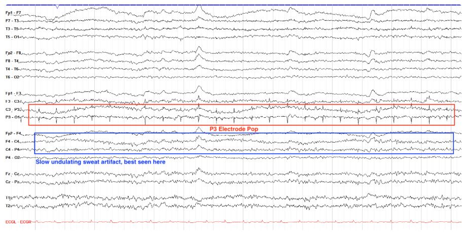
        Is the P3 finding below cerebral or an artifact (and if so, what kind)?

        Electrode Pop (and sweat)[…]

        This page shows periodic positive phase reversals at P3, but these are in fact just electrode pop, from a slightly dislodged P3 electrode. Note that there is really not much of a field beyond the local P3 area (though you can see a bit in F3-C3), and there is no disruption of the background otherwise. There is also some sweat artifact on this page, seen as very slow undulating activity across the tracing; sweat artifact arises due to the electrical potential of the sodium chloride in sweat.

        EEG
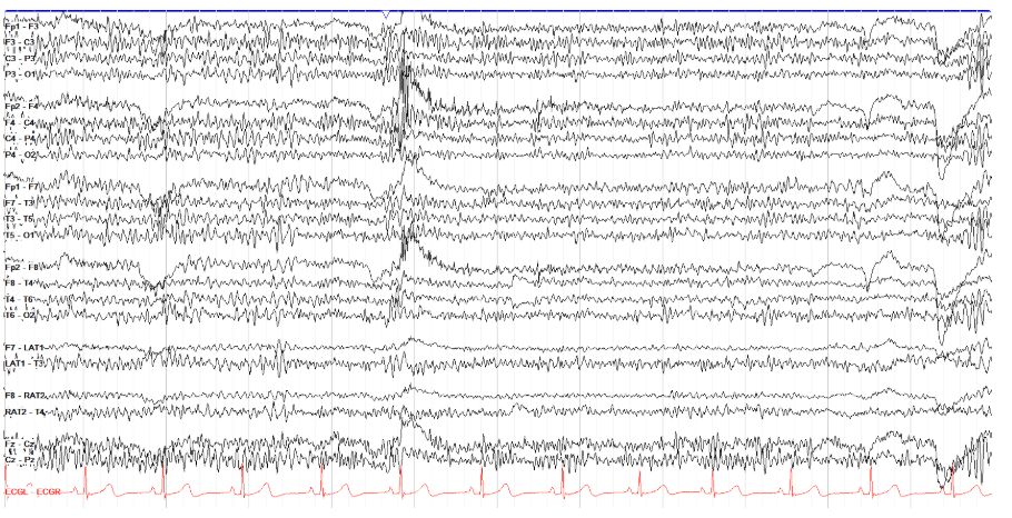
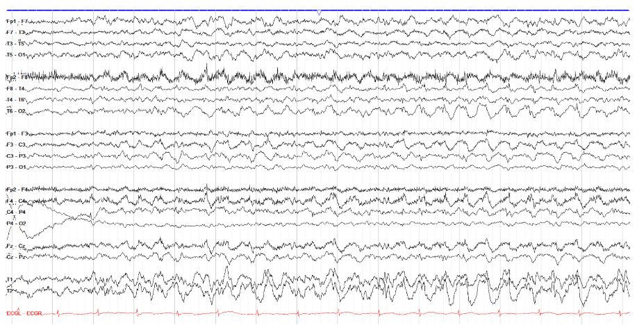
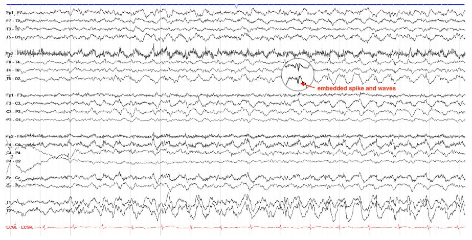
        Is the tracing below cerebral or artifactual (and if so, what kind)?

        Cerebral; this is generalized intermittent rhythmic delta[…]


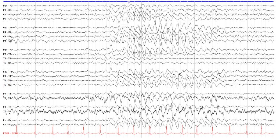
        While this page looks sort of similar to hypoglossal artifact, that tends to have a more synchronized appearance across the entire page, and may come with some myogenic artifact of chewing or other mouth movements. Here, though, we have generalized intermittent rhythmic delta activity (GIRDA), which is usually evidence of diffuse and nonspecific cerebral dysfunction, but this particular page also has embedded spike and waves throughout, suggesting cortical hyperexcitability and enhanced seizure potential.

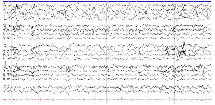
        EEG
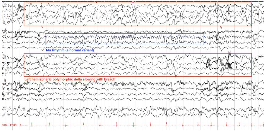
        There are two prominent findings on this page; where is the abnormal finding, and where is the normal variant?

        Right Mu rhythm, and left hemispheric slowing with breach[…]

        This page shows prominent high amplitude, sharply contoured delta slowing throughout the left hemisphere, more prominent in the parasagittal than temporal chain. This is consistent with an underlying focal dysfunction of that region and breach artifact. There is mu rhythm seen in the right parasagittal region, marked by arch-like alpha activity that's often sharply contoured.

        EEG
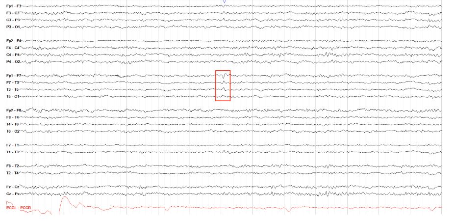
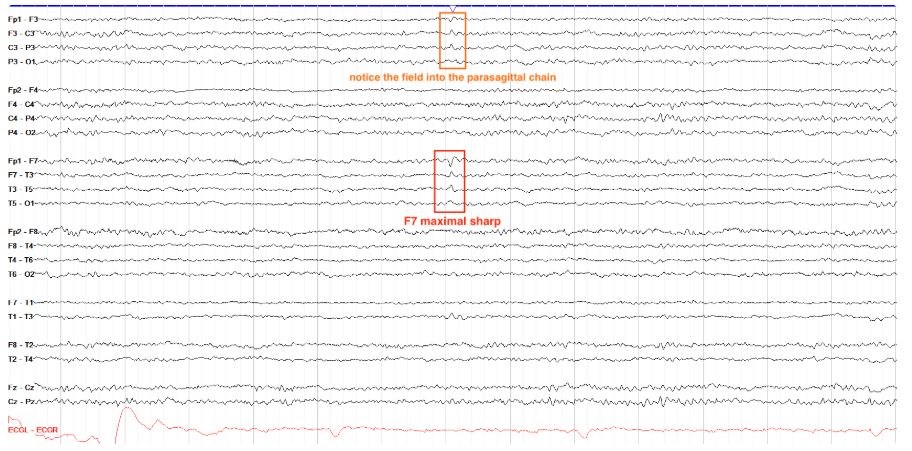
        Is the discharge marked below a wicket or an epileptiform discharge?

        Epileptiform Discharge[…]


        This discharge is in the left temporal region, where wickets are expected to be seen. However, this particular discharge does not have the classic arch-like morphology of a wicket, instead showing a spike and slow wave appearance that is more in line with an epileptiform discharge. Furthermore, it has a field into the left parasagittal chain that would not be present with wickets.

        EEG
        Are the marked waveforms below wickets or epileptiform discharges?

        Wickets[…]


        These waveforms are sharply contoured, somewhat arcuate in appearance, come in bursts of about a second, and they do not have an aftergoing slow wave. All of these characteristics are consistent with wicket waves. Note that these wickets are coming both bilaterally and unilaterally, which is expected.

        EEG
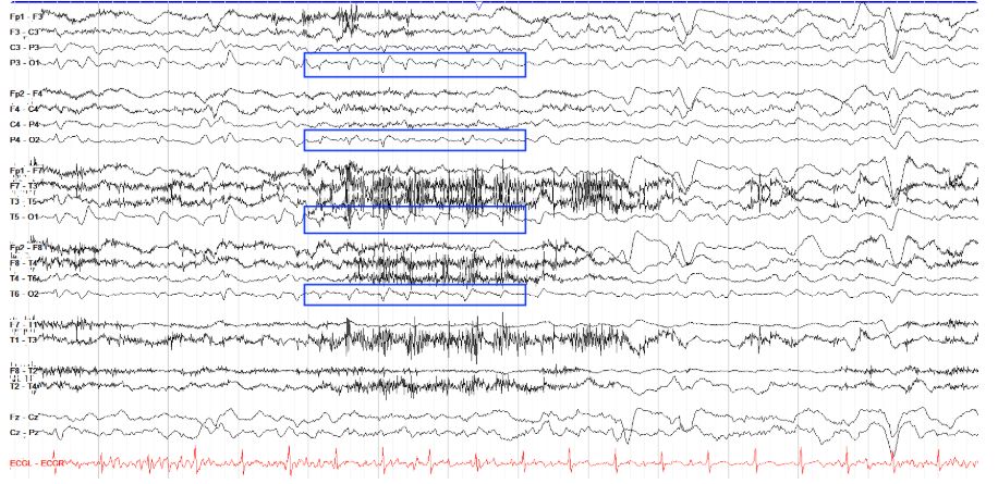
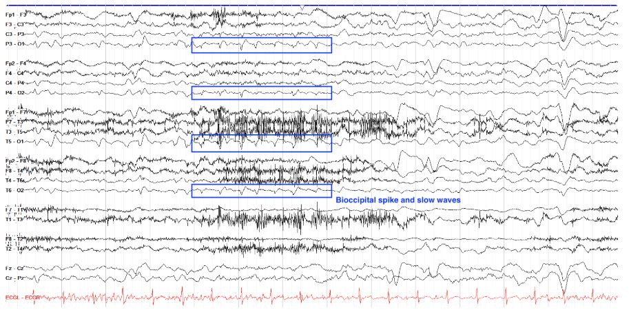
        Are the waveforms marked below lambda waves or epileptiform discharges?

        Bioccipital epileptiform spike and waves[…]


        The marked waveforms, while relatively symmetric as you'd expect with lambda waves, are otherwise wholly different. First off, they have a negative polarity while lambdas are positive. Second, the marked waves have the classic epileptiform morphology of a spike and slow wave, although some show this more clearly than others. Don't let them fool you just because there are a series of them; epileptiform discharges can come in nonevolving runs as you see here.

        EEG
        Are the marked waveforms BETs or epileptiform discharges?

        Epileptiform Discharges[…]


        This is actually an easy question, as you can see a clear PDR of 9-10 throughout this page. Therefore, the marked discharges cannot be BETS, as BETS are only present in sleep by definition. In looking at the marked waveforms, however, they are also too high amplitude and last a little too long to be BETS, which should be very small (<50µV) and short.

        EEG
        Which normal variant is shown below?

        Mu[…] rhythm


        This page has intermittent periods of bilateral sharply contoured, arch-like alpha activity over the parasagittal regions without associated disturbance of the background. This is consistent with mu rhythm, the idling activity of the sensorimotor cortex that goes away when you think about movement or actually move an extremity.

        EEG
        Is the finding below abnormal or a normal variant (and if so, which one)?

        Wickets[…]

        This tracing shows multiple intervals of brief bitemporal or unitemporal sharply contoured nonevolving activity with an arch-like morphology, consistent with wickets. Note that epileptiform activity tends to disturb the backgroud more, often including a field centrally that is not present here.

        EEG
        Is the finding below abnormal or a normal variant (and if so, which one)?

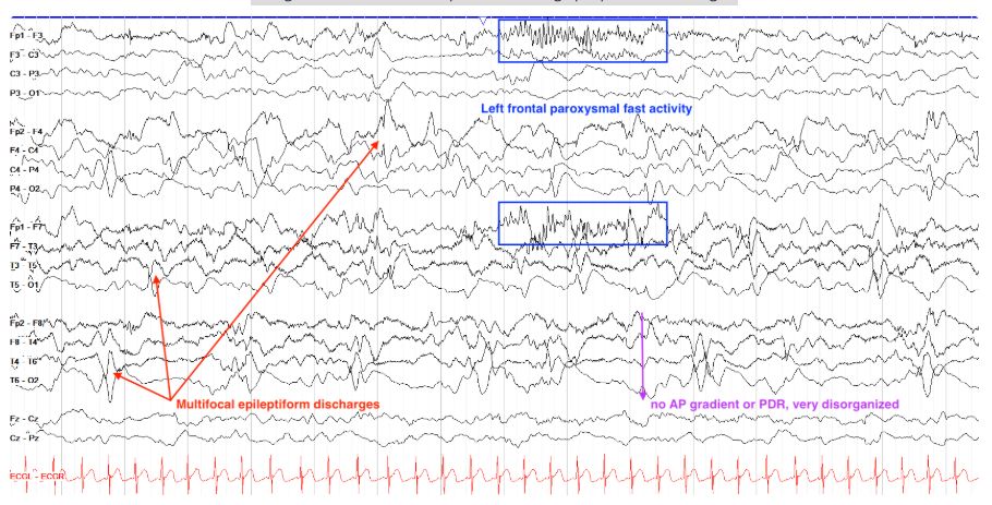
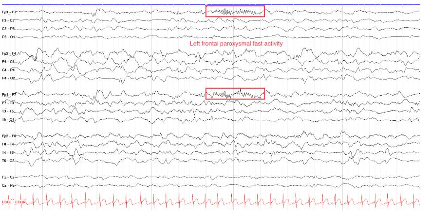
        Abnormal; L frontal paroxysmal fast activity (epileptiform finding)[…]

        Far from a normal variant, this is a highly abnormal tracing from a patient with Lennox Gastaut Syndrome.
        Don't mistake the marked box for 14 and 6 positive spikes; even though they are indeed about 14 Hz, they are in the wrong area (14 & 6 spikes tend to be posterior) and come with too many other surrounding abnormalities including a highly disorganized background with multifocal phase reversing epileptiform discharges.

        EEG
        Is the tracing below abnormal or a normal variant (and if so, which one)?

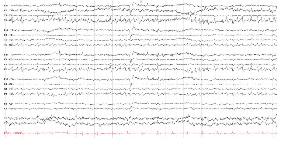
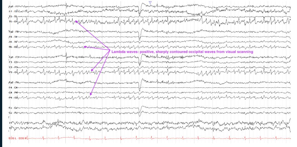
        Lambda Waves[…]

        This tracing shows symmetric occipital, sharply contoured positive waves that look very similar to POSTS that you'd expect in sleep. However, due to the presence of an eye blink in the middle of the page, along with a lot of frontal myogenic activity, you know this patient is awake. Therefore, these POST-like waves are in fact lambda waves; this patient was reading for several hours, during which time she had nearly continuous lambda wave activity because lambdas arise from visual scanning.

        EEG
        Is the pattern in the parasagittal chains below abnormal or a normal variant (and if so, which one)?

        Mu[…] rhythm


        This page has periods of both bilateral and independent unilateral parasagittal arch-like, sharply contoured alpha activity maximal over the centroparietal region. This is consistent with mu rhythm, the idling activity of the sensorimotor cortex. Note that while some periods of mu are better formed than others, the activity never evolves or really changes in appearance.

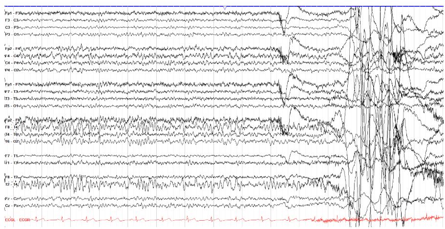
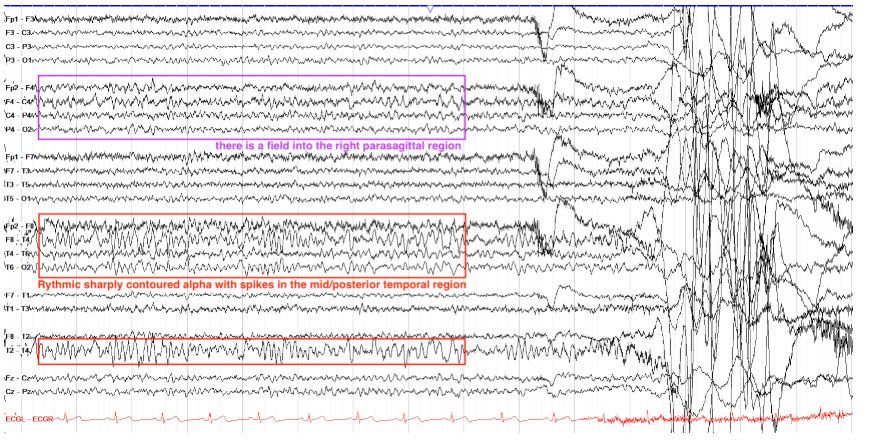
        EEG
        Is the finding in the right temporal chain below abnormal or a normal variant (and if so, which one)?


        Abnormal; this is a portion of a right temporal seizure[…]


        While this activity might look kind of similar to mu, it is off in the wrong distribution--this is in the right temporal region--and has too broad a field, moving into the right parasagittal chain. Mu does not do either. You may also consider wickets, but this morphology does not have the arch-like activity of wickets, and is too prolonged; also, again, it has a disruptive field into the parasagittal chain that wickets typically don't. We'll look at this full seizure, focusing on its evolution in time and location, in the seizures section.

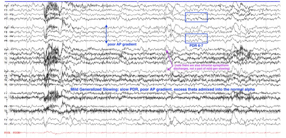
        EEG
        Does the tracing below show generalized slowing and, if so, how would you grade it?


        Mild generalized slowing[…]

        This tracing is marked by a mildly slow PDR of 6-7 Hz, along with a notably less organized AP gradient. Much of the usual architecture of a normal study is here though, including eye blinks and variability across the page. We see some excess theta admixed into the normal and expected alpha activity, but no major delta activity. The area marked by a purple arrow may appear to be frontal delta activity, but these are in fact epileptiform spike and slow waves (note the notch-like spike before each large slow wave), discussed in the epileptiform activity section.

        EEG
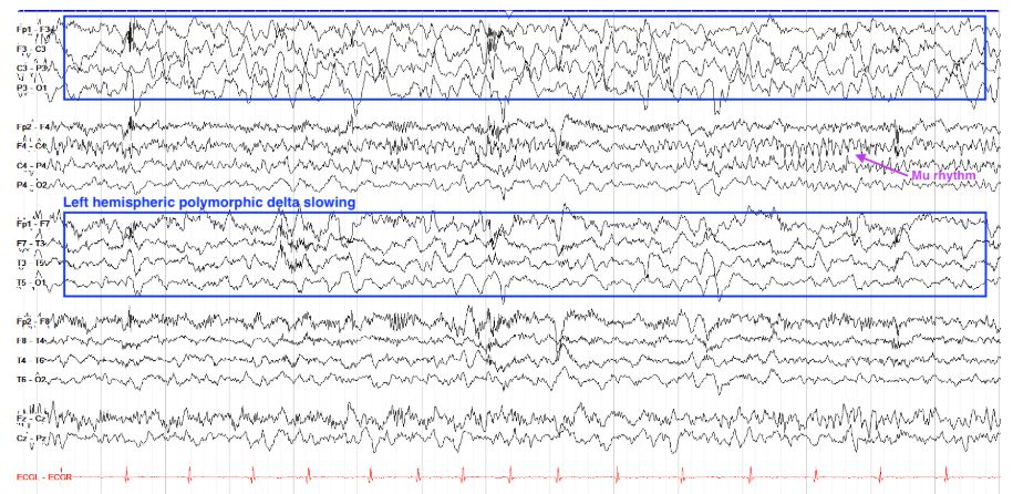
        How would you classify the slowing below?


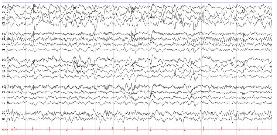
        Left Hemispheric Polymorphic Delta[…] Slowing


        This tracing shows persistent high amplitude delta slowing over the left hemisphere. Given the high amplitude particularly over the parasagittal chain, this may represent not just slowing but breach, which is seen after brain surgery and discussed more below. Note also the periods of mu rhythm--the resting alpha frequency of the sensorimotor cortex that is arch-like in morphology, over the right parasagittal chain; mu rhythm goes away with thoughts of or initiation of movement.


        EEG
        Find the rhythmic activity on this tracing.

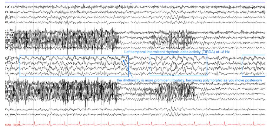
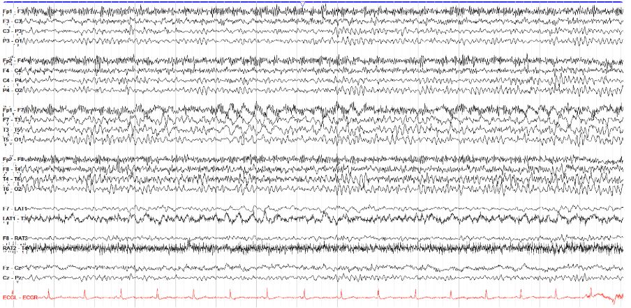
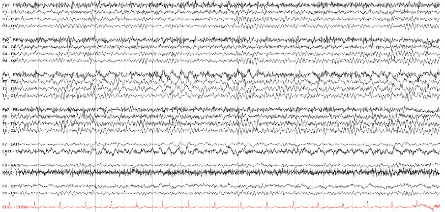
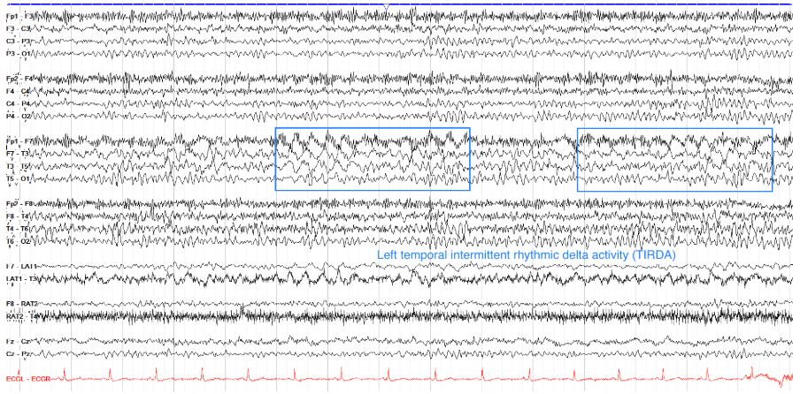
        Left temporal intermittent rhythmic delta activity[…] 


        This tracing shows intermittent runs of approximately 3 Hz delta activity over the left temporal region, more prominent in the anterior than posterior temporal region. Because these intervals of 3Hz delta last at least 2 seconds, they complete the 6 cycle requirement for rhythmic delta activity. Note that the delta is very apparent in the lateral temporal leads below the traditional parasagittal and temporal chains--a reminder to always keep an eye on all of your available chains on EEG.

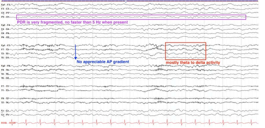
        EEG
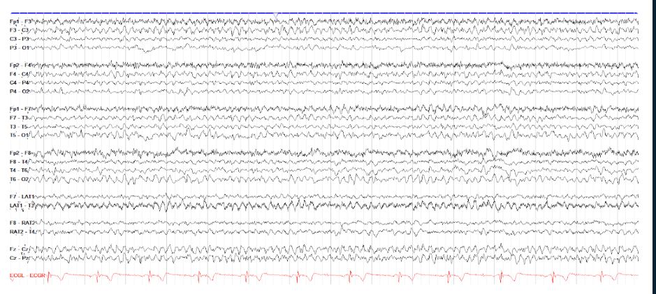
        Grade the slowing on the tracing below.

        Moderate Generalized Slowing[…]

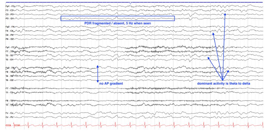
        Recall that when assessing background you should look for a PDR, AP gradient, synchrony, symmetry, continuity, reactivity, and clear wave morphology. On this page, we have a continuous tracing that is synchronous and symmetric. However, there are only fragments of a PDR on both sides, and when present it is no faster than 5 Hz. Thus we have at least mild generalized slowing here. However, there is also no AP gradient--realistically, the page would look much the same if we flipped it upside down. Furthermore, we don't have much alpha activity, with the page mostly theta and delta activity that is relatively monotonous in appearance from beginning to end. Overall, then, this would be considered moderate generalized slowing.

        EEG
        Categorize the slowing below.

        Persistent Right Hemispheric Polymorphic Delta[…] Slowing on a background of Mild Generalized[…] Slowing


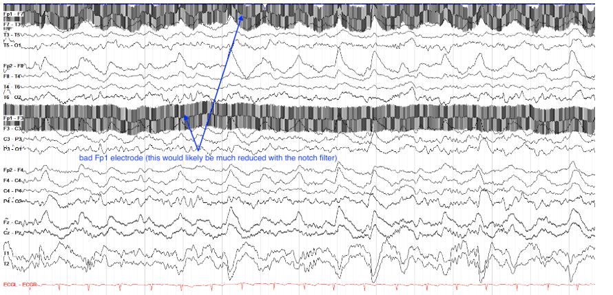
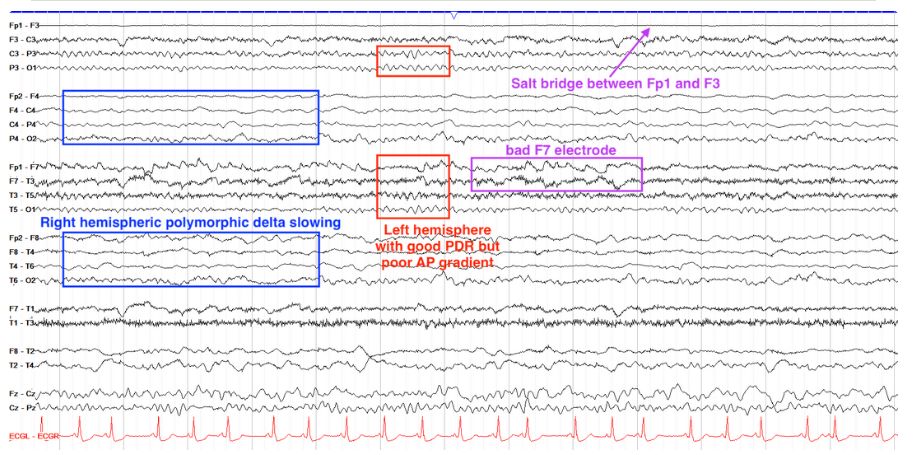
        This is a complicated tracing so let's look at one side at a time. First note that on the left side you have a clear PDR of 8-8.5 Hz, which is in the normal range. However, the AP gradient on the left is poor, with excess slow activity in the frontal regions. Note also that the F7 electrode is bad (note how Fp1-F7 and F7-T3 move in exact opposition to each other, a sign of a bad F7 electrode) and Fp1 and F3 are too close to each other (they've formed a salt bridge, seeing the same voltage as each other and thus cancelling each other out on bipolar, leading to a flat line; on an average montage, they'd have identical appearing lines).

        Moving to the right side, we see a persistent / continuous, somewhat low amplitude delta activity in the frontal regions, and fragments of a PDR in the occipital leads. There is even less of an AP gradient and normal morphology than on the left side. As such, we say this tracing has persistent right hemispheric polymorphic delta slowing on a background of mild generalized slowing.


        EEG
        Classify the slowing below.

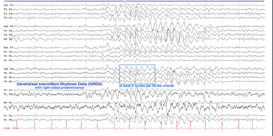
        Generalized Intermittent Rhythmic Delta[…]


        This tracing shows a burst of generalized high amplitude delta activity at about 3Hz, lasting several seconds and thus meeting the ACNS criteria for rhythmicity (aka requires 6 cycles). The delta activity is generalized but more prominent on the right side, particularly in the temporal chains. We would thus classify this as generalized intermittent rhythmic delta activity (GIRDA) with right-sided predominance. Recall that GIRDA is similar to FIRDA prognostically in that it suggests nonspecific and diffuse cerebral dysfunction.


        EEG
        Find the slowing below.

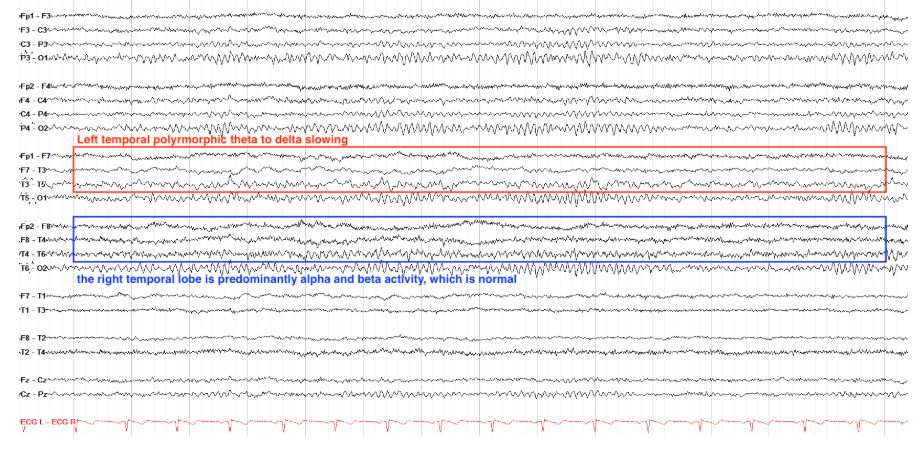
        Left Temporal Polymorphic Theta / Delta[…] Slowing


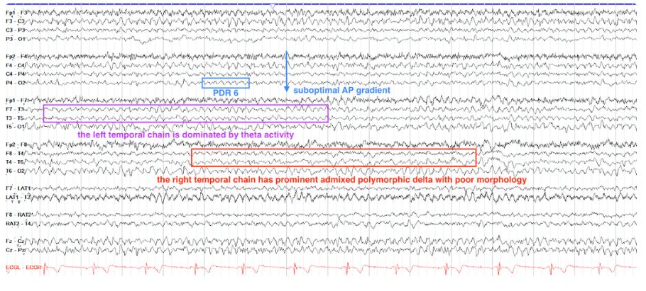
        Here we see a good PDR throughout of 10 Hz, but note while the right temporal region is alpha and beta activity, the left temporal region has a kind of "sloppier" appearance with admixed slower frequencies from theta and delta. In a normal awake adult, you should basically never see theta or delta activity. The slowing here is not rhythmic and seems to run across the entire page, so we'd classify it as focal left temporal persistent polymorphic theta to delta slowing.

        EEG
        Find the slowing below.

        Left Temporal Polymorphic Theta / Delta Slowing[…]
        EEG
        Find the slowing below.

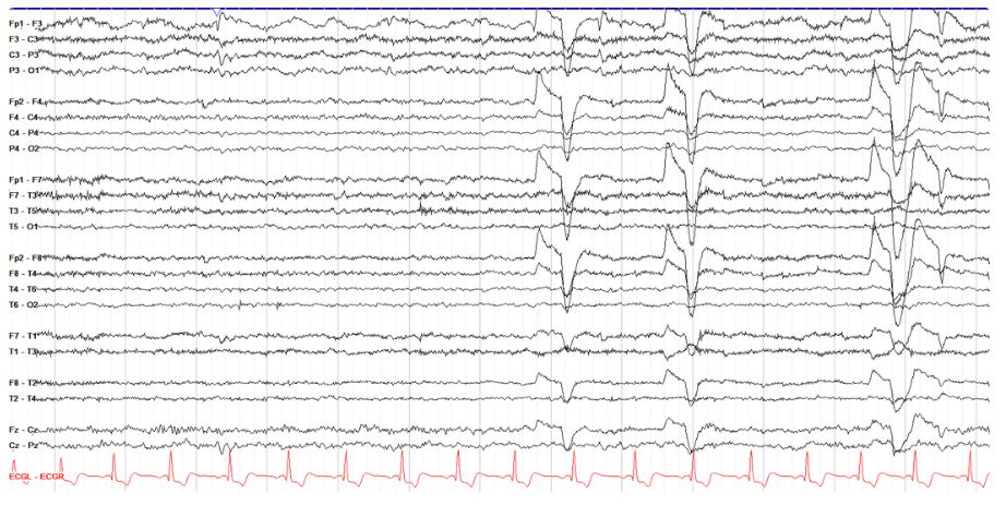
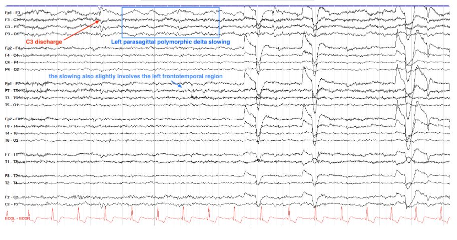
        Left parasagittal[…]

        Here we see continuous polymorphic delta slowing involving essentially the whole left parasagittal chain, as well as more subtly the left frontotemporal region. Also of note here are several negative phase reversals over C3 with spike and slow wave morphology, consistent with epileptiform discharges.

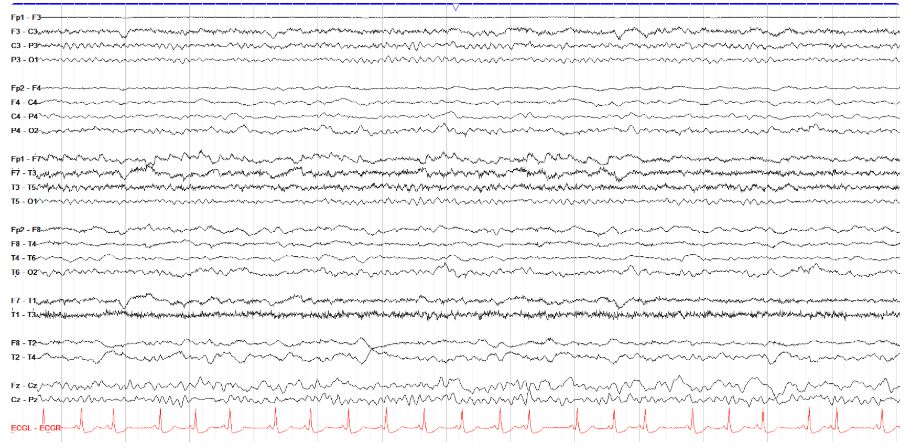
        EEG
        How would you categorize the tracing below?

        Mild Generalized Slowing with Focal Polymorphic Left Temporal Slowing[…]

        This tracing shows diffuse theta activity with a slow PDR and relatively poor AP gradient, consistent with mild generalized slowing. however, there is also a polymorphic focal delta slowing over the right temporal region. Especially when you're starting to read EEGs, this sort of slowing-on-slowing can be hard to see.

        EEG
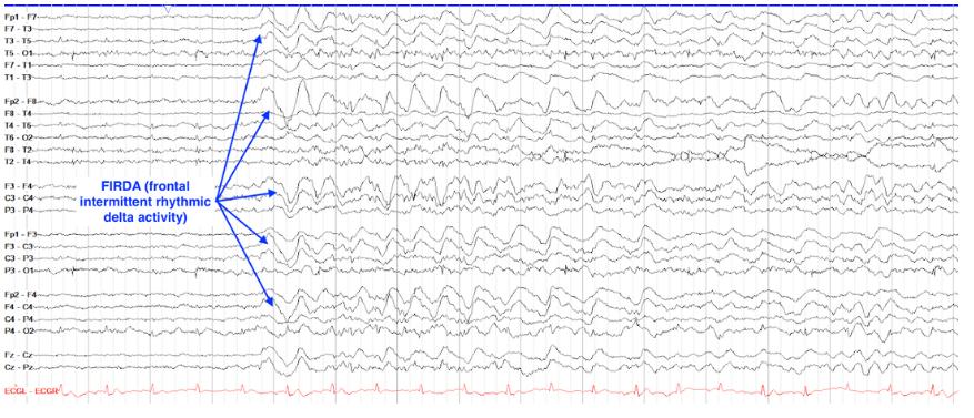
        Categorize the slowing below.

        Mild Generalized Slowing with FIRDA[…]

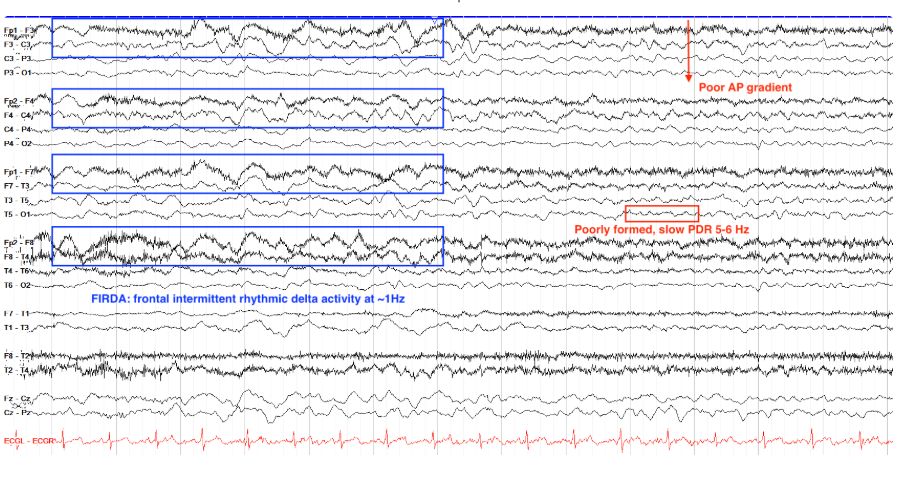
        Starting with the background here, we see a relatively poor AP gradient and slow, often poorly formed PDR. This is consistent with mild or even the beginnings of moderate generalized slowing. We also see, in the first half of the page, higher amplitude and somewhat rhythmic delta activity in the frontal regions that goes on for at least 6 cycles, consistent with FIRDA albeit not the cleanest example as such.


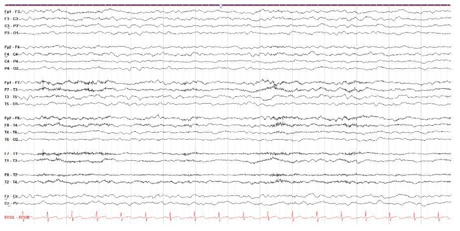
        EEG
        Grade the generalized slowing below.


        Moderate Generalized Slowing[…]

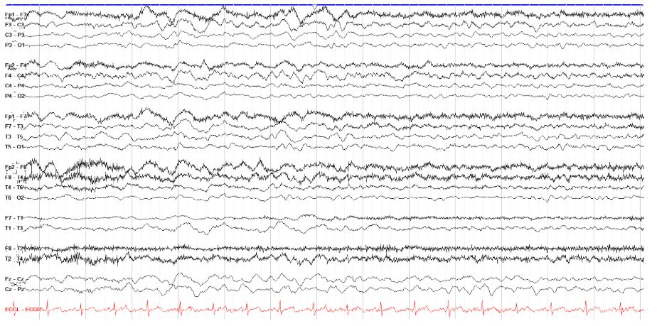
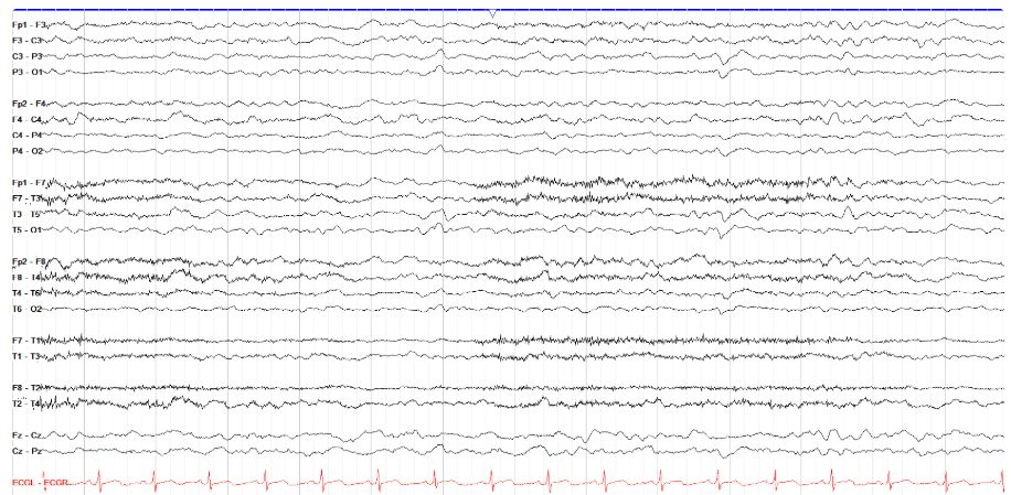
        This tracing has no clear AP gradient, a PDR that is mostly absent and, when seen, is no faster than 5 Hz, and is predominated by theta to delta activity. All of this is consistent with moderate generalized slowing. Recall that mild generalized slowing retains a PDR and AP gradient but the former is slowed and the latter is suboptimal, and that severe generalized slowing usually has discontinuity, poor reactivity/variability, and mostly delta activity that is often low amplitude.


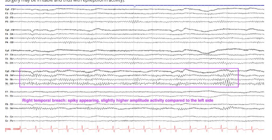
        EEG
        Which of the following is seen on the tracing below?

        Right Temporal Breach[…]

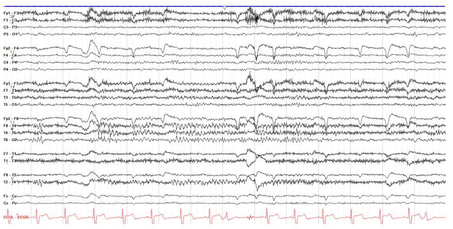
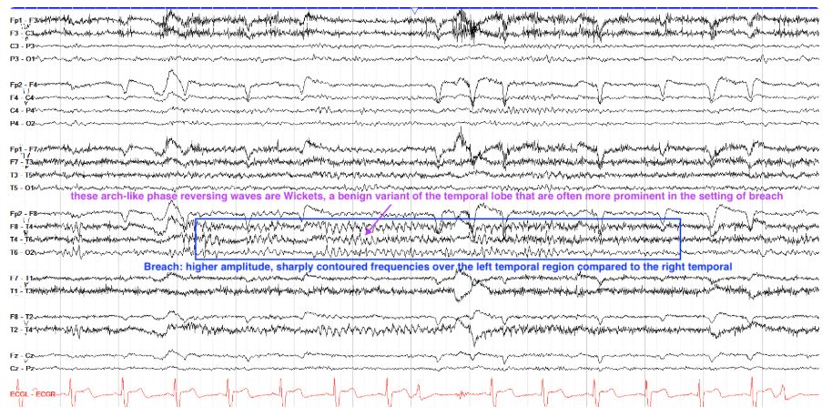
        This tracing has a normal background with a PDR of about 12 Hz. The right temporal region, however, has higher and more sharply contoured morphology than does the left temporal or other regions, consistent with breach artifact as is seen after brain surgery due to the residual skull defects. Within the breach rhythm we see very nicely formed wickets, a benign finding in the temporal lobes that are marked by arch-like, phase reversing waves that come in runs. They should not be mistaken for epileptiform discharges, and are often more easily seen within breach.

        EEG
        Localize the discharge below.

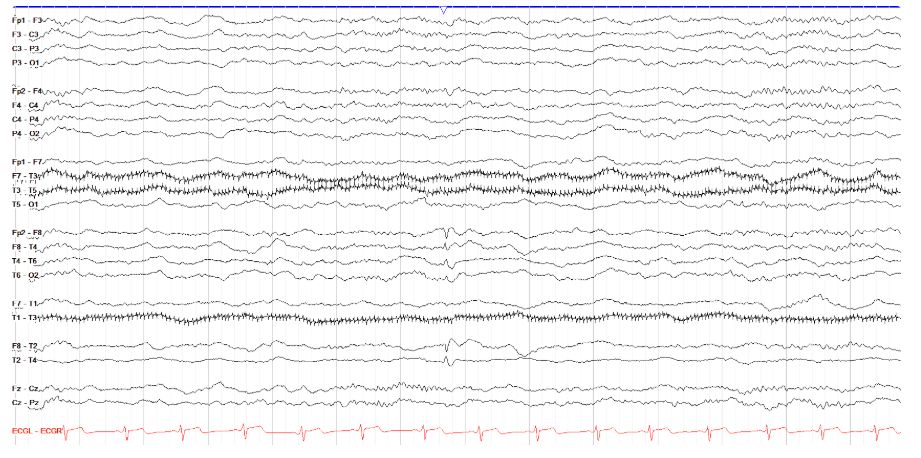
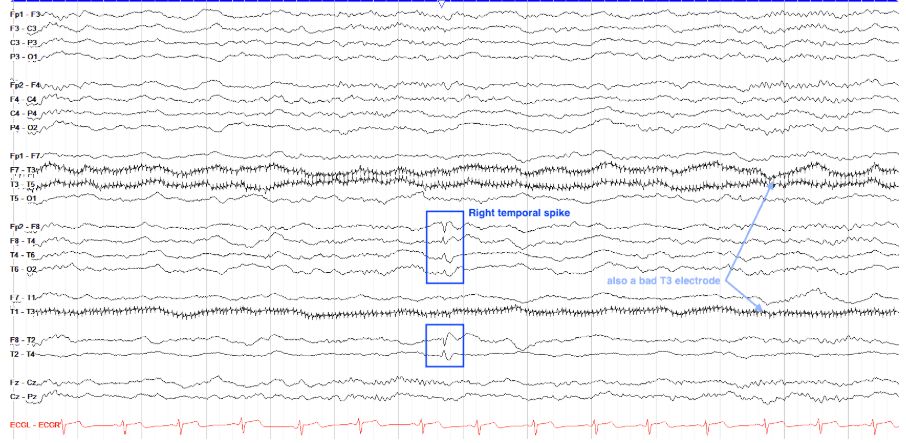
        Right Anterior Temporal Spike and Wave[…]

        This bipolar tracing shows a phase reversing spike and slow wave in the right anterior temporal region. Note the phase reversal (the point at which two tracings "point to one another") between Fp2-F8 and F8-T4, but also at F8-T2 and T2-T4. This puts the discharge between F8 and T2, in the anterior temporal region. Other notable findings on this tracing are a bad T3 electrode, seen as repetitive fast artifact over the T3 leads, and several low amplitude sleep spindles suggesting this is in stage II sleep.

        EEG
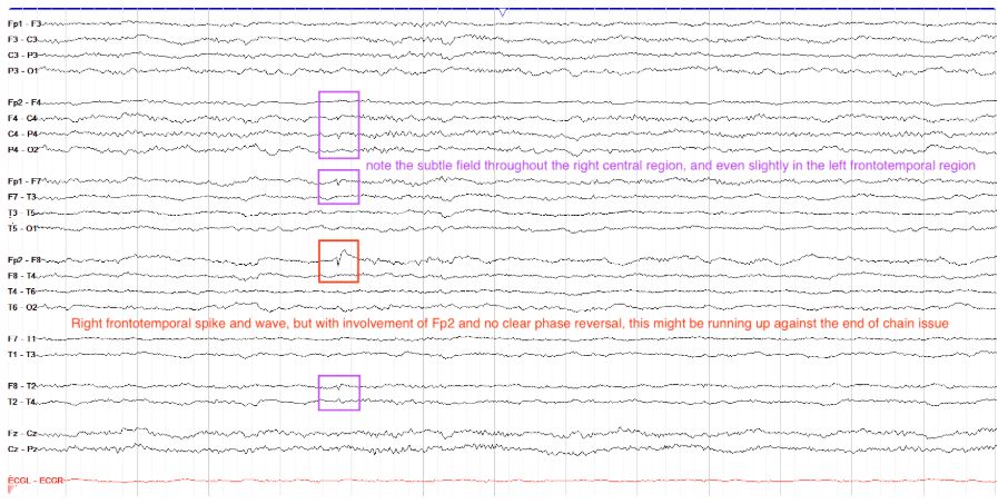
        Localize the discharge below.

        Right Frontotemporal / Frontal Spike and Wave[…]

        This tracing shows a clear spike and wave discharge at Fp2-F8. However, despite this being a bipolar montage there is no phase reversal. This is a good example of the end of chain issue, where there is nothing anterior to Fp2 to which to compare its voltage. It is possible that the maximal discharge, where the phase reversal would be, is actually anterior to or deep to Fp2, but because there isn't an electrode there to measure it, we can't see the full extend of what would be the phase reversal on scalp EEG. Also note the good field in the right frontocentral region, with some reflection to the left frontal/frontotemporal region.

        EEG
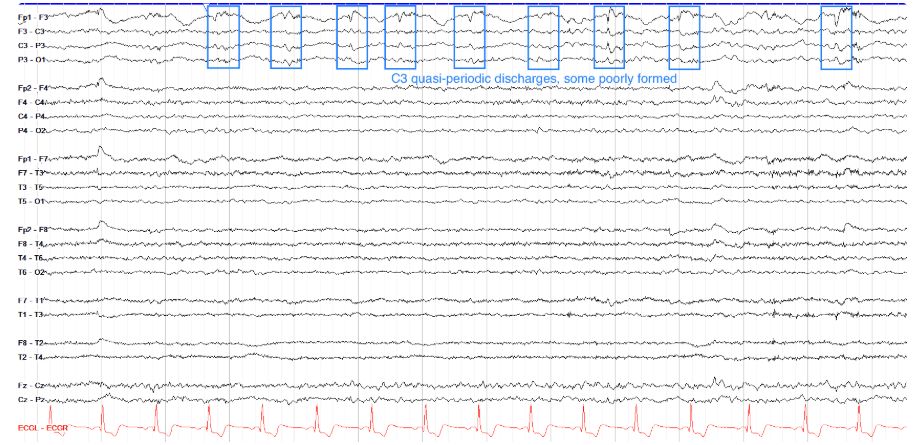
        Does the following tracing show a periodic pattern?

        yes at C3[…]


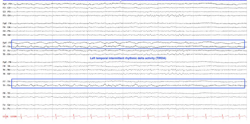
        EEG
        Find the rhythmic activity on this tracing.

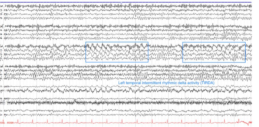
        Left temporal intermittent rhythmic delta activity[…]


        This tracing shows intermittent runs of approximately 3 Hz delta activity over the left temporal region, more prominent in the anterior than posterior temporal region. Because these intervals of 3Hz delta last at least 2 seconds, they complete the 6 cycle requirement for rhythmic delta activity. Note that the delta is very apparent in the lateral temporal leads below the traditional parasagittal and temporal chains--a reminder to always keep an eye on all of your available chains on EEG.

        EEG
        Find the epileptiform activity on this tracing.
        note: there are multiple findings


        • R temporal sharps[…]
        • L temporal spikes[…]
        • R frontal spikes[…] and paroxysmal fast activity[…]


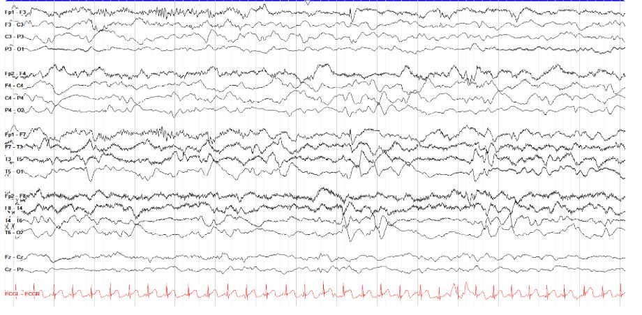
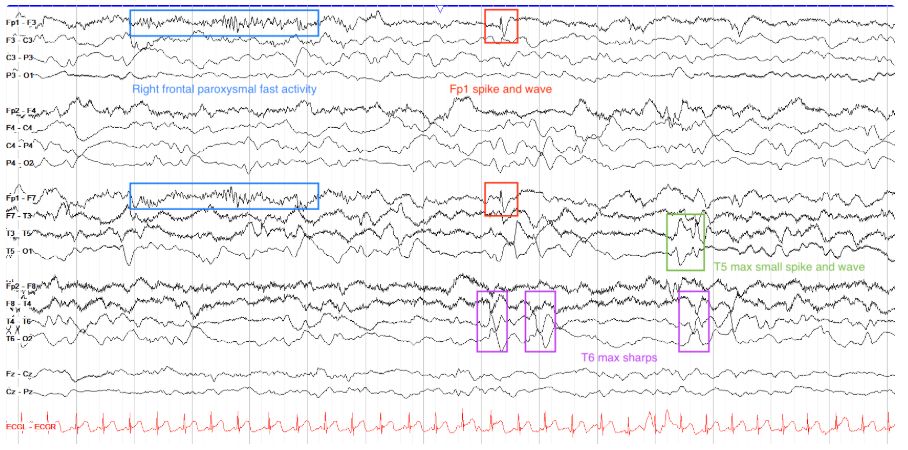
        This is a complicated tracing. First note the state of the patient, who appears to be in slow wave sleep (relatively synchronized high amplitude delta activity diffusely, no eye blinks or other evidence of being awake). There are multiple epileptiform discharges on the page, including a run of right frontal paroxysmal fast activity, right frontal spike and waves (Fp1 max), right mid-temporal sharps (T6 max, although poorly formed), and left temporal spike and waves. Note the field of the right temporal sharps into the right parasagittal chain. This kind of tracing--disorganized with multifocal discharges--is typical for Lennox Gastaut Syndrome.

        EEG
        Is the discharge below most consistent with an epileptiform discharge, artifact, or nonepileptiform abnormality?

        Triphasic Wave[…]

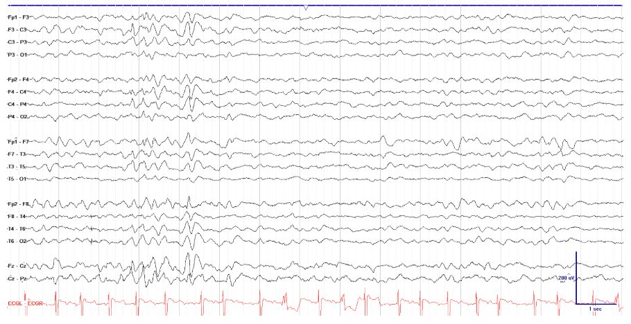
        Here we see a three-phased generalized discharge with slight right hemispheric and bifrontal predominance, with a slight anterior to posterior lag in onset and in which each each phase is slightly longer than the preceding one. These are all hallmarks of triphasic waves, which are not epileptic in nature and are most often seen with toxic-metabolic encephalopathy.

        EEG
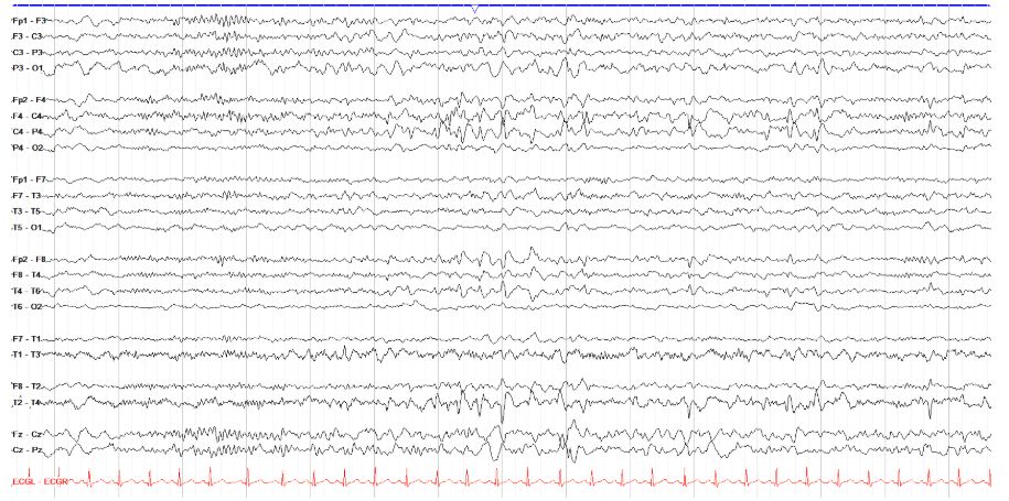
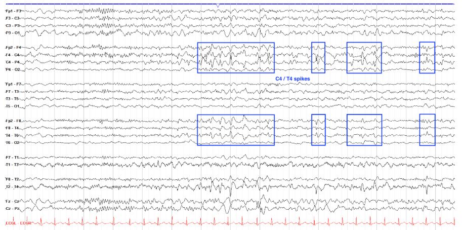
        Localize the epileptiform activity on this tracing.

        T4 spikes[…]


        Here we have multiple, very well formed phase reversing spike and slow wave discharges that point toward T4 and C4, with slightly better formed morphology over the temporal than central region. These would thus be right centrotemporal spikes. Note the field into the left central region, and also that in the right parasagittal chain, the frequent negative phase reversals can appear somewhat similar to electrode artifact, but you don't see evidence of such artifact elsewhere on the page.

        EEG
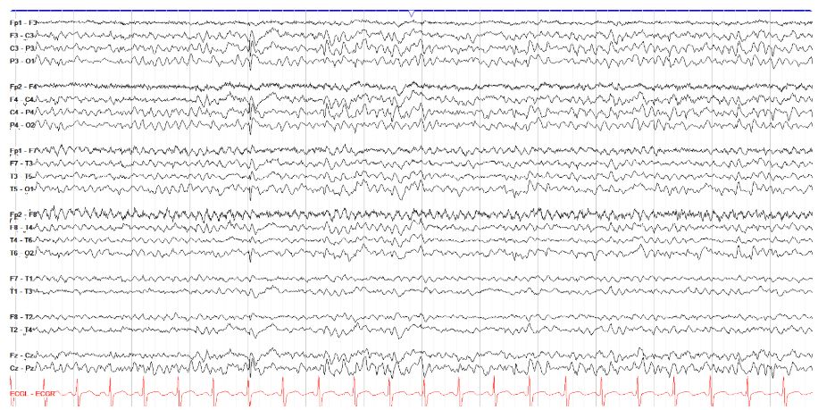
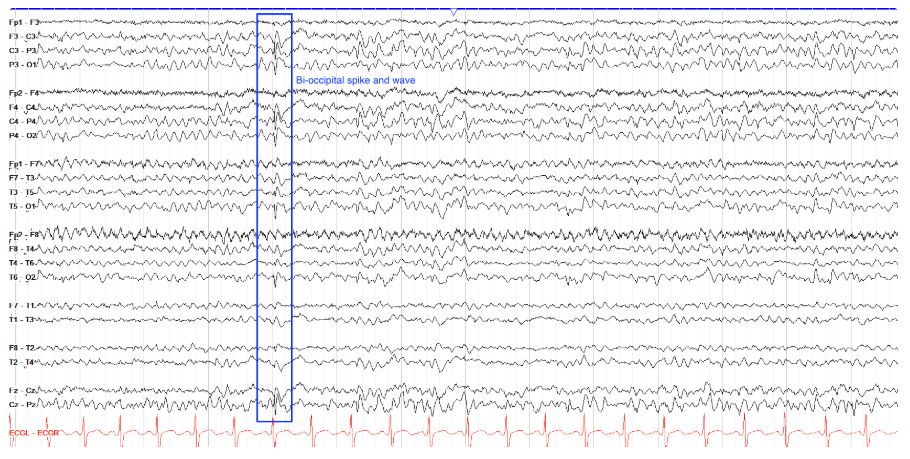
        Localize the interictal discharge(s) below.

        Bilateral occipital spike and waves[…]


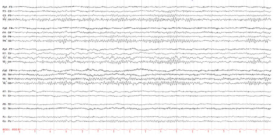
        First off, note that this tracing has mild generalized slowing, with a PDR not exceeding 7.5 Hz. There are multiple relatively high amplitude spike and slow waves in the bilateral occipital regions, fairly symmetric in amplitude, with a field extending anteriorly throughout the parasagittal, temporal and central chains.

        EEG
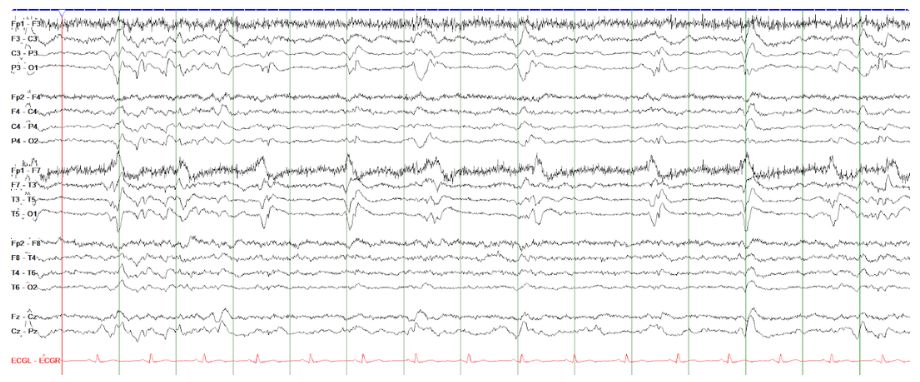
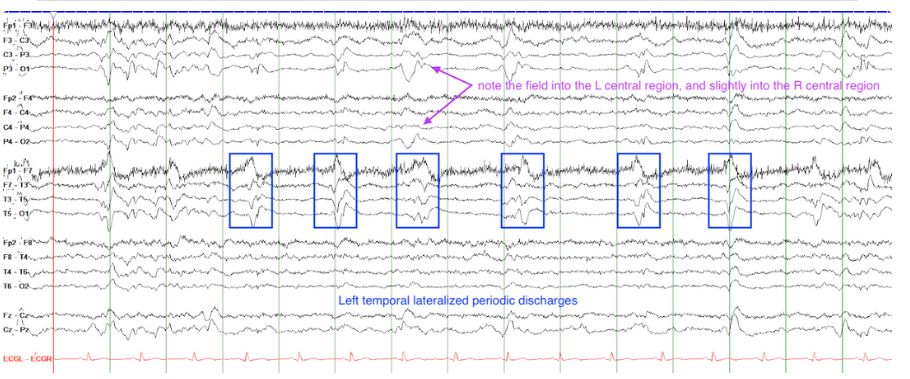
        With which infection is the following tracing classically associated?

        PLEDs (LPDs) often seen with HSV encephalitis[…]

        This tracing shows left temporal lateralized periodic discharges (LPDs), also called periodic lateralized epileptiform discharges (PLEDs). This is an epileptogenic pattern commonly, but not exclusively, seen with HSV encephalitis. Recall that for both periodic and rhythmic patterns, a cycle of at least 6 is required by ACNS classification. Note the field of the discharges into the left central regions, and slightly into the posterior right central region, but the clear area of maximal amplitude is the left temporal region.


        EEG
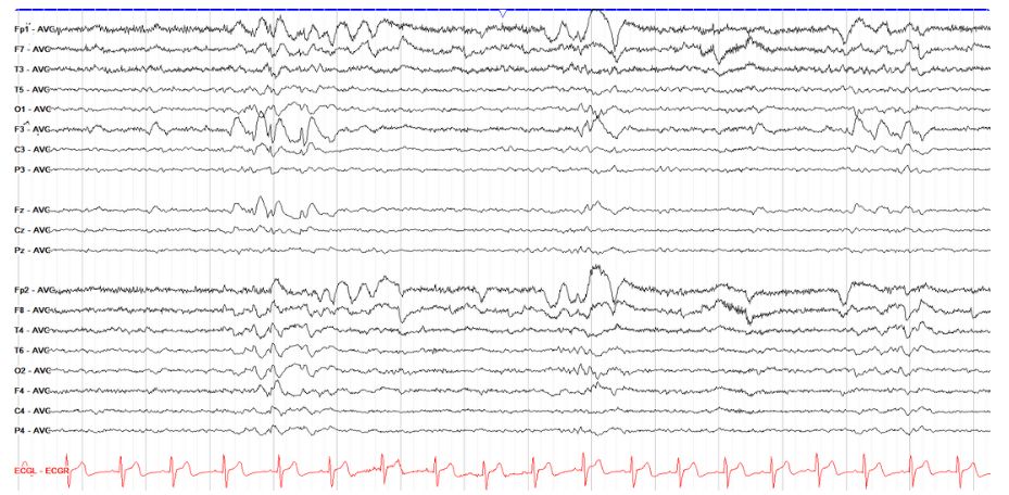
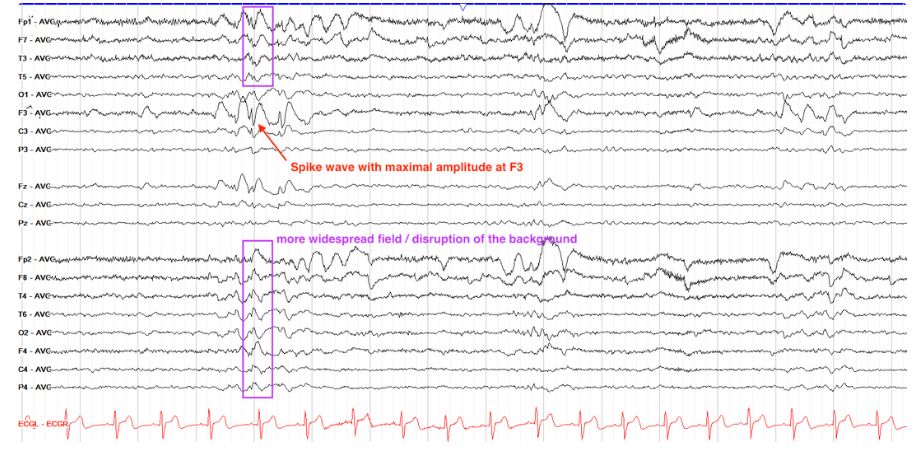
        Localize the interictal discharge(s) below.

        Left frontal spike[…]

        This page shows an average tracing, so recall that you aren't looking for a phase reversal here, just for the point of maximal amplitude for a sharp, spike, or spike wave discharge. As such, F3 has a clear spike and slow wave as marked, with a more subtle field from that spike throughout the rest of the electrodes. Average montages are useful to determine the point of maximal amplitude for a discharge if a phase reversal is somehow unclear.

        EEG
        Localize the interictal discharge(s) below.

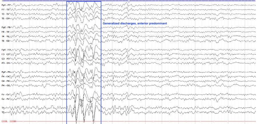
        Generalized[…] with anterior predominance


        This was kind of a trick question (sorry?), as these discharges are, actually, not localizable. They are generalized discharges, albeit with some anterior/frontal predominance, which is very common in generalized epilepsies. Even though the discharges are much higher amplitude frontally, you can see the field extend with clear (much lower amplitude) spikes all the way back in the occipital regions. The discharges have a spike or polyspike prior to each slow wave, and those spikes are fairly (though not completely) symmetric in morphology and amplitude.


        EEG
        Localize the interictal discharge(s) below.

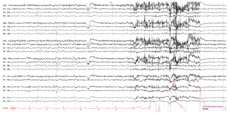
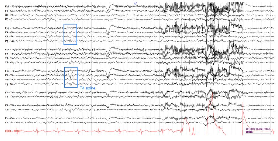
        Right mid-temporal, T4 max[…]


        This awake tracing (note the eye blink around the middle of the page, and frontal muscle artifact in the second half of the page), shows a clear phase reversing sharp maximal at T4, with a field into the right central region.

        EEG
        Seizure or not?

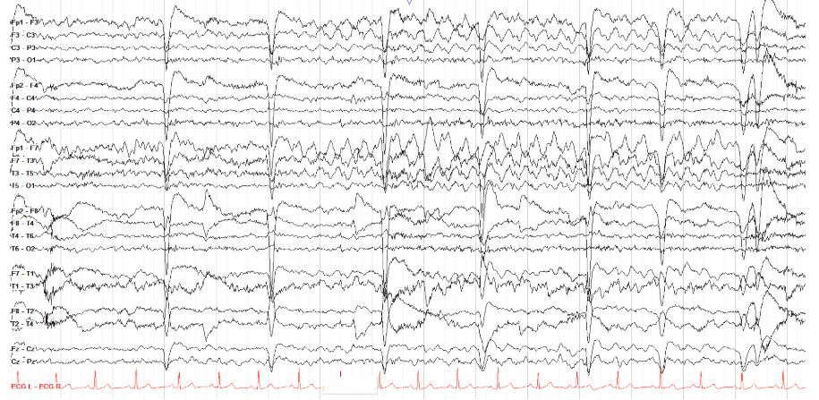
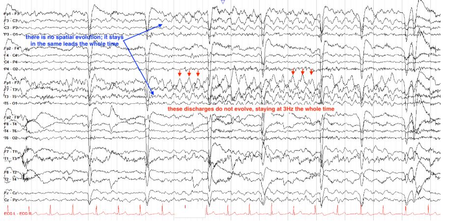
        Not a seizure! L frontotemporal rhythmic delta with embedded spikes[…]


        This is not a seizure, as it does not evolve either temporally or spatially. It starts with 3 Hz spike wave activity that is maximal temporally with a field into the parasagittal chain, and it ends that same exact way. It also appears to be slightly less than the ten second requirement for seizure, although there may be some subtle lead-in in the first few seconds of the page so that is debatable. The lack of evolution, however, is clear and thus this is just an interictal run.

        EEG
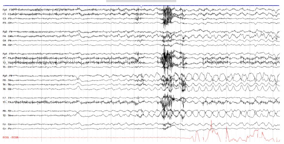
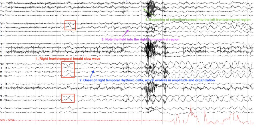
        Find the seizure onset.

        Right frontotemporal[…] region


        This seizure begins with a herald slow wave in the right frontotemporal region, which leads into subtle rhythmic delta of the right temporal region that evolves in amplitude and morphology, and within a few seconds is quite prominent with a field / spread into the right frontocentral region. Shortly thereafter, there is some reflection into the left frontotemporal region.

        EEG
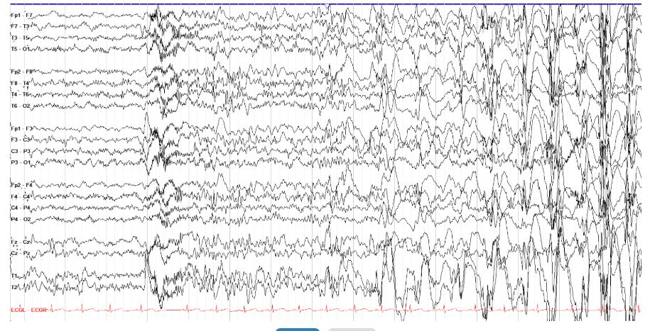
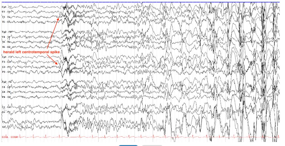
        Is the seizure below focal or generalized?

        Focal[…], with a left centrotemporal herald spike and secondary generalization[…]

        This is a subclinical seizure that may at first appear generalized in onset, but if you look closely you can see a left centrotemporal spike (note the clear positive spike temporally and prominent phase reversal centrally) immediately before onset of generalized fast activity leading into sharply contoured 6-7 Hz activity that progresses to very high amplitude generalized, frontally predominant spike wave activity at about 1 Hz, which on the second page slows into a rather abrupt offset.

        EEG
        Is this seizure focal or generalized?

        Generalized Tonic Seizure[…]

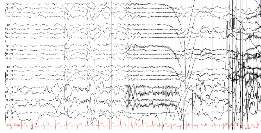
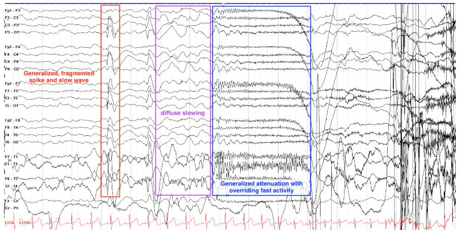
        This seizure begins with a generalized albeit somewhat fragmented generalized spike and wave discharge (note that the discharge is seen in all the chains with similar morphology in them all), with subsequent slowing for several seconds before onset of generalized attenuation with overriding fast activity, the hallmark appearance of a tonic seizure.

        EEG
        Localize this seizure's onset.

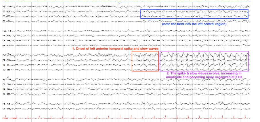
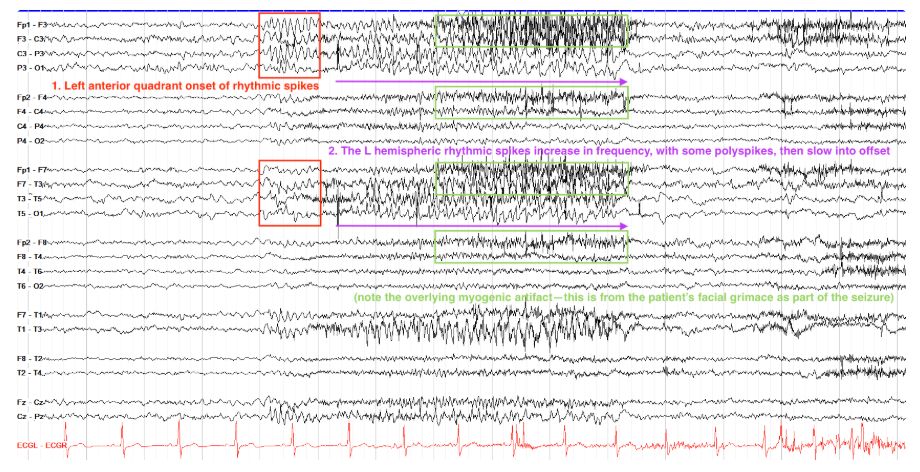
        Left Anterior[…] Quadrant


        This seizure starts with a second of rhythmic spikes best seen over the left frontocentral regions, but with simultaneous albeit less well formed spikes over the left anterior temporal region. With the frontal regions of both the parasagittal and temporal chains being involved, we'd call this a left anterior quadrant onset seizure. Note that this seizure continues to evolve via higher amplitude spikes waves that increase in frequency for several seconds, then slow prior to offset; this seizure remains focal to the left hemisphere, but there's associated muscle artifact over the right hemisphere.


        EEG
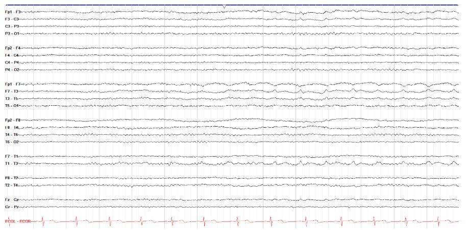
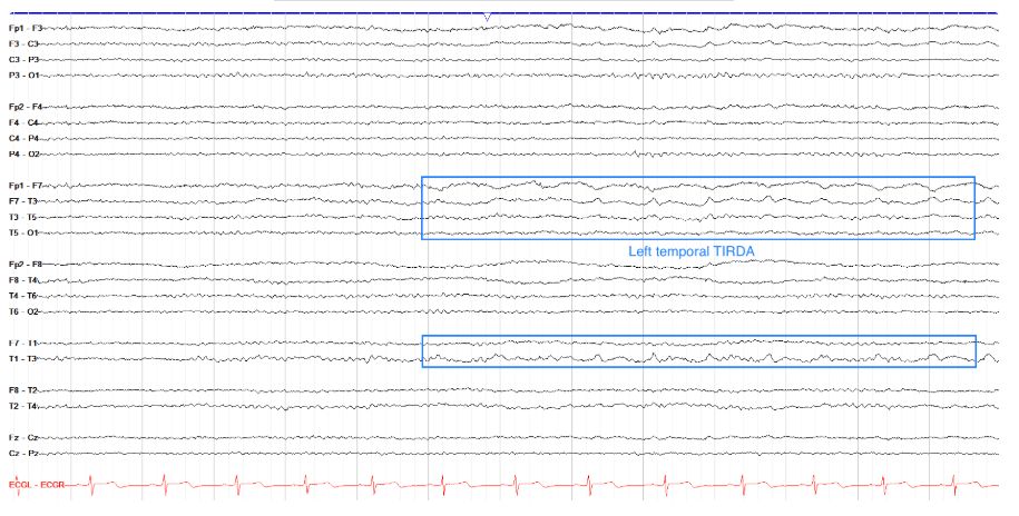
        Does this tracing show a seizure?

        no - shows L Temporal TIRDA[…]

        This page shows anterior left temporal sharply contoured delta activity at about 1 Hz, but there is no evolution that we can see on this page so it doesn't fit criteria for seizure. This is likely an example of left temporal intermittent rhythmic delta activity (TIRDA), an epileptiform finding. The caveat to this is that we don't see the following page--if there is some evolution in frequency or distribution of this activity, seizure would return as a possibility.

        EEG
        Does this tracing show a seizure?

        No, this is eye flutter artifact[…]


        This tracing shows about seven seconds of bifrontal rhythmic activity. Note, however, that while there might appear to be evolution here, the change is very localized to the frontal leads, and their morphology is not consistent with typical epileptiform spike wave activity. This is actually a pretty classic example of eye flutter. Also remember that seizures require a least ten seconds of evolution if there is no clinical correlate, so even if this were epileptifrom in nature, it still wouldn't be categorized as a seizure unless there was a clinical correlate.


        EEG
        Localize, if possible, the seizure onset below.

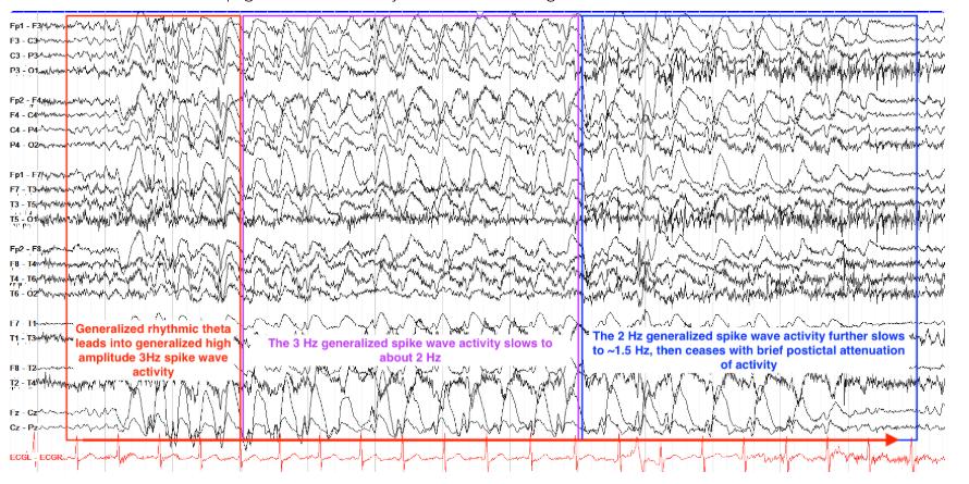
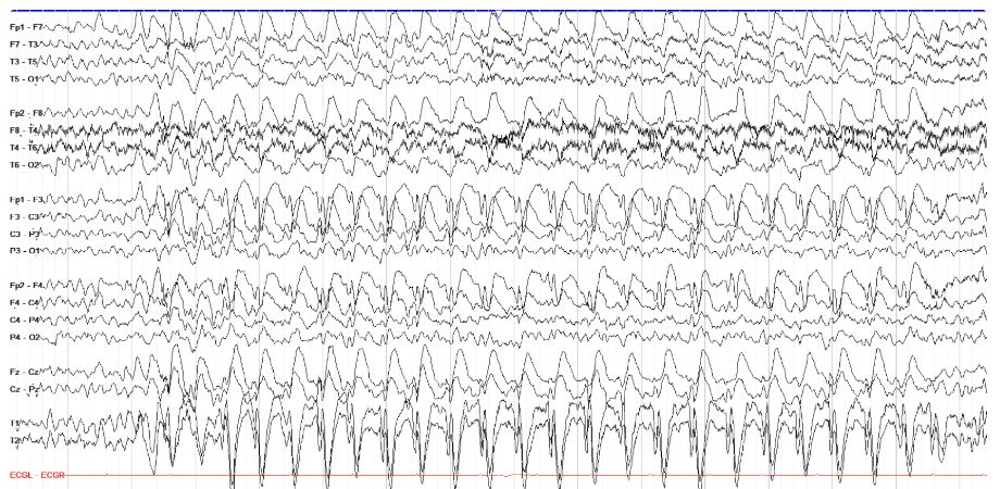
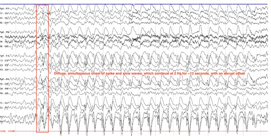
        Generalized Seizure[…]
        This seizure is not localizable, as it is generalized. This particular seizure was subclinical, with no outward clinical signs, but electrographically is marked by a sudden onset of generalized spike and slow waves (with frontal predominance) that continue at about 2Hz for 12 seconds, before slowing suddenly with an abrupt offset, and no clear postictal changes that we can see in the last second of the page.


        EEG
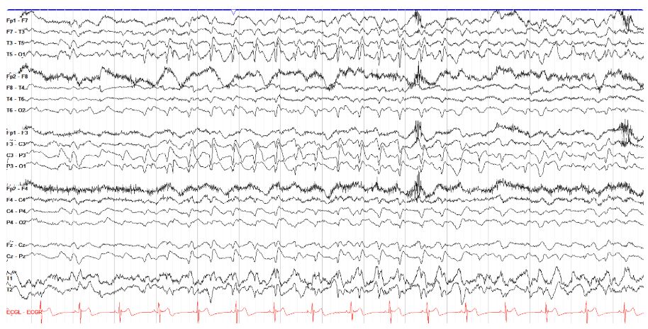
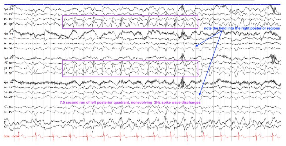
        Does this tracing show a seizure, interictals, or artifact?

        Interictals[…]


        Here we see prominent left posterior quadrant spike and slow wave discharges, at about 2 Hz and with a prominent field into the right posterior region. However, they only last for 7.5 seconds and are non evolving, and thus do not fit criteria for a seizure. Recall that technically seizures require evolution, and should be longer than 10 seconds. There are, of course, exceptions to this rule; myoclonic jerks, absence seizures, and tonic seizures, for example, are often less than ten seconds long but are still considered seizures as long as there is a consistent clinical correlate (seen on video) to electrographic epileptiform activity.

        EEG
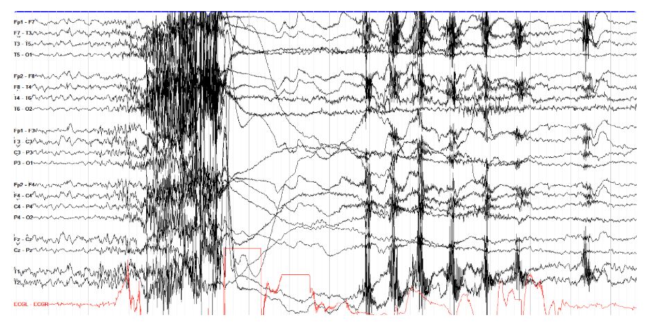
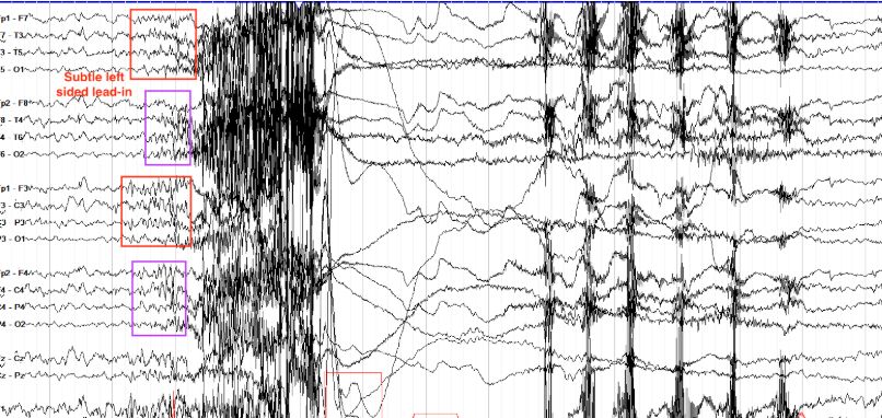
        Lateralize the seizure onset below.

        Left Hemispheric[…]
        Here we see a tonic clonic seizure that might at first look rather generalized in onset, but note the half a second of sharply contoured fast activity over the left hemisphere (in red boxes) prior to the onset of similar activity in the right hemisphere (in purple boxes). So while this seizure has very rapid generalization, it does arise from the left hemisphere.

        Note also that in terms of evolution, we see that fast spike activity on the left, then right side, then diffuse myogenic artifact from which arises several seconds of diffuse slowing and attenuation, followed by generalized polyspike and waves with overriding myogenic artifact, which slow from 1-2Hz to <1Hz prior to offset.

        A 41-year-old man has focal seizures with rising epigastric aura, oral/manual automatisms, and impaired awareness; EEG shows anterior temporal epileptiform discharges and MRI shows hippocampal atrophy, localizing to the temporal[…] lobe and indicating mesial[…] temporal sclerosis as the underlying etiology
        A 20-year-old woman with 2-year history of stress-triggered whole-body shaking, normal MRI and routine EEG, and monitoring-unit captured generalized onset myoclonic to bilateral tonic-clonic seizures with generalized spike discharges on ictal EEG is most consistent with genetic[…] generalized epilepsy syndrome, and her episodes therefore represent generalized[…] onset seizures.
        A 15-year-old girl with a morning generalized tonic-clonic seizure, frequent morning arm jerks causing her to drop objects, sleep deprivation as a trigger, and EEG showing photic-provoked (photosensitive) generalized spike-wave discharges has a generalized epilepsy syndrome consistent with juvenile[…] myoclonic epilepsy, and her seizures are therefore generalized[…] onset