CSF DX

CSF DX

Subspecialties · 29 cards

SIH - Pathophysiology and Symptoms

Spontaneous intracranial hypotension (SIH) is the modern name for spontaneous spinal CSF leak[…], which by definition excludes iatrogenic causes such as LP, epidurals, or spine surgery, as well as traumatic dural tears like whiplash or chiropractic injury.

Mechanistically, CSF drains from the intracranial compartment, creating downward traction on dura-innervating nerves and producing the orthostatic headache[…] that is the signature symptom.

Per the Monro-Kellie[…] doctrine, intracranial veins engorge to compensate for lost CSF volume, stretching cranial nerves and causing facial pain, numbness, and diplopia[…].

CSF pressure changes also disturb the inner ear's endolymph and perilymph, producing vestibulo-auditory[…] symptoms such as tinnitus and vertigo.

Classically, SIH headache is worse upright[body position], worse with Valsalva[…], worse in the second half of the day[…], and improves when supine[…].

The pain is often described in a coat hanger[…] distribution radiating from the posterior head into the neck and trapezii, with associated nausea, vomiting, dizziness, photophobia, or diplopia[…].

Severe complications include brain sagging dementia[…] (mimicking bvFTD), bibrachial amyotrophy[…] (mimicking ALS with UMN/LMN findings), superficial siderosis[…] (predominantly infratentorial with hearing loss and ataxia), and spinal cord herniation.
SIH - Diagnosis and SEEPS MRI Findings

SIH is diagnosed by demonstration of a spinal extradural CSF leak[…], OR by SEEPS MRI findings plus one of [low LP opening pressure ≤6, spinal meningeal diverticulum, improvement after epidural blood patch], OR by orthostatic headache plus 2 of the B criteria[…].

Notably, the majority[most or minority?] of CSF leak patients have a normal[normal or abnormal?] opening pressure on LP.

The SEEPS mnemonic stands for Subdural fluid collections, Enhancement (smooth diffuse pachymeningeal), Engorged venous structures, Pituitary hyperemia, and Sagging of brain[…].

Subdural collections may be unilateral or bilateral; always consider CSF leak when subdurals are atraumatic[…].

Pachymeningeal enhancement is of the outer[inner or outer?] dural layer and is diffuse[…] because leaked CSF disperses uniformly around the dura — this contrasts with leptomeningeal[…] enhancement (pia + arachnoid), which tends to be more focal[…].

Engorged venous structures show downward bowing of the transverse sinuses[…] and outward bowing of the superior sagittal sinus[…].

Pituitary hyperemia appears as pituitary enlargement with reduced height of the suprasellar cistern[…] and leftward bowing on sagittal MRI.

Brain sag shows reduced suprasellar cistern (from sagging of the 3rd ventricle[…] floor), reduced mammillopontine[…] distance, and effaced prepontine[…] cistern from brainstem sag — sometimes with cerebellar tonsillar descent mimicking Chiari[…] malformation.
SIH - Risk Factors and Mimics

SIH risk factors include connective tissue disorders[…] (Marfans, Ehlers-Danlos), disc osteophytes[…] in the spine, bariatric surgery[…] (which widens the spinal CSF/paraspinal vein pressure gradient and promotes CSF-venous fistula[…] formation), and prior intracranial hypertension[…] that can spontaneously open a leak at the skull base or spine.

SIH mimics include chronic vestibular migraine[…] (trigeminal afferents activated), Chiari[…] malformation (tonsillar ectopia, Valsalva-worsened), and neurogenic orthostatic hypotension or POTS[…].
SIH - Radiographic Evaluation Algorithm

The SIH imaging algorithm begins with MRI brain with and without contrast[…], optionally with a radioisotope cisternogram[…] — looking for SEEPS findings.

The Bern[…] score quantifies the probability of finding a CSF leak on myelography based on MRI brain findings.

Step 2 is MRI full spine without contrast[…], looking for secondary spinal extradural CSF (evidence a leak exists but not necessarily its location), best seen on T2 STIR (fat-suppressed)[…] sequences.

If an extradural collection IS present, perform prone or decubitus[…] positioning followed by digital subtraction myelography (DSM)[…] OR dynamic CT myelography[…].

If NO extradural collection is present, you must rule out a CSF-venous fistula[…] by getting decubitus DSM with cone-beam CT, OR decubitus CTM with a photon-counting[…] scanner if available.

If still nothing is found, rule out a slow CSF-venous fistula[…] and leaking meningeal diverticula with delayed CTM[…] OR MR myelogram with intrathecal contrast[…].
SIH - Spine Imaging and Localization

The main goal of spinal MRI in SIH is to rule out a fast[rate?]-leaking dural tear; if absent, patients can still have slow[…]-leaking CSF-venous fistulas or meningeal diverticula.

The position of an extradural collection guides localization: ventral extradural CSF suggests a ventral[…] dural tear, while dorsal/lateral extradural CSF suggests a posterolateral[…] dural tear.

Ventral dural tears are characteristically caused by osseous spicules[…] (osteophytes, calcified discs) penetrating the dura, while posterolateral dural tears are associated with connective tissue disorders[…] that weaken the dura.
SIH - Radioisotope Cisternography

Radioisotope cisternography uses intrathecal injection of indium-111 disodium pentate[…], then assesses how quickly the tracer migrates up the spinal canal and through the intracranial space.

Patients with SIH show slower or incomplete[…] ascent of radiotracer compared to normal.
SIH - Advanced Myelography (DSM vs CTM)

Digital subtraction myelography (DSM) involves LP injection of iodine contrast under continuous fluoroscopy[…], with digital subtraction removing bone and dense structures.

DSM pros: real-time visualization[…] and high temporal resolution at 1-2 frames per second, allowing discrimination from tiny adjacent structures.

DSM con: CSF leaks superimposed over contrast-enhanced structures (like meningeal diverticula[…]) can be missed — cone-beam CT[…] helps with this.

Dynamic CT myelography also uses LP iodine injection, then multiple CT scans.

Pros include cross-sectional detail[…] and better identification of anatomical lesions like bony spicules.

Con: lower[…] spatial and temporal resolution compared to DSM.
SIH - Positioning and CSF-Venous Fistulas

If a ventral dural tear is suspected, position the patient prone[…] so the tear faces up; if posterolateral, position decubitus[…] so the tear faces toward the imaging direction.

The characteristic dural tear finding on dynamic CTM or DSM is a split contrast column[…] — which delayed CTM typically misses due to contrast washout[…].

The majority of CSF-venous fistulas occur in the thoracic[…] spine.

Photon-counting[…] CT — a next-generation scanner using direct x-ray detection — is faster, better, and excellent for detecting CSF-venous fistulas and dural tears.

Patients without extradural CSF on spine MRI usually have a CSF-venous fistula[…], which is more common than a leaking meningeal diverticulum.

DSM and dynamic CTM workup of these are best done decubitus[…].
SIH - Treatment

SIH treatment depends on leak classification. Type 1a is a ventral dural tear of the thecal sac[…], treated with surgery[…].

Type 1b is a posterolateral dural tear of the proximal nerve root sleeve[…], treated with a targeted epidural blood patch (EBP)[…].

Type 2 is a leaking meningeal diverticulum[…], treated initially with CT-guided EBP[…] and surgery if that fails.

Type 3 is a CSF-venous fistula[…], treated with transvenous embolization[…].

If no leak is found, give an empiric nontargeted EBP[…].

Medications include caffeine[…] up to 2 days/week (vasoactive effects) and, for comorbid migraine, any migraine med — but avoid topiramate[…] because its CSF-lowering effect worsens hypotension.

EBP mechanism: blood is drawn IV, a small amount discarded to prevent coagulation during injection, then injected into the epidural[…] space — creating a tamponade[…] effect that prevents CSF leakage from the intrathecal compartment, allowing the dura to repair itself.

EBP side effects include infections[…] (meningitis, arachnoiditis, subdural abscess), cauda equina syndrome[…], and rebound intracranial hypertension[…] — treated with a trial of acetazolamide[…].

Post-EBP, patients should lie supine[…] and avoid activities that increase ICP (cough, strain, bending, lifting) for several weeks.
IIH - Pathophysiology and Associations

  • IIH is driven by a positive feedback cycle: obesity[…] raises central venous pressure, which raises ICP[…], which compresses transverse dural venous sinuses[…] causing stenosis, which further worsens venous hypertension and ICP.
  • MRI shows transverse sinus stenosis[…] in an overwhelming majority (>90%) of IIH patients.
  • IIH is associated with obesity[…], PCOS[…] or other Uro/OBGYN/Endocrine disease, and OSA[…].
  • Importantly, OCPs[…] are NOT associated with IIH (common misnomer).
  • Implicated medications include vitamin A[…] products for acne, tetracyclines and fluoroquinolones[…], lithium[…], withdrawal from chronic steroid[…] use, and growth hormone[…].
IIH - Diagnosis (modified Dandy criteria)
  • The modified Dandy[…] criteria require: signs/symptoms of increased ICP, no localizing findings except possible CN6 palsy[…] causing binocular diplopia, LP opening pressure ≥25[…] mmHg, MRI/MRV without structural lesion or CVST[…], and no other cause identified
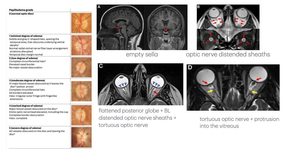
  • IIH symptoms include (3) headache, papilledema, and tinnitus[…]; papilledema can progress from blurry vision and visual obscurations to vision loss[…]
  • Associated MRI/MRV features include (6) empty sella, optic nerve sheath distention, flattened posterior globe + tortuous optic nerve, optic nerve protrusion into the vitreous, and transverse venous sinus stenosis[…]
  • Papilledema is graded on the Frisen[…] scale: Grade 0 is no edema; Grade 1 is partial C-shaped circumferential edema; Grade 2 is complete circumferential edema; Grade 3 adds ≥1 blood vessel obscured leaving the disc[…]; Grade 4 adds ≥1 major BV obscured on the disc[…]; Grade 5 is complete obscuration[…] of BVs on the optic disc
  • Optical coherence tomography (OCT)[…] provides noninvasive, high-resolution quantification of papilledema
IIH - Visual Testing
  • The most common IIH visual field defects include enlargement of the blind spot[…], inferior nasal step[…], peripheral constriction, and arcuate defects from retinal nerve fiber bundle[…]-type loss.
  • Visual acuity is typically preserved[…] until late disease, making visual fields and OCT the primary monitoring tools.
IIH - Treatment and Fulminant IIH
  • All IIH patients need weight loss[…] with lifestyle intervention; GLP-1 agonists[…] are great with comorbid DM/obesity.
  • First-line ICP-lowering medication is acetazolamide[…]; alternatives include topiramate[…] (also helps migraine and weight loss) and furosemide.
  • Procedural options for worsening papilledema or vision loss include optic nerve sheath fenestration (ONSF)[…], venous sinus stenting (VSS)[…], and CSF shunting (VP or LP shunt)[…] for refractory cases.
  • Fulminant IIH presents with very severe vision loss[…] over days to weeks[…] of symptom onset and requires urgent surgical intervention[…] (ONSF, VSS, or shunt) rather than waiting for medical therapy.
Atraumatic Cranial CSF Leaks - Clinical Picture

Atraumatic cranial CSF leaks present with unilateral[…] clear fluid dripping from the nose (rhinorrhea[…]) or ear (otorrhea[…]), often with positional worsening (bending forward, Valsalva).

Unlike rhinitis, CSF rhinorrhea does not respond[…] to ipratropium nasal spray.

These leaks carry risk of meningitis[…] from ascending infection through the dural defect.

Atraumatic cranial leaks are most commonly a complication of IIH[…], in which elevated ICP erodes through the skull base.
Atraumatic Cranial CSF Leaks - Diagnosis and Treatment

Fluid is collected from the nose or ear and tested for β-2 transferrin[…], which is highly specific for CSF.

High-resolution CT[…] identifies bony defects; if multiple defects are present, a CT cisternogram[…] localizes the active leak site.

Treatment first reduces high ICP[…] via VSS, ONSF, or shunt — otherwise repaired leaks will recur.

Anterior skull base defects (facing nasal cavity) are repaired via endoscopic endonasal[…] approach; lateral skull base defects (facing ear) are repaired via transmastoid or middle fossa[…] approach.
Persistent HA after CSF Normalization - Pathophysiology and DDX

Likely MOA is central sensitization[…], where pain-sensitive structures like dura, vessels, and trigeminal afferents become hypersensitized after prolonged abnormal CSF pressure.

DDX of persistent HA after normalization includes central sensitization, migraine[…] (IIH patients are 6x[…] more likely to have comorbid migraine), rebound intracranial hypertension[…] (worse lying down, classically 1-3 days post-treatment of SIH), medication overuse headache[…], and inadequately treated primary disease (residual SIH leak or persistent elevated ICP).
NPH - CSF Physiology

CSF is secreted by the choroid plexus[…] in the lateral, 3rd, and 4th ventricles at approximately 500 mL/day[…], with a total CSF volume of 150 mL[…] (so the entire volume turns over ~3x daily).

CSF flows from lateral ventricles → foramen of Monro[…] → 3rd ventricle → cerebral aqueduct[…] → 4th ventricle → foramina of Luschka/Magendie → subarachnoid space.

Reabsorption occurs via arachnoid granulations[…] into the dural venous sinuses, with additional contribution from glymphatic and lymphatic[…] pathways.
NPH - Pathophysiology

Major NPH theories include CSF outflow resistance[…] leading to ventricular accumulation, reduced CSF absorption[…] at arachnoid granulations, and glymphatic dysfunction[…] impairing waste clearance.

Idiopathic NPH overrepresentation of enlarged head circumference[…] suggests a genetic/developmental contribution to pathogenesis.

Stretched periventricular corticospinal[…] fibers from the leg cortex contribute to the characteristic gait dysfunction.
NPH - Diagnosis

NPH classically presents as a triad of gait dysfunction, cognitive impairment, and urinary incontinence[…] — neither necessary nor sufficient for diagnosis.

The first and cardinal symptom is gait dysfunction[…], classically magnetic, broad-based, and shuffling.

Cognitive impairment is typically executive dysfunction[…] with impaired attention, psychomotor slowing, and planning/organizing difficulty.

Imaging shows ventriculomegaly out of proportion to atrophy, with a key shunt-responsive feature being DESH (disproportionately enlarged subarachnoid space hydrocephalus)[…].

Diagnosis is supported by large-volume LP (tap test)[…] or extended lumbar drain[…] showing symptomatic improvement after CSF removal.

Other supportive features include Evans' index >0.3[…] and a callosal angle <90 degrees[…].
NPH - Treatment and Newer Imaging

Shunt placement[…] is performed as soon as possible to avoid permanent damage — VP, VA, or LP configurations.

Overdrainage complications include orthostatic headache[…] (worse standing, improves supine) and subdural hematoma.

Newer imaging: Phase-contrast MRI[…] shows increased flow through the cerebral aqueduct; Time-SLIP[…] visualizes real-time CSF movement and absence of reflux from 3rd to lateral ventricle.

DTI-ALPS[…] measures diffusion along periventricular white matter tracts at the centrum semiovale — a low[…] ALPS index correlates with worse NPH.

Intrathecal gadolinium[…] imaging shows reflux of contrast from subarachnoid space into the lateral ventricles, causing transependymal enhancement.

MR elastography[…] quantifies tissue stiffness.
CSF Disorder Terminology

Ventriculomegaly[…]: large ventricles on imaging without symptoms.

Symptomatic hydrocephalus[…]: impaired CSF dynamics from high ICP — ventriculomegaly that IS symptomatic.

Hydrocephalus ex vacuo[…]: large ventricles, sylvian fissures, and subarachnoid space due to decreased parenchyma[…].

Benign enlargement of subarachnoid spaces[…]: a common developmental variant in kids with macrocephaly[…], usually incidental and not requiring follow-up.
Childhood Hydrocephalus - Treatment

Initial treatment is EVD[…] for 1-2 weeks until CSF dynamics recover; in neonates a ventriculosubgaleal shunt[…] is preferred due to EVD risk.

If no recovery, a permanent shunt[…] is placed as in adults.

For cerebral aqueduct stenosis, web, or postinfectious hydrocephalus, endoscopic 3rd ventriculostomy (ETV)[…] is performed, often with choroid plexus cauterization[…] to reduce CSF secretion.
Practice Q - SIH Headache Phenotype

The most common SIH symptom is an orthostatic headache[…] that worsens when upright[…] and improves when lying flat[…].

SIH should be strongly considered in new daily persistent headache that worsens with Valsalva[…] maneuvers and steadily worsens through the second half of the day[…].
Practice Q - SIH Imaging Signs and Mechanisms

Smooth, non-nodular diffuse pachymeningeal enhancement[…] is essentially pathognomonic for SIH.

Effacement of the suprasellar and prepontine cisterns in SIH is best explained by CSF volume depletion[…] causing downward brain sag.

An underlying spontaneous spinal CSF leak should be considered in atraumatic subdural hematoma[…], especially with headache or features of intracranial hypotension.
Practice Q - SIH Workup and Treatment Decisions

In high SIH suspicion with no visible extradural CSF on spinal imaging, the most likely explanation is a CSF-venous fistula[…] draining CSF directly into a paraspinal vein without a detectable collection.

The most likely cause of a ventral dural tear in SIH is puncture by an osseous spicule[…].

For DSM of a suspected ventral dural tear, position the patient prone[…] to best visualize ventral contrast pooling.

Transvenous embolization[…] is effective specifically for CSF-venous fistulas, but not for other CSF leak types.

Topiramate should be avoided as preventive in patients with comorbid migraine and SIH because its carbonic anhydrase[…] activity reduces CSF production and intracranial pressure.

The most common EBP adverse event is back pain[…], occurring in up to 80% shortly after the procedure.
Practice Q - IIH Clinical Pearls
  • The only localizing neurologic sign consistent with IIH is CN6 palsy[…], caused by increased ICP stretching the nerve.
  • The most characteristic IIH visual field change is localized retinal nerve fiber bundle-type loss[…], often as arcuate defects with or without enlarged blind spot.
  • The earliest visual field defect in papilledema is enlargement of the blind spot[…] on automated perimetry.
  • The MRI finding most consistent with IIH is empty sella[…]; hydrocephalus or space-occupying lesions suggest secondary causes.
  • The most serious complication of untreated IIH is vision loss[…], often irreversible if not treated promptly.
  • The main modifiable risk factor is obesity[…]; weight loss is associated with disease remission.
  • In IIH patients with worsening papilledema and early vision loss despite failed weight loss, the best next step is initiation of acetazolamide[…].
Practice Q - CSF Leak Diagnostics

The expected response to ipratropium nasal spray in CSF rhinorrhea is no change[…], distinguishing it from rhinitis-related rhinorrhea.

The most reliable lab test for confirming CSF in nasal or ear fluid is β-2 transferrin[…].

When high-resolution CT shows multiple bony defects, the next study to localize an active CSF leak is CT cisternogram[…].
Practice Q - Headache in CSF Disorders

The most common symptom in IIH or SIH is headache[…], present in the vast majority.

The most common headache phenotype associated with IIH is migraine type[…] — daily/constant pain with photophobia and nausea.

The most consistent etiology of new persistent headache after SIH treatment — especially when worse lying down and starting 1-3 days post-treatment — is rebound intracranial hypertension[…].
Practice Q - NPH Clinical Pearls

The most common first symptom of idiopathic NPH is gait dysfunction[…].

Overrepresentation of enlarged head circumference[…] in idiopathic NPH supports a possible genetic contribution.

The most typical cognitive impairment at presentation is executive dysfunction[…], including impaired attention, psychomotor slowing, and planning difficulty.

A radiographic feature predictive of shunt-responsiveness is DESH[…] on imaging.

The most suggestive symptom of CSF overdrainage in adults with a ventricular shunt is orthostatic headache[…].