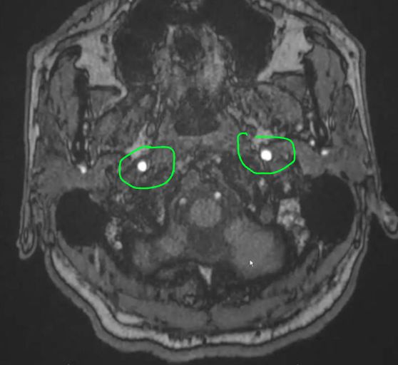
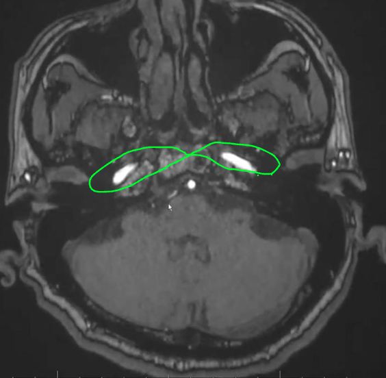
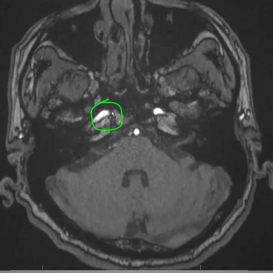
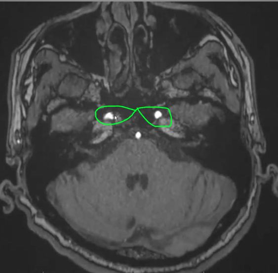
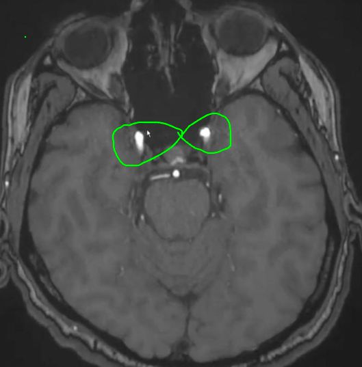
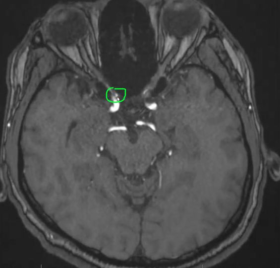
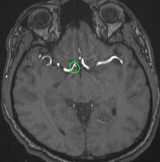
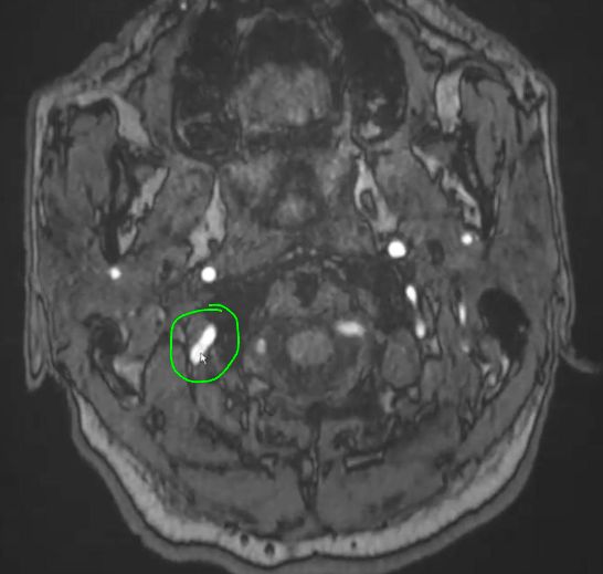
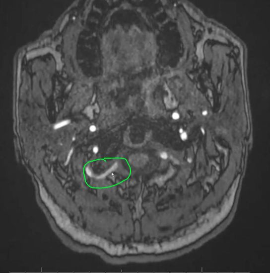
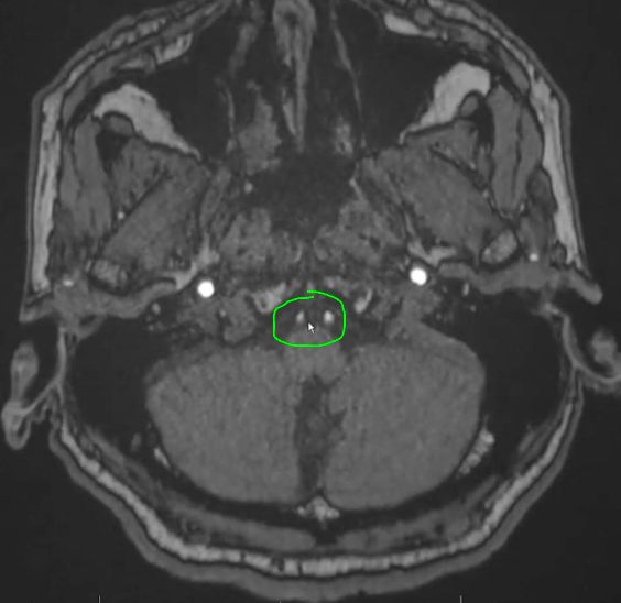
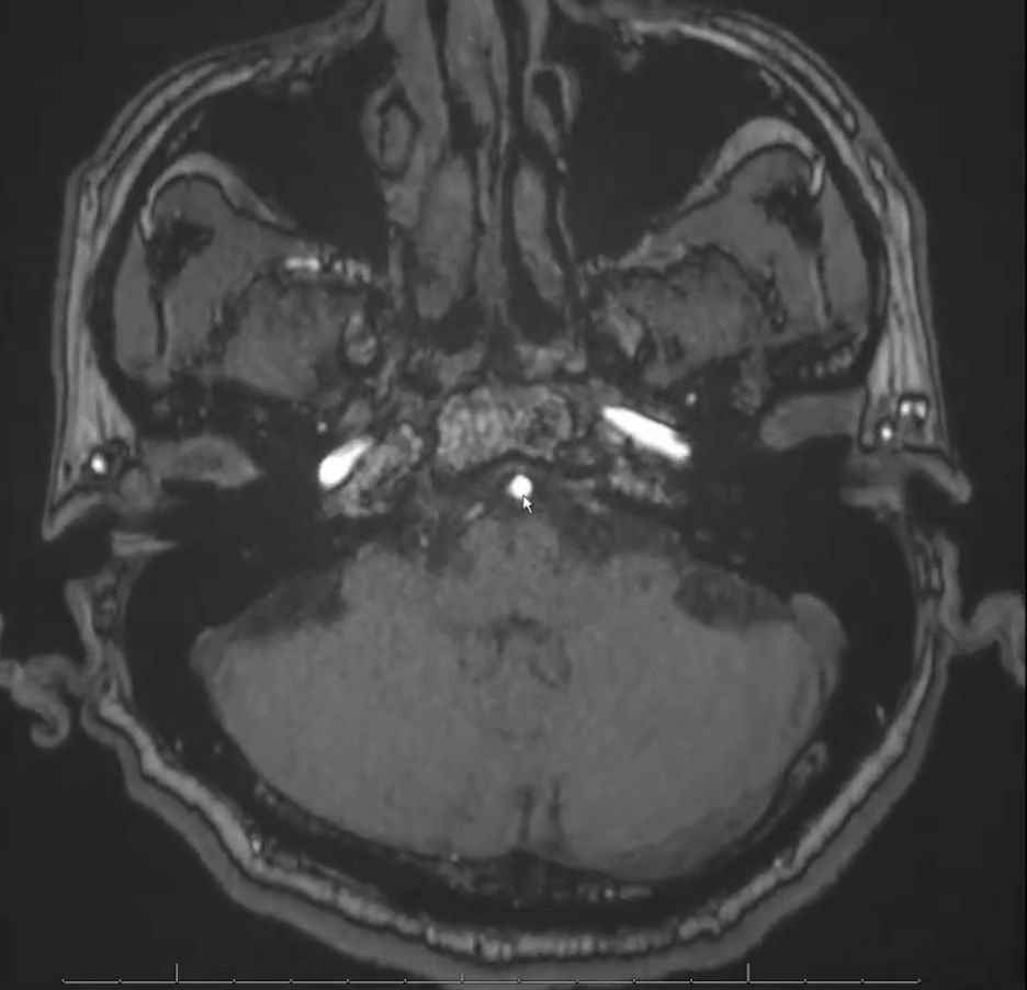
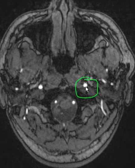
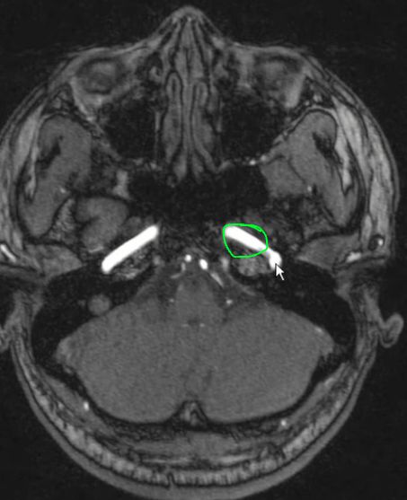
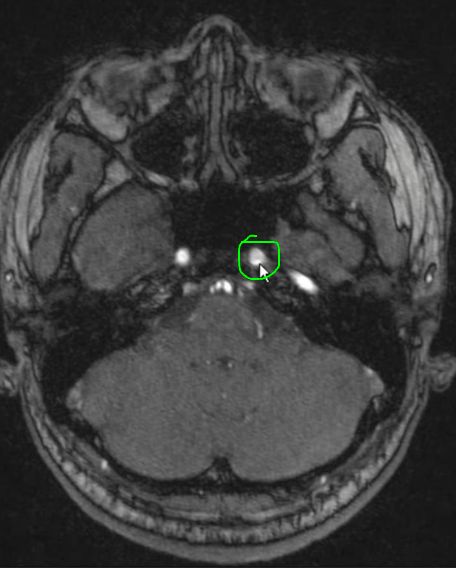
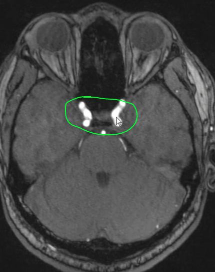
This is the cervical ICA[…] > entering the carotid canal > petrous part of the temporal bone[…] > traverses the petrous canal[…] > to become the petrous ICA[…]
This is the petrous ICA[…]
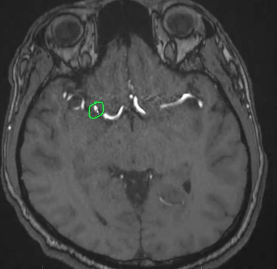
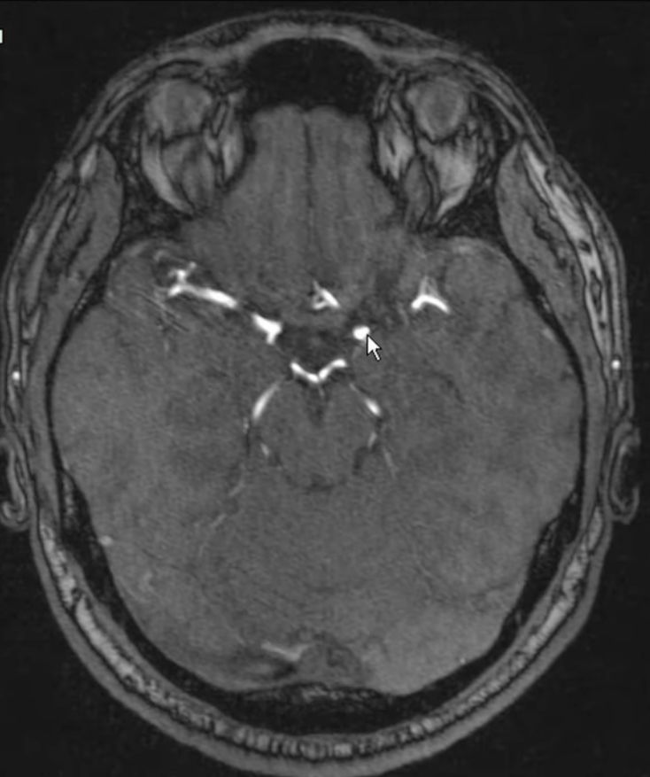
These are the petrous ICAs[…] after traversing the petrous canal[…] of the temporal bone/skull base prior to moving anterior[which direction?]ly and up before becoming the cavernous ICAs[…]
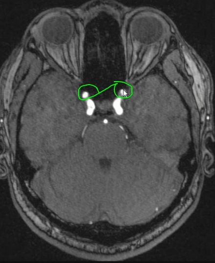
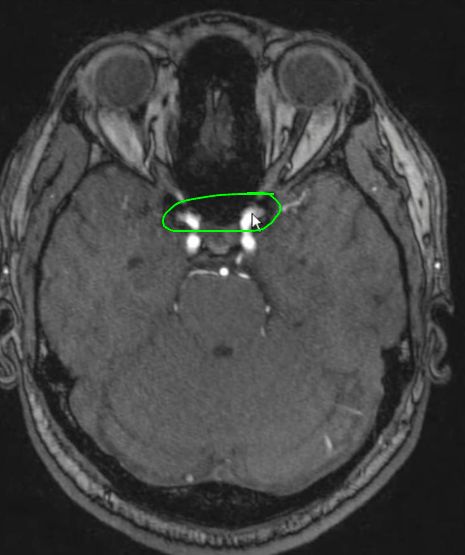
These are the cavernous ICAs[…] prior to exiting the cavernous sinus[structure] and prior to giving off the ophthalmic[…] artery looping back and becoming the supraclinoid ICAs[…]
- the cavernous ica is the 4th segment of the ICA
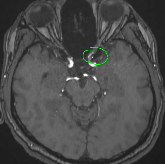
This is the supraclinoid ICA[…] giving off the ophthalmic artery[…] just after exiting the cavernous sinus[…]
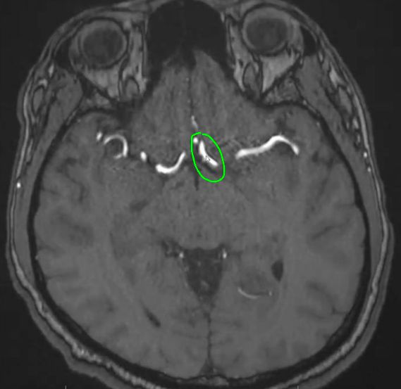
The ICA C7 segment divides into 2 vessels; this is the ACA[…]A1[…] segment
The C7 ICA divides into 2 vessels; this is the MCA[…]M1[…] segment which then enters the sylvian fissure[…]
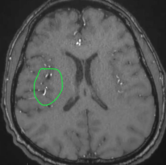
These are the distal branches of the MCA[…] traveling through the Sylvian Fissure[…]
This vessel is the Acomm[…] connecting the bilateral ACAs[…]
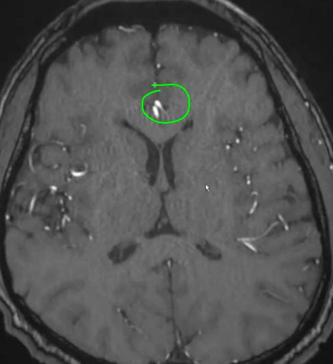
These are the distal branches of the ACAs[…]
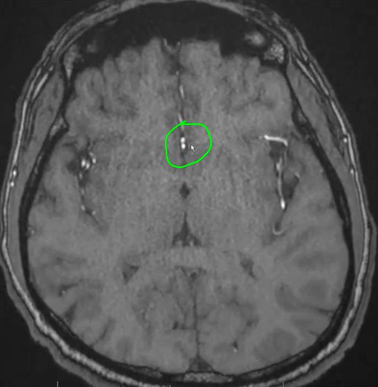
These are the distal branches of the ACAs[…] traveling in the Falx Cerebri[…]
This shows the ophthalmic[…] artery branching off the supraclinoid ICA[…] on the left
This is the A1[segment]ACA[artery] on the left
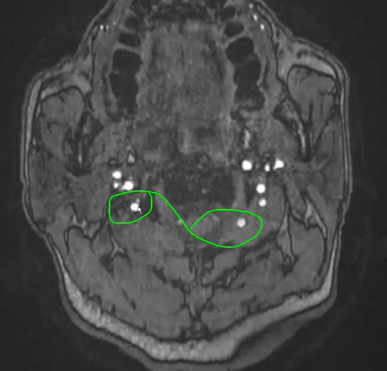
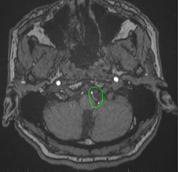
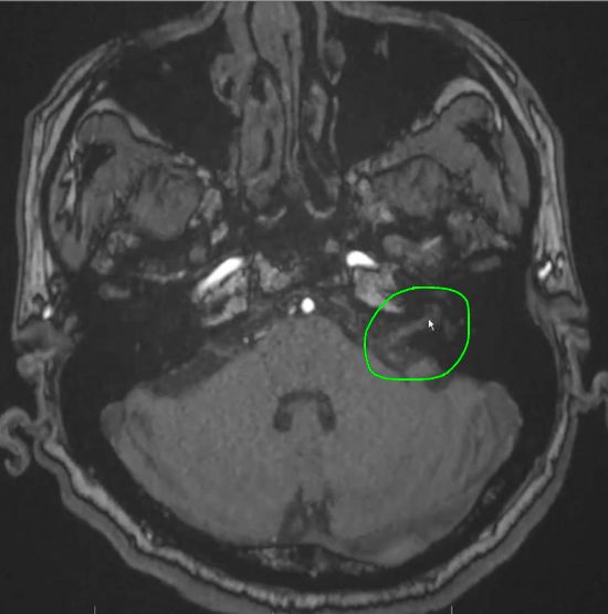
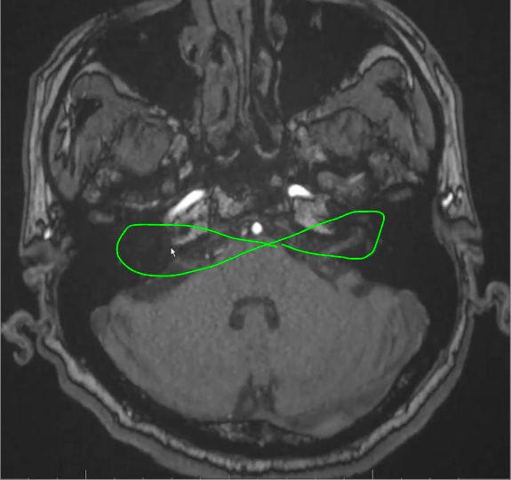
These are the Vertebral Arteries[…]
This is the vertebral artery[…] (cervical[…] segment) ascending via transverse foramina[…] of the cervical vertebrae[…]prior to turning into and entering through the foramen magnum[…] of the skull base[…]
This is the vertebral artery[…] (cervical[…] segment) > immediately prior to entering via the foramen magnum[…] into the intracranial space
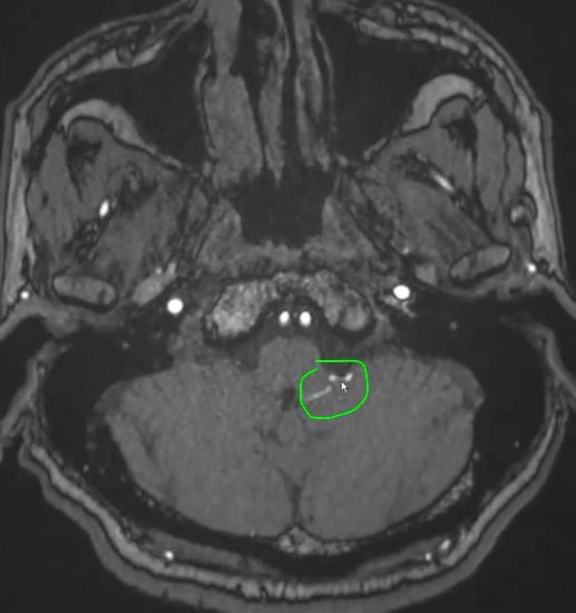
These are the intracranial[…] segments of the vertebral arteries[…]
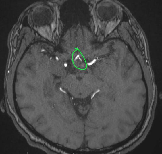
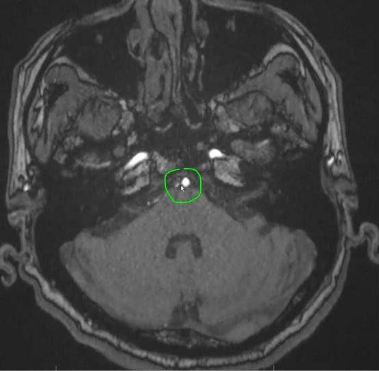
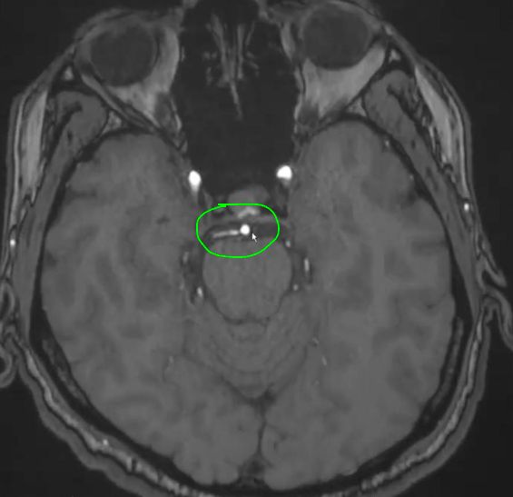
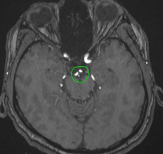
This is the basilar artery[…] at the brainstem level around the pontomedullary junction[…] formed by the union of the two vertebral arteries[…] after ascending along the anter[…]ior surface of the medulla[brainstem structure]
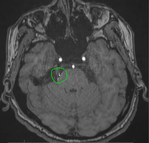
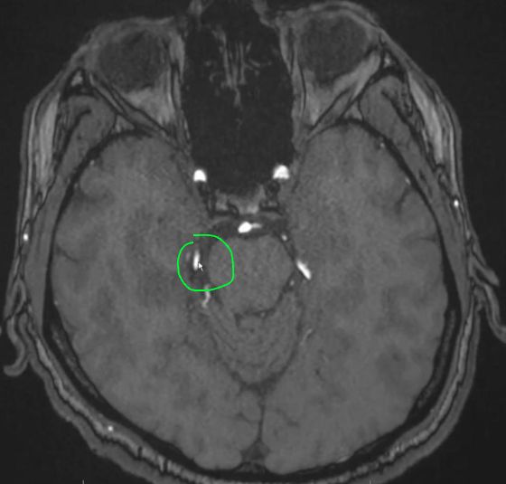
This is the PICA[…] branching off the left Vertebral Artery[…]
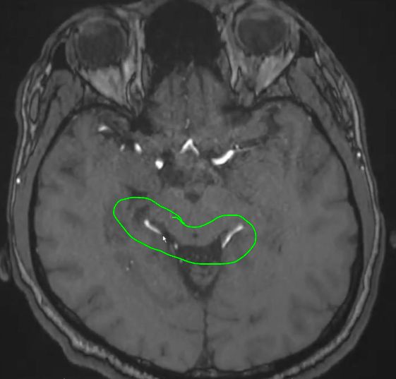
This is the PICA[…] that branched off the left vertebral artery and now supplying the inferior cerebellum[…]; it has a swirly course
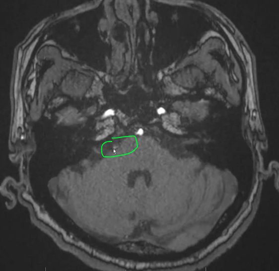
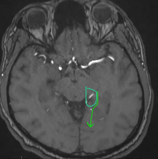
This first branch coming off the basilar artery[…] pictured here is the AICA[…]
This is the AICA[…] at the level of the IAC[…] which previously branched off the basilar and is coursing along the cerebellum
These canals more adequately visualized on the left side are the Internal Auditory Canals (IACs)[…]
The second to last branch of the Basilar[…] artery shown here is the SCA[…]
This was the second to last branch of the basilar coming off as what's now shown as a the SCA[…]
This is right after the tip of the basilar[…] artery as it branches into the two PCAs[…]
This is the PCA[…] on the right side after having branched off the basilar[…] artery
These are the PCAs[…] more distally and we know this because they have circumferentially moved around the midbrain[structure?] to move posteriorly where they will then move to perfuse the occipital[…] lobes
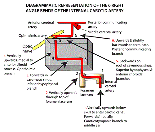
Name the ICA segment and number shown: cervical (C1)[…]
Name the ICA segment and number shown: this is the junction between the cervical (C1)[…]and petrous (C2)[…]segments moving through thepetrous[…]canal of thetemporal[…]bone after the ICA has arisen through thecarotid[…]canal
Name the ICA segment and number shown: The petrous ICA becomes the lasserum (C3)[…] ICA as it gets into the foramen lacerum[…] - we can ID it by knowing that it just moved through the entire petrous[…] canal and taking a right angle anteriorly
Name the ICA segment and number shown: cavernous (C4)[…]
Name the ICA segment and number shown: supraclinoid (C5)[…]
Name the ICA segment and number shown: ophthalmic (C6)[…] - we can ID this segment by knowing it's the part where the ICA starts to bend posterior[…]ly
Name the ICA segment and number shown: communicating (C7)[…] - we can ID this segment by knowing it's the part where the ICA gives off Pcomm[…] artery and bifurcates into the ACA[…] and MCA[…]