CTH

CTH

Radiology · 345 cards · 164 labeled figures

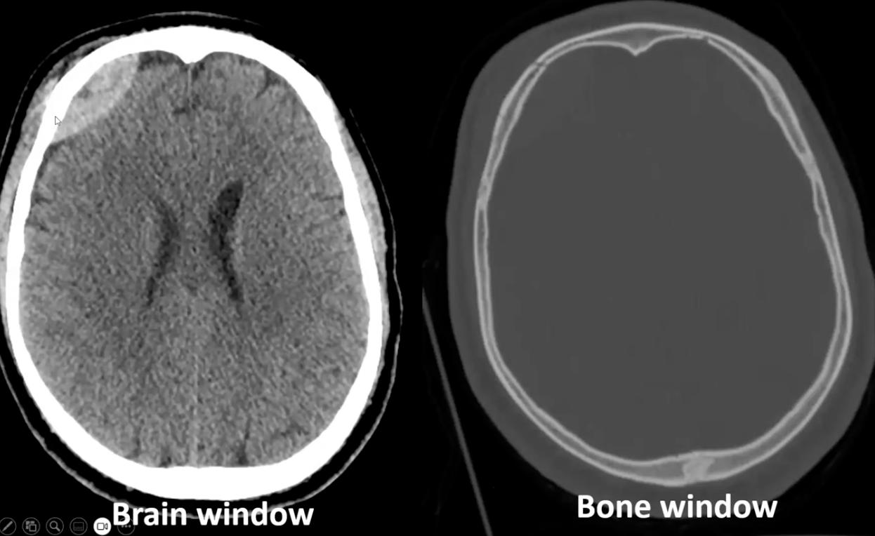
Explain why the L image is bright, R image dark using the principles of CT imaging & photons
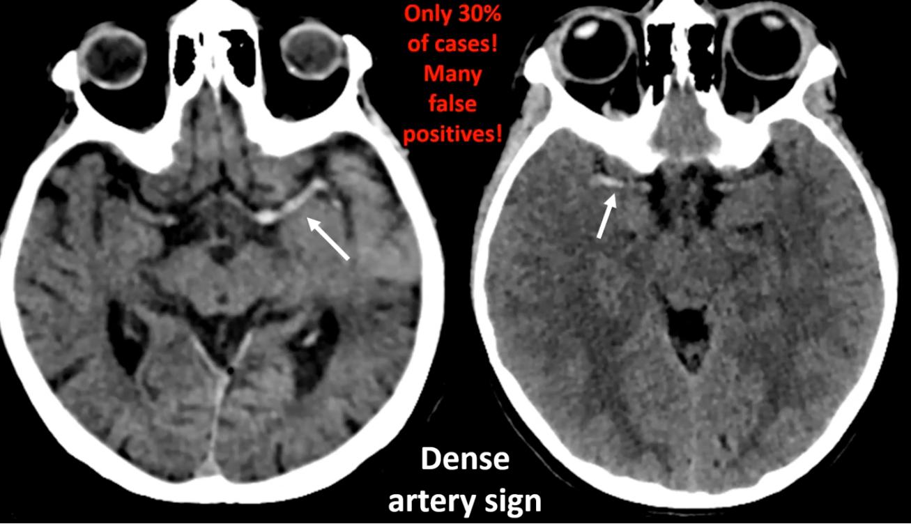
left = acute clotted hyperdense blood (absorbs photons), right = stroke/edema which is made of water which is hypodense (lets photons pass through)[…]
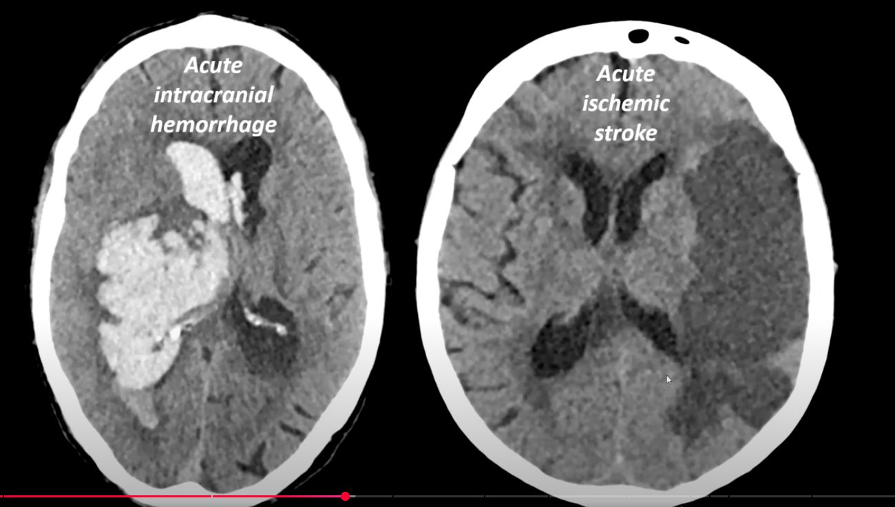
Cursor at bottom - this shows a subgaleal hematoma[…]
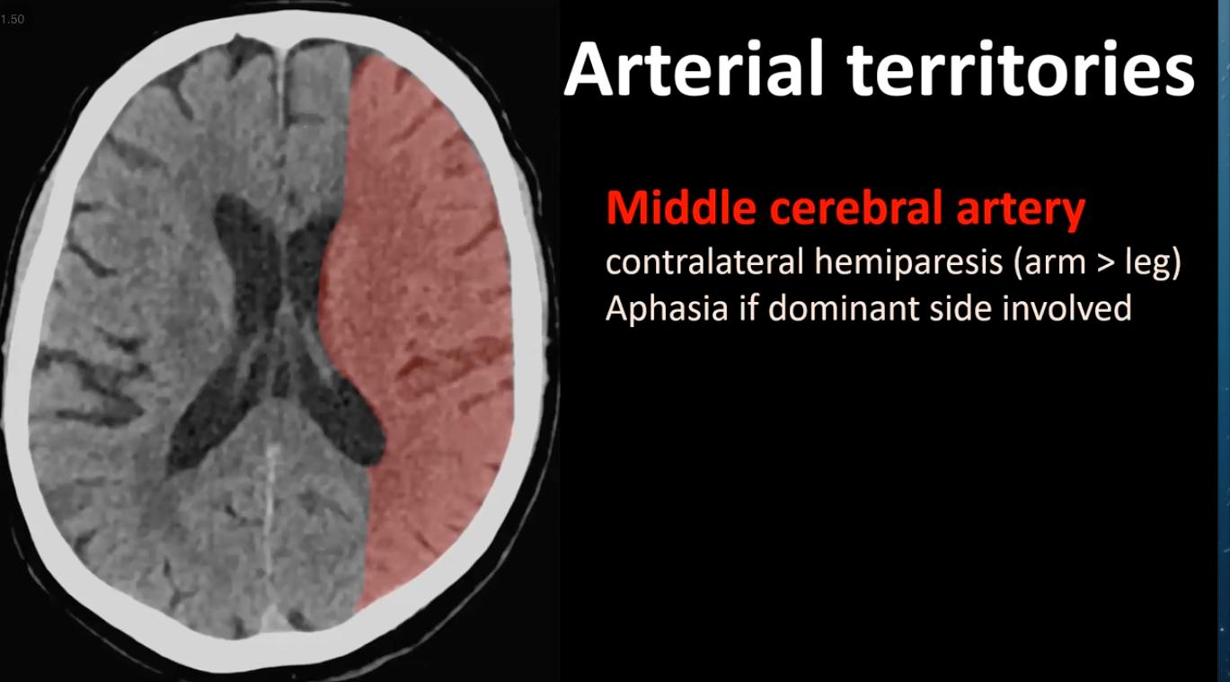
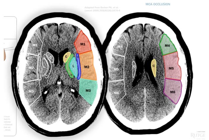
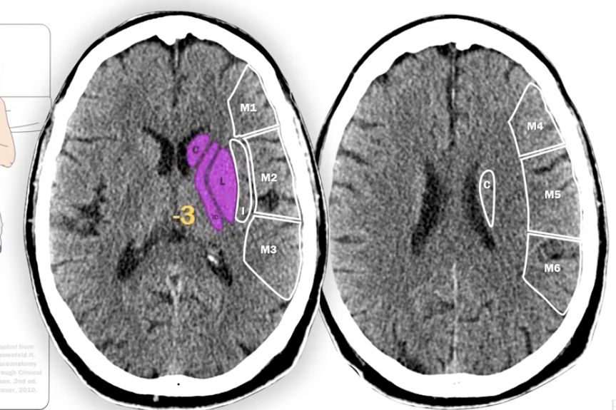
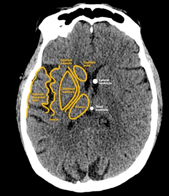
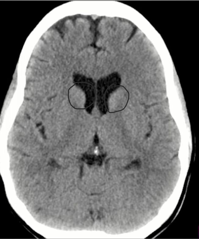
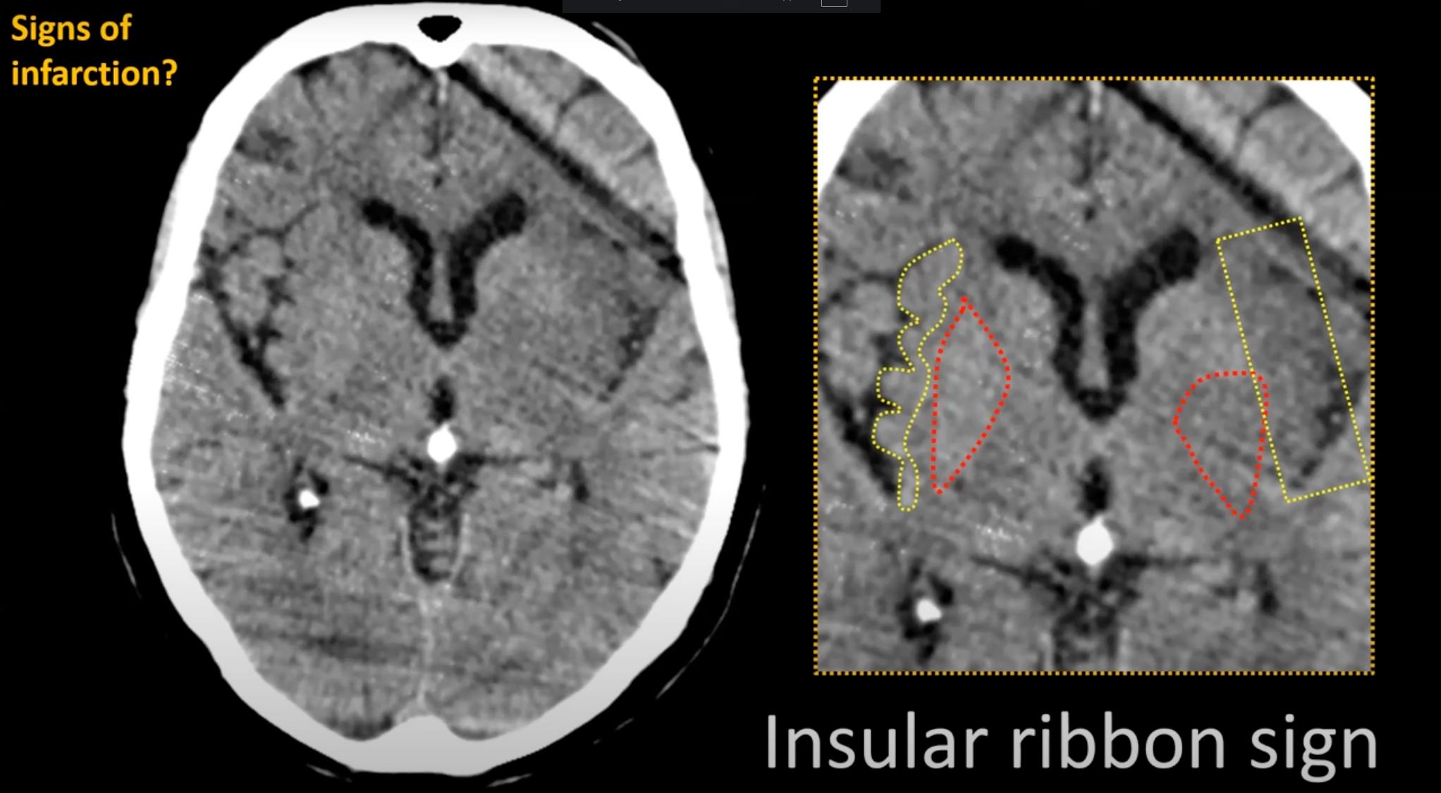
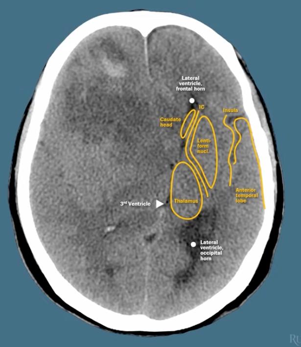
The ASPECTS[…] Score used for assessing patients with MCA[…] ischemic stroke for intervention divides the territory into 10[…] segments and 1 point is subtracted for each hypodense segment:
  • Caudate Head[…]
  • Lentiform Nucleus[…]
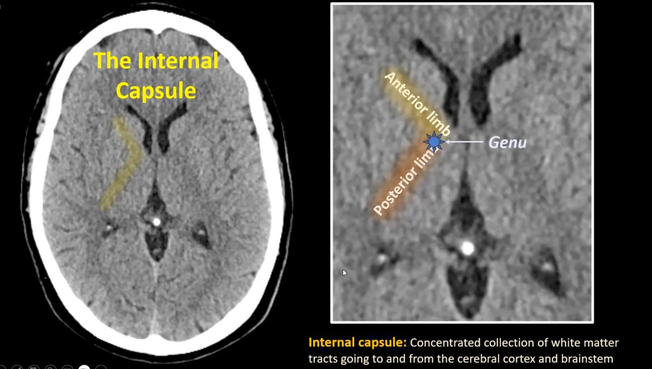
  • Internal Capsule[…]
  • Insula[…]
  • M1[…]M2[…]M3[…]M4[…]M5[…]M6[…] segments of cortex
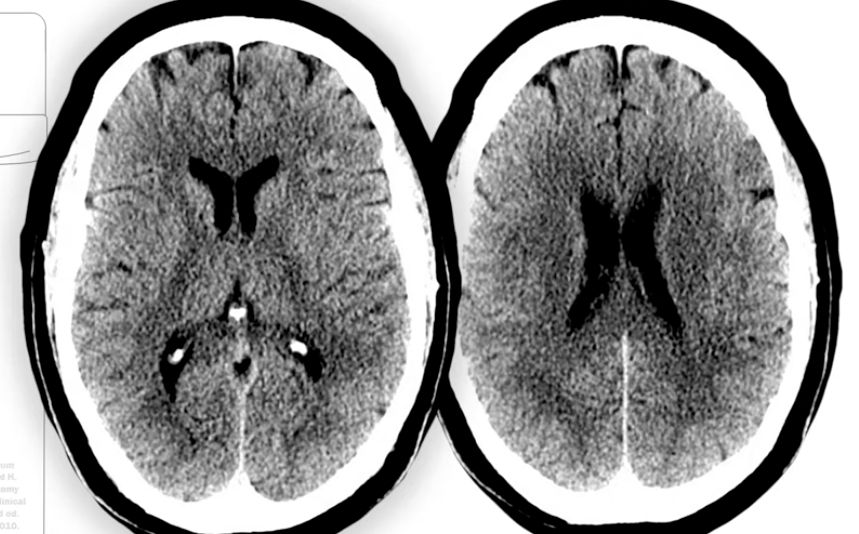
What is the ASPECTS Score?
10[…]

normal CTH
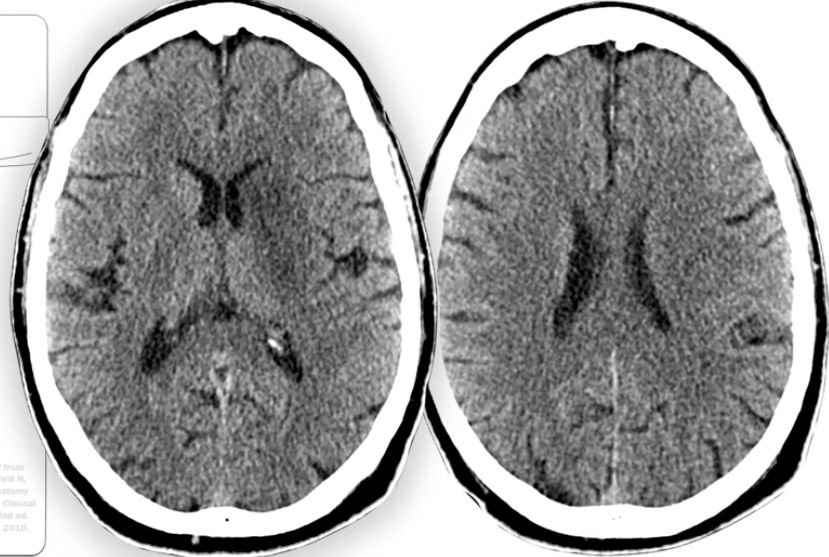
What is the ASPECTS Score?
7[…]
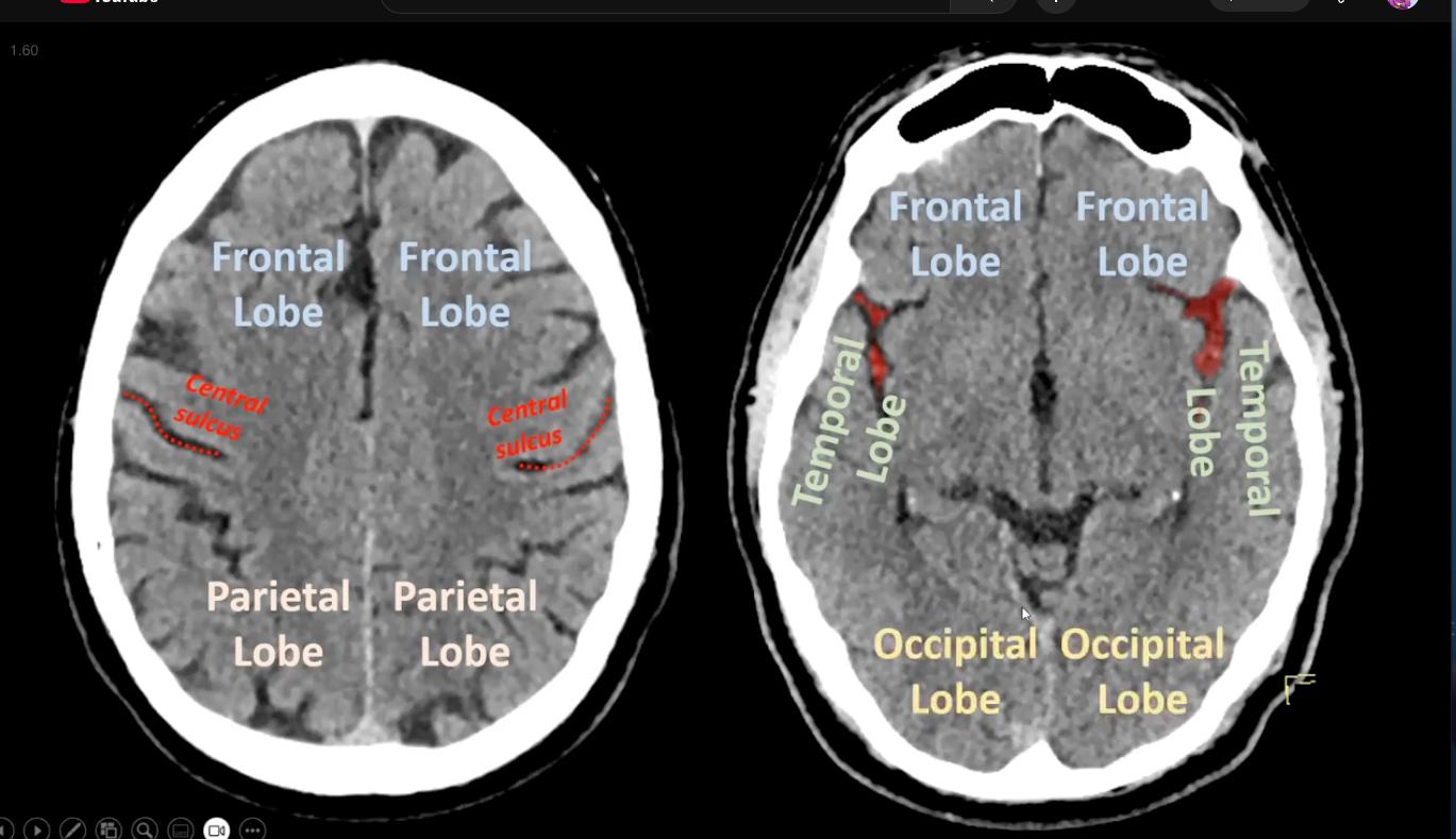
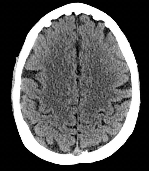
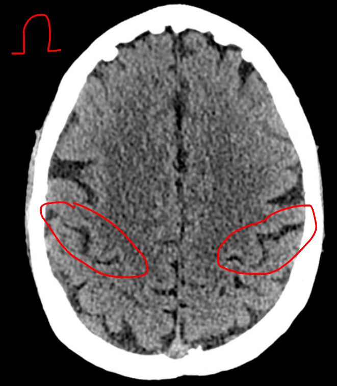
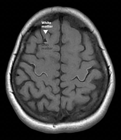
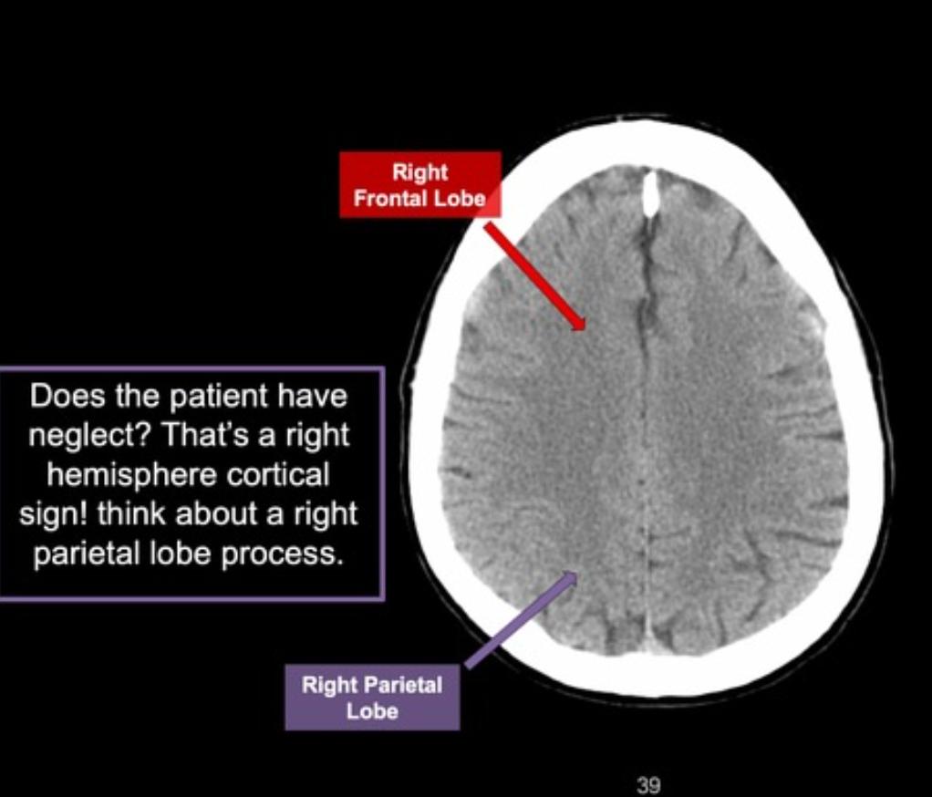
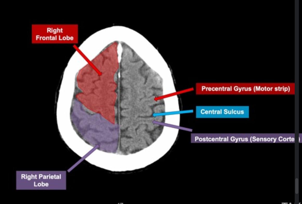
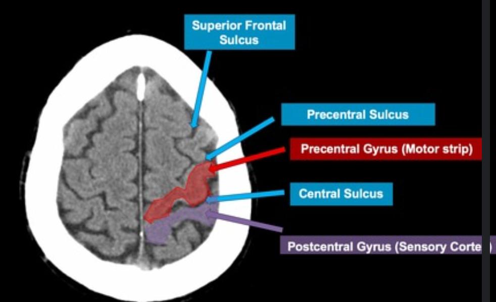
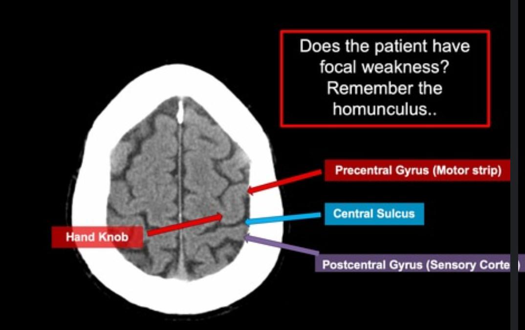
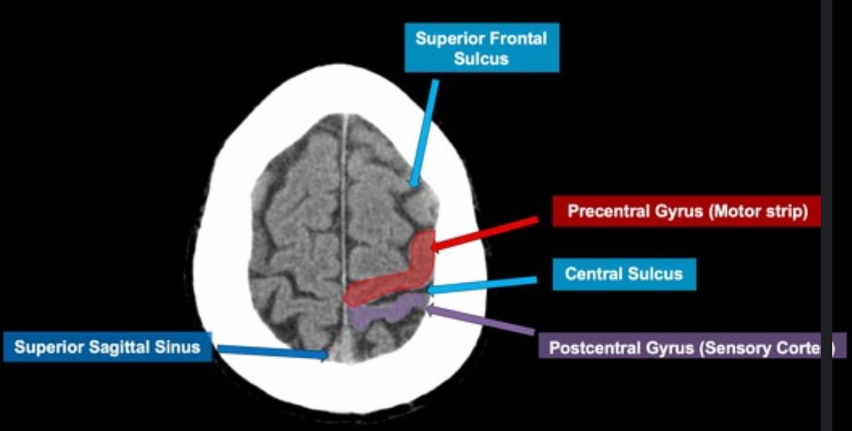
Where is the central sulcus and how do you know?

makes "Omega" sign[…]
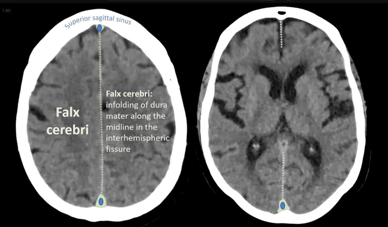
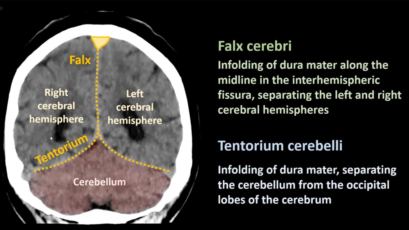
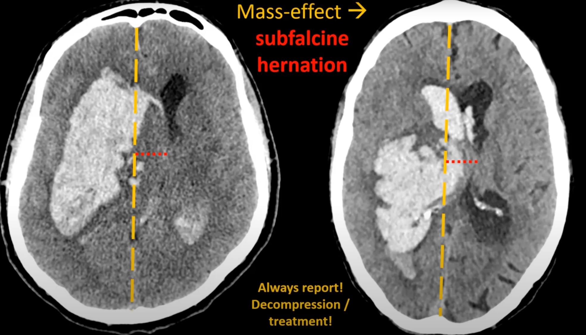
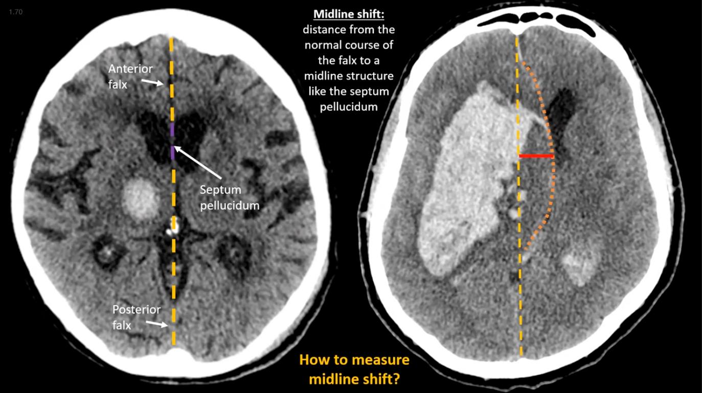
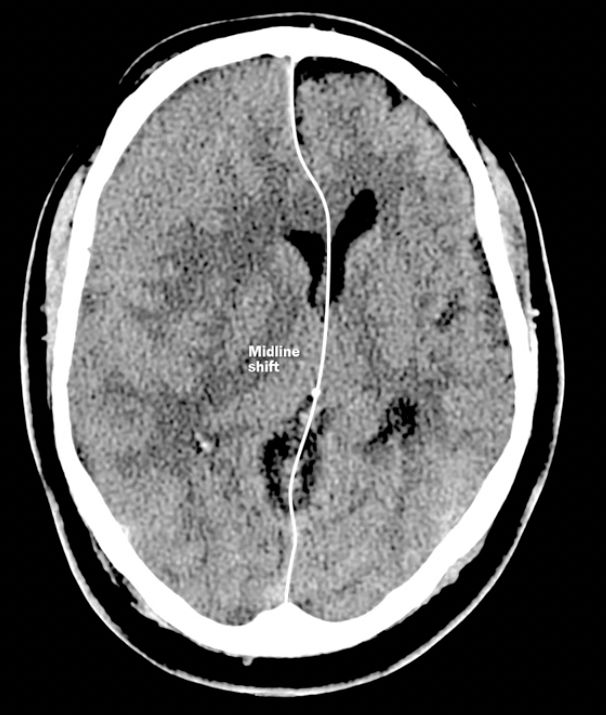
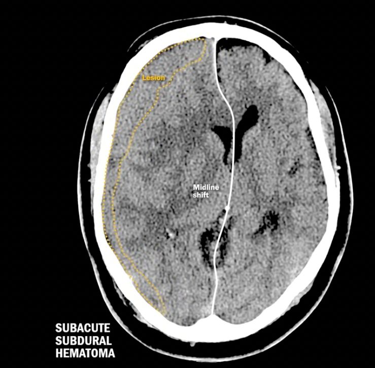
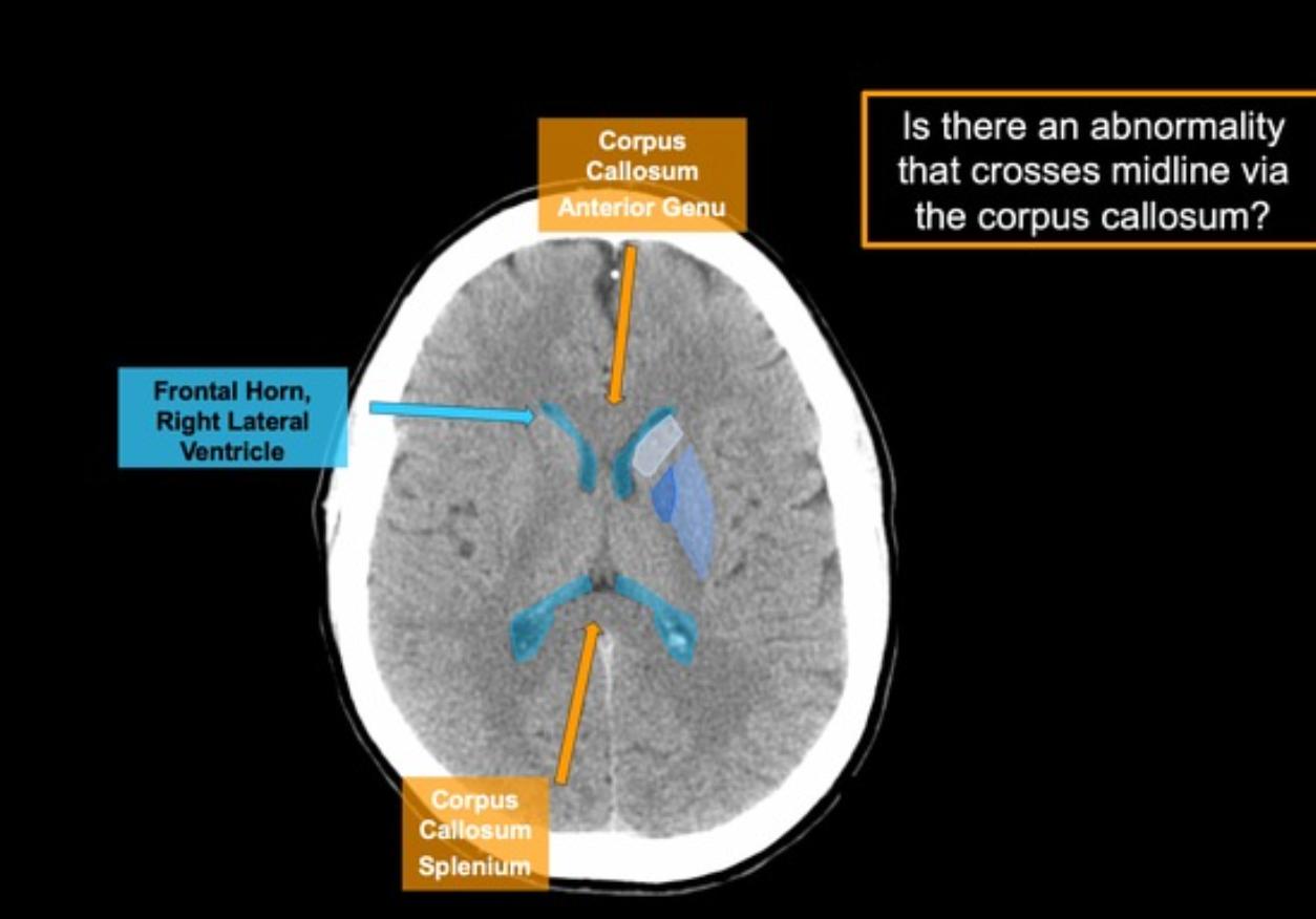
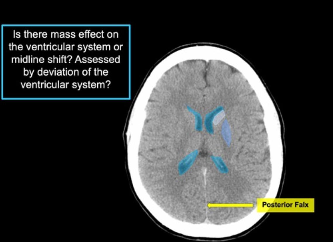
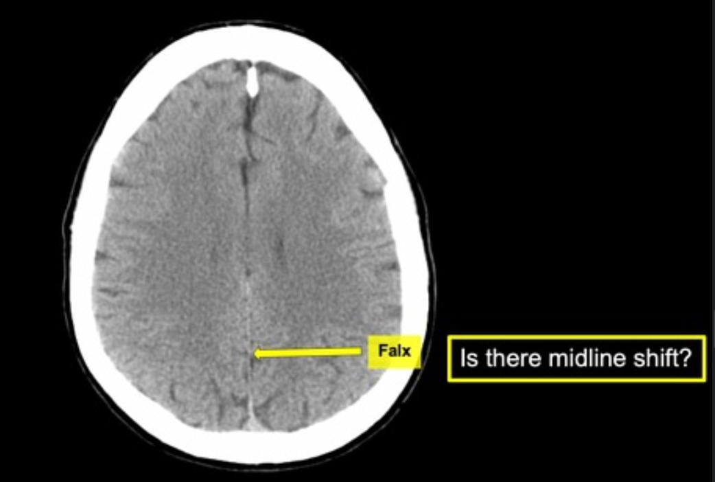
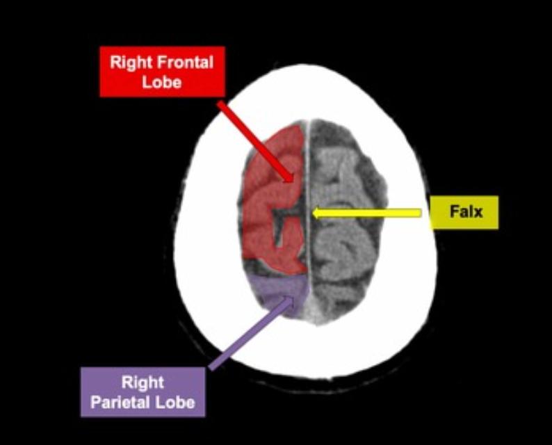
To find midline for determining MLS, draw a line from the
  • falx cerebri[…]
  • thru the septum pellucidum[…] in the middle of the lateral ventricles[…]
  • to the confluence of venous sinuses[…]
  • this image shows a R SDH[…] w/MLS to L

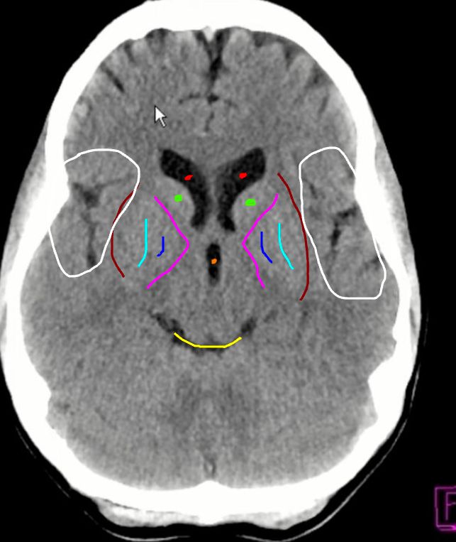
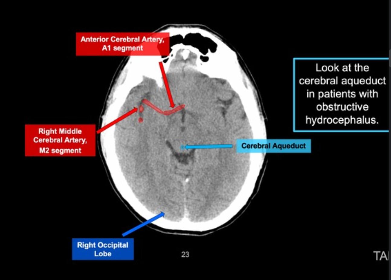
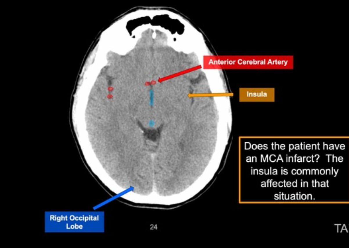
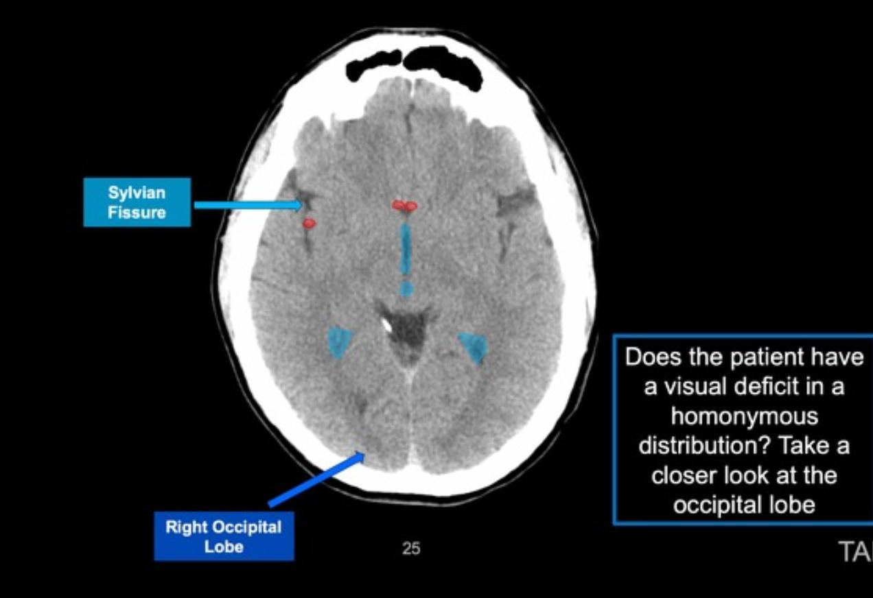
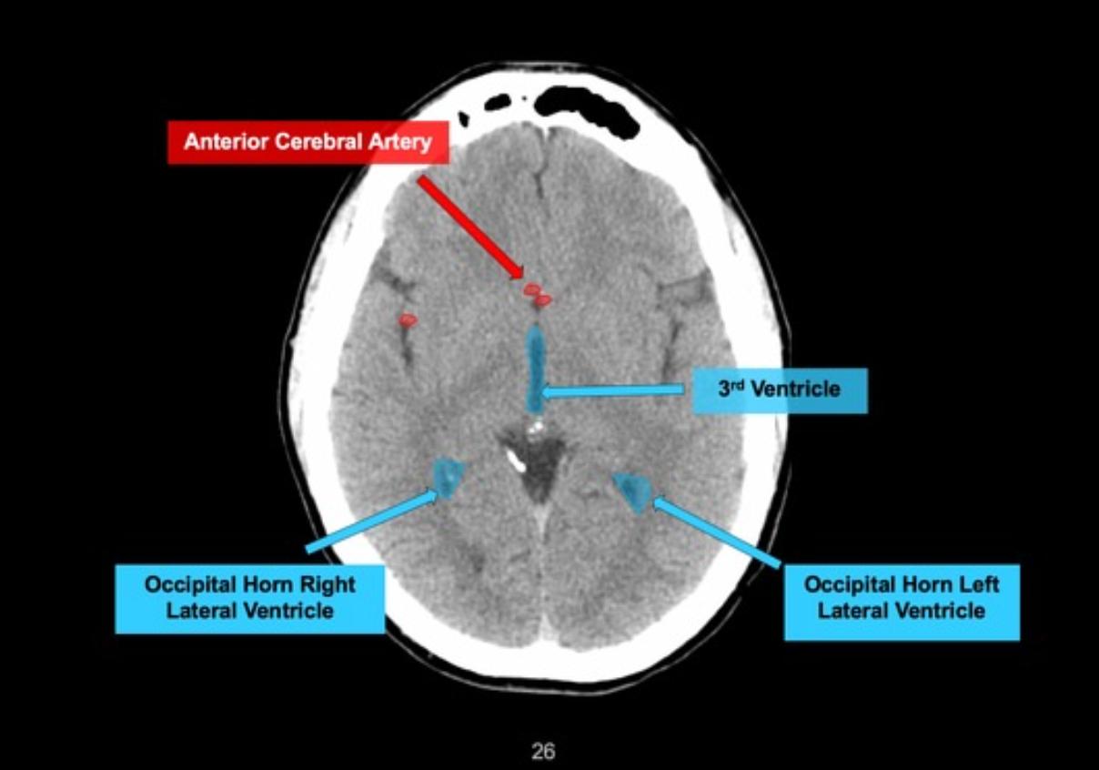
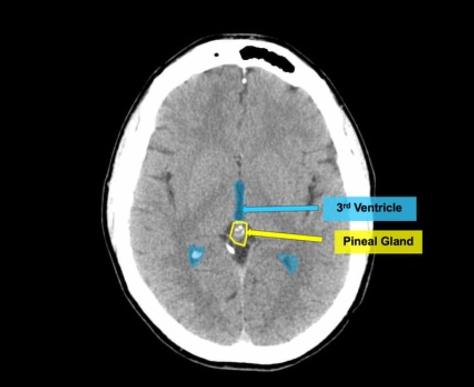
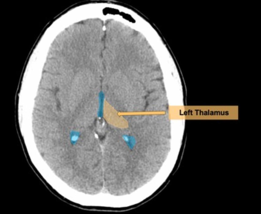
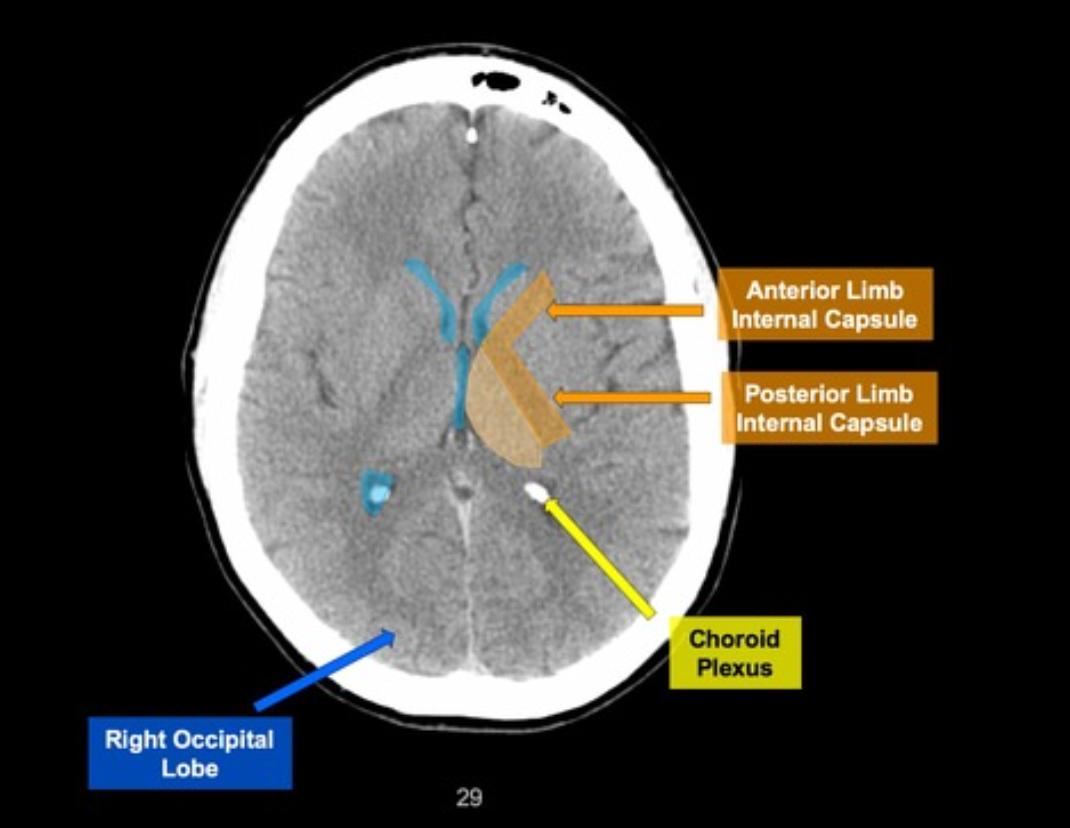
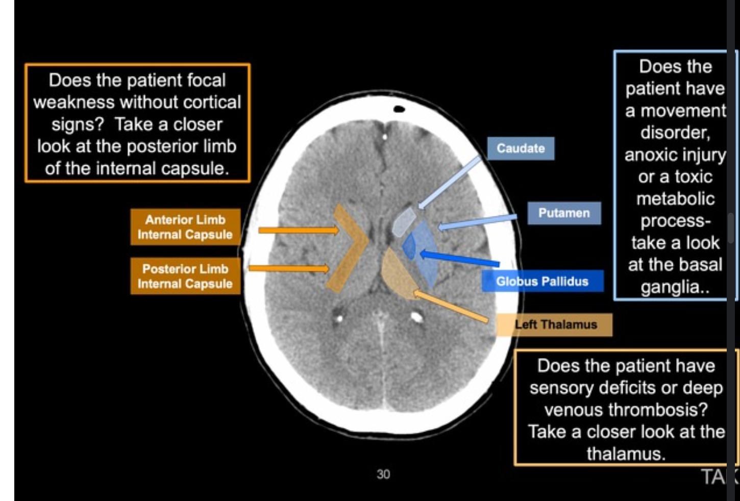
CTH Anatomy
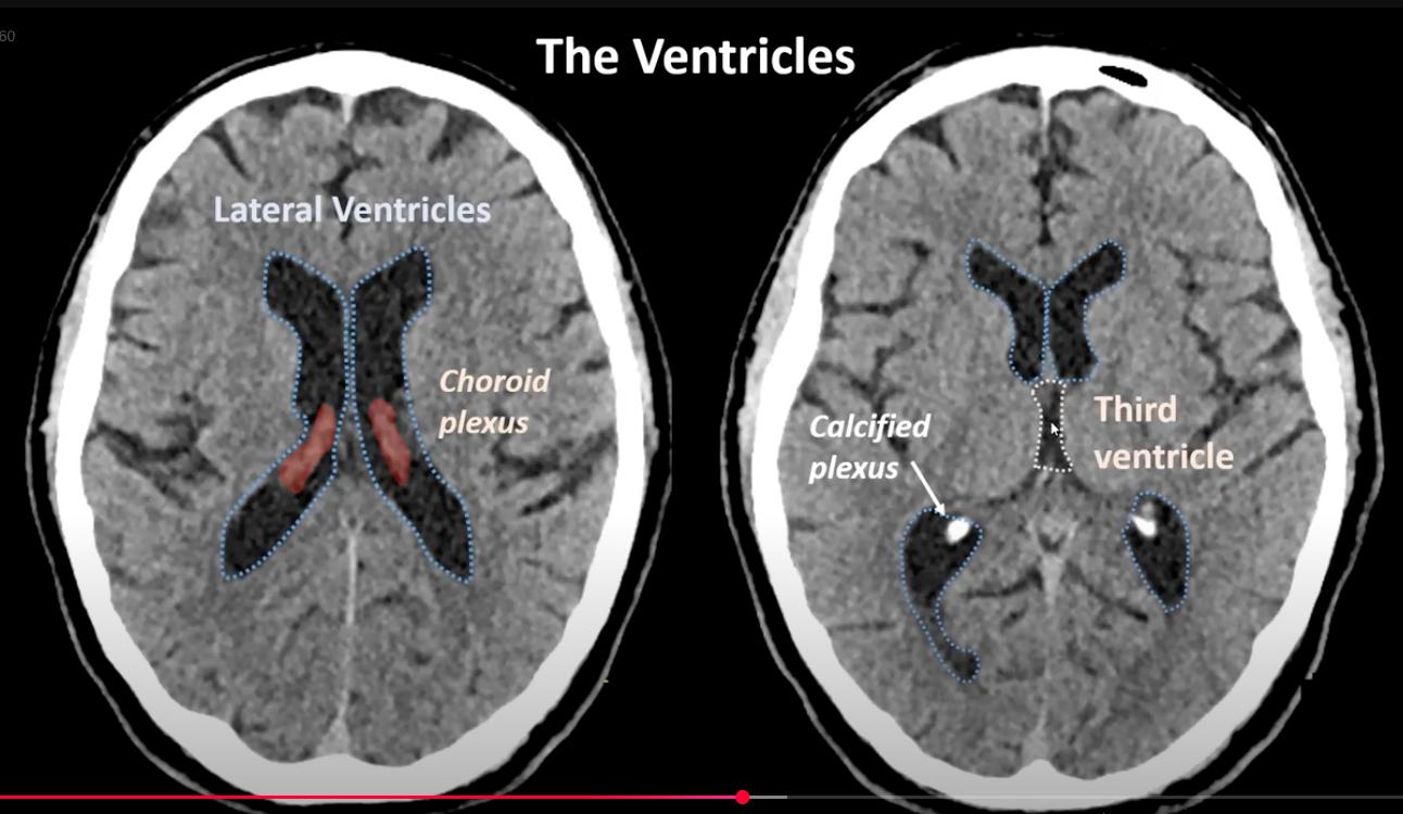
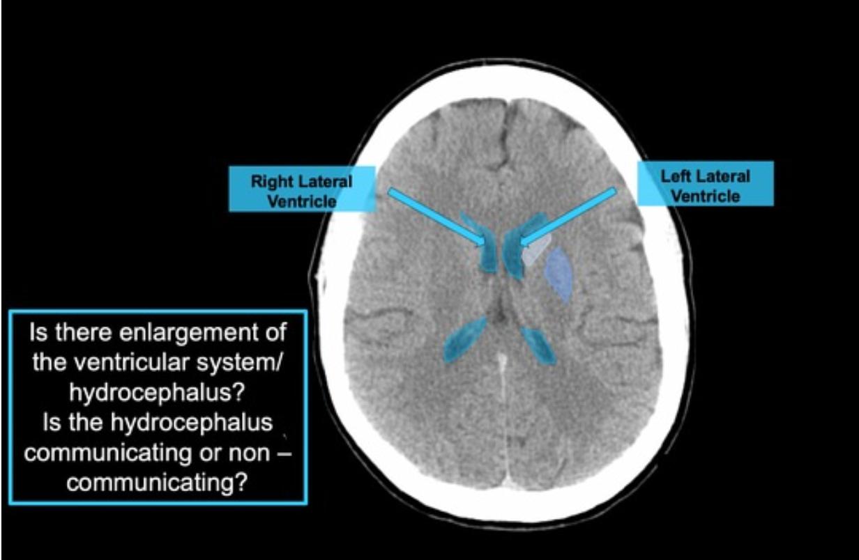
  • red: frontal horn, lateral ventricles[…]
  • yellow: occipital horn, lateral ventricles[…]
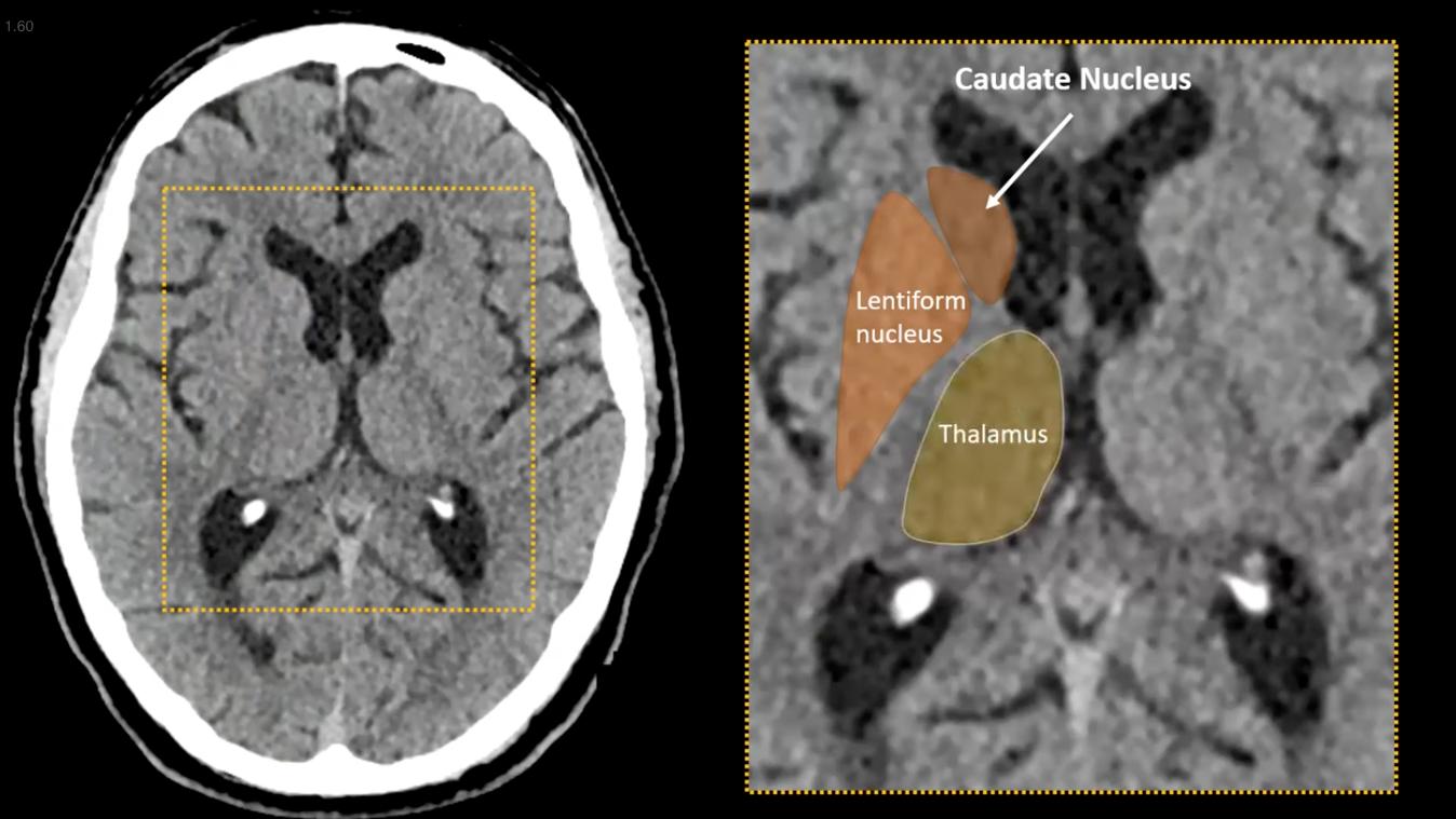
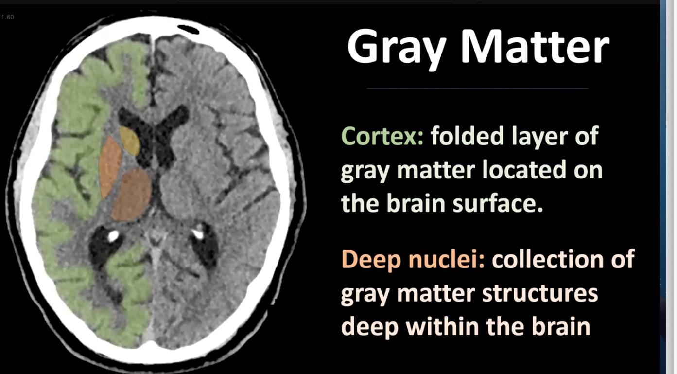
  • light green: head of caudate[…]
  • dark pink: anterior limb/genu/posterior limb IC[…]
  • dark blue: Gpe/i[…]
  • light blue: putamen[…]
  • dark red: external capsule[…]
  • white: insula[…]

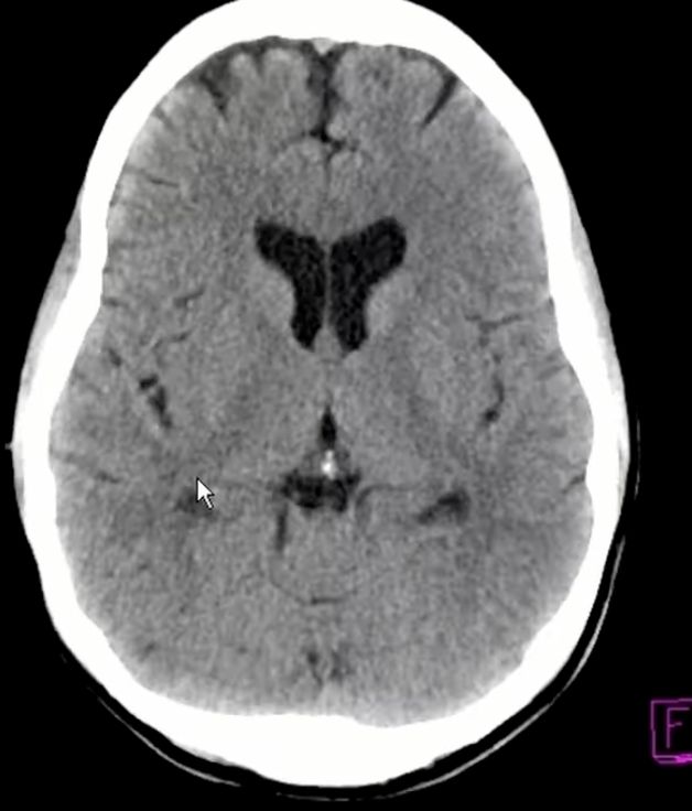
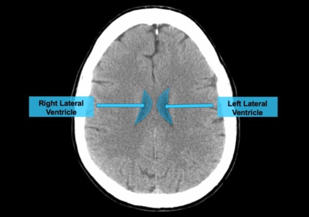
CTH Anatomy
  • these are the occipital horns, lateral ventricles[…] surrounded by the tail[…] of caudate nucleus

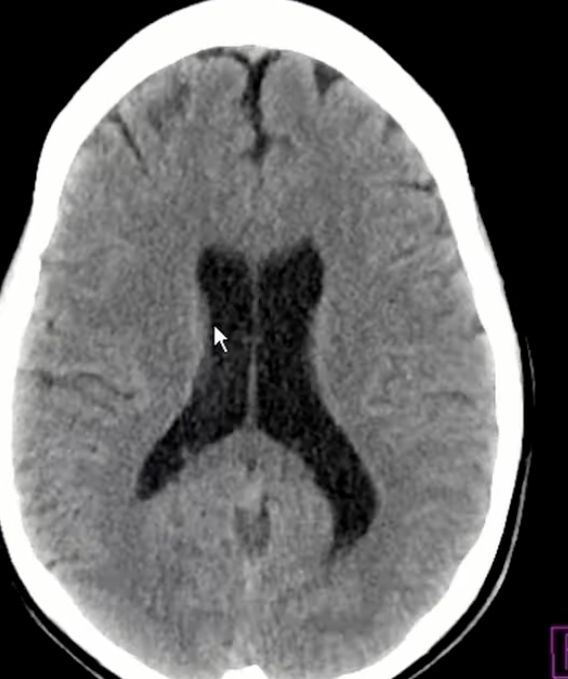
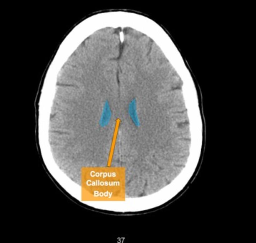
CTH Anatomy
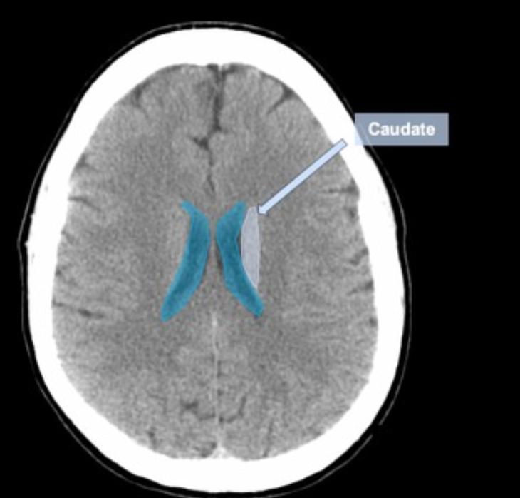
  • the body[…] of caudate nucleus runs lateral to these lateral ventricles[structures?]

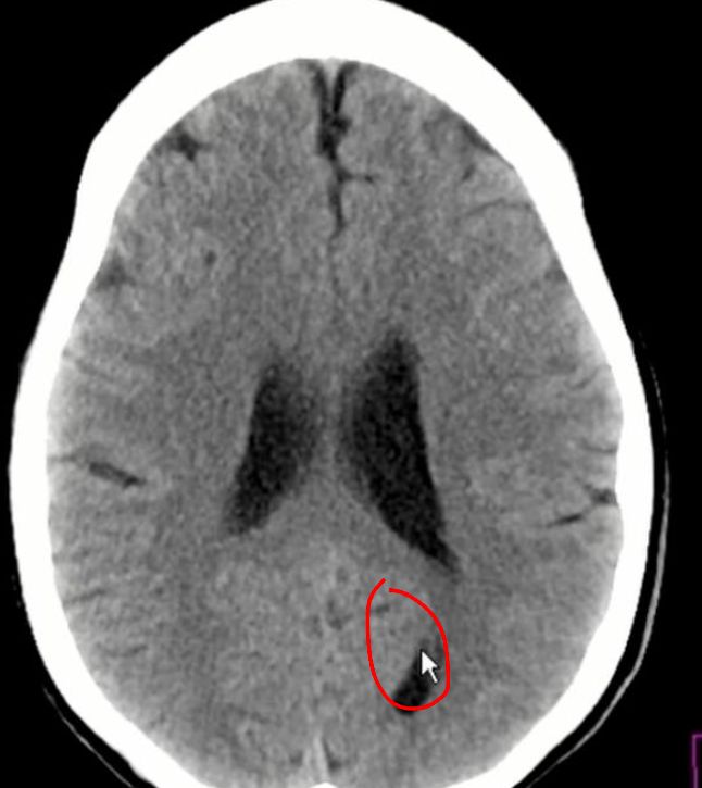
CTH Anatomy
  • this is occipital horn, lateral ventricle[…]

CTH Anatomy
  • caudate nucleus
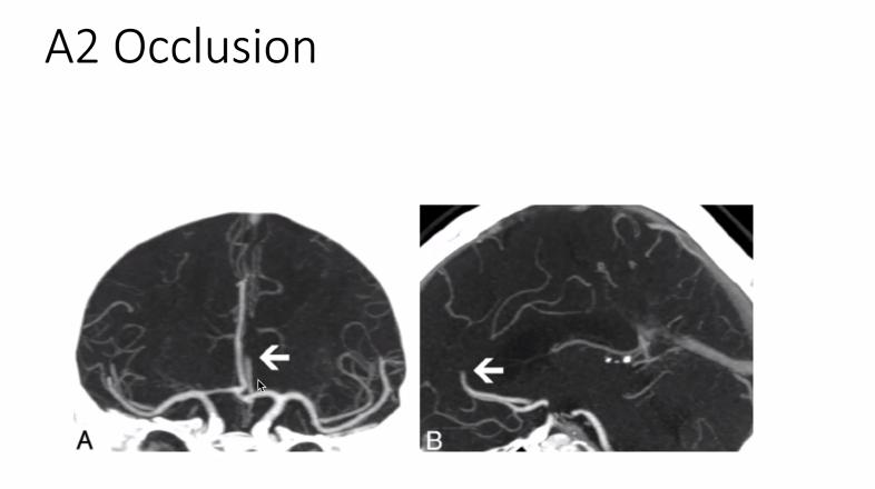
    • arterial supply: A1[…] or A2[…] segment of ACA[…]

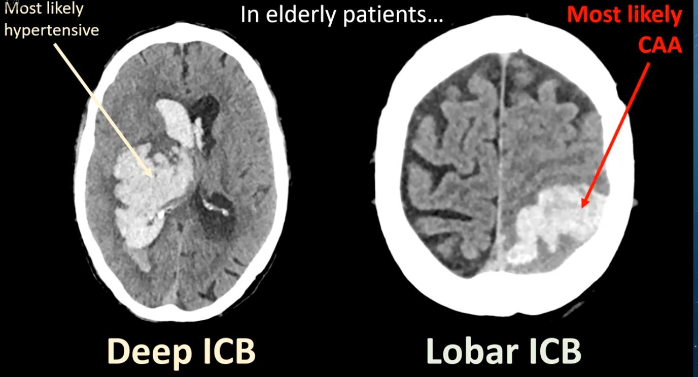
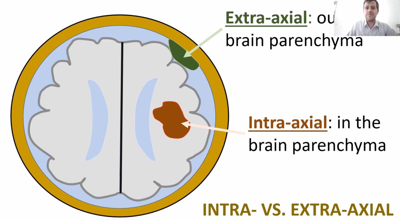
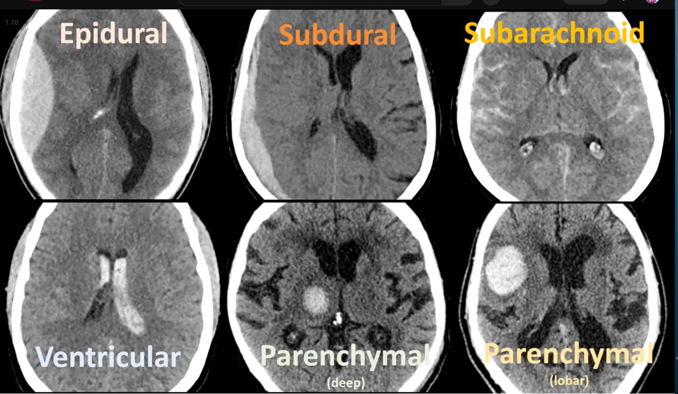
Intraaxial vs Extraaxial
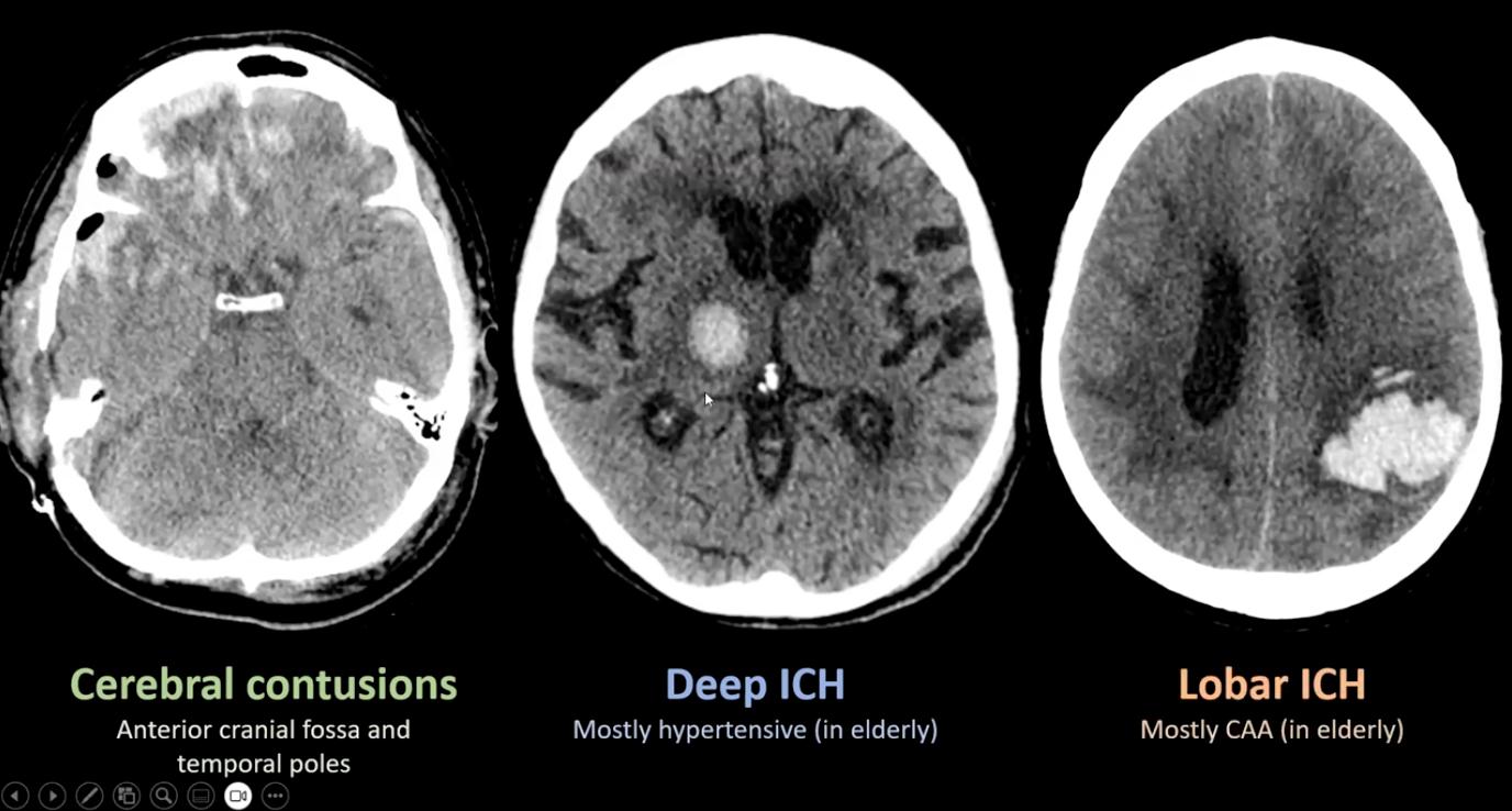
  • Intraaxial[…]: within the brain parenchyma
  • Extraaxial: outside the brain parenchyma[…]
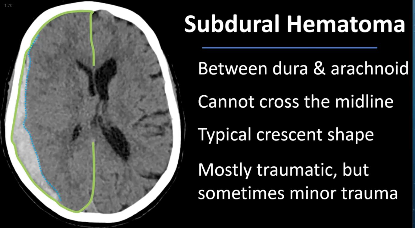
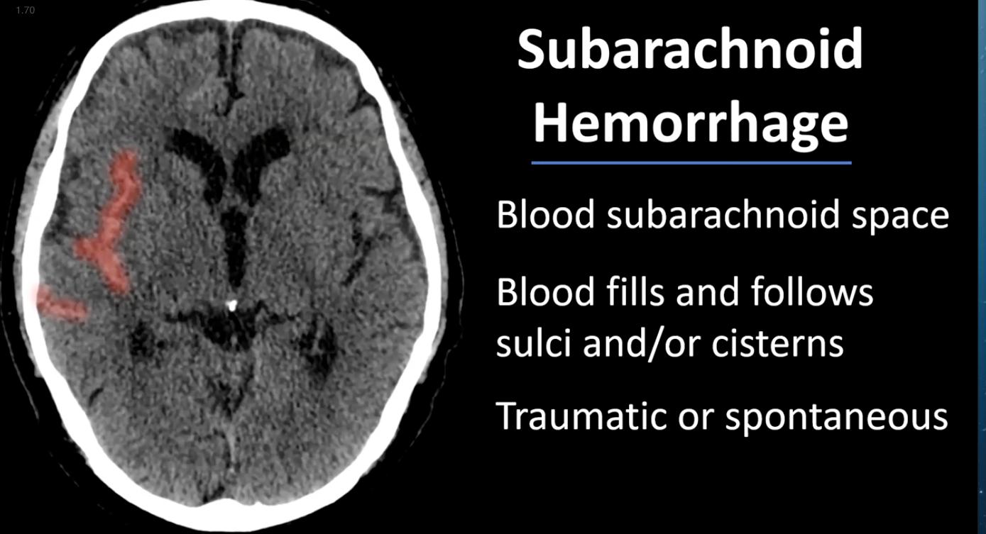
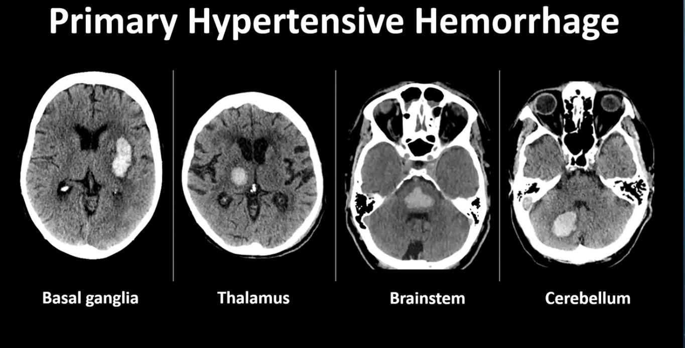
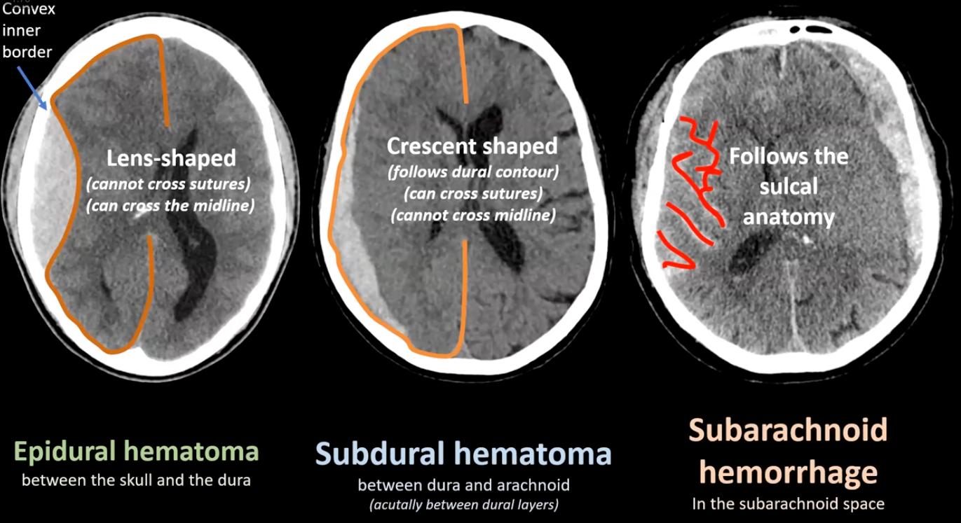
    • Most common extraaxial hemorrhage is subarachnoid[…] hemorrhage, followed by subdural[…] and epidural[…] hematomas
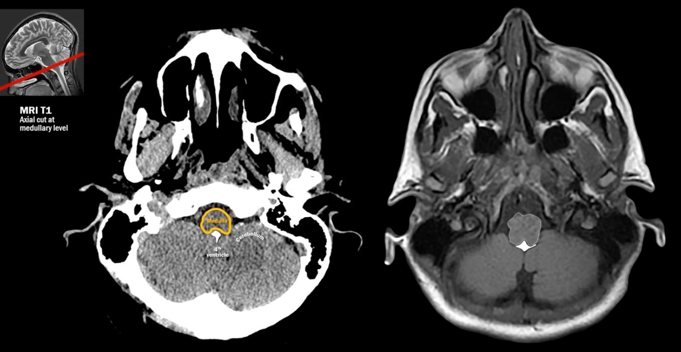
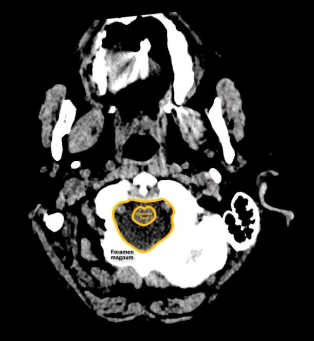
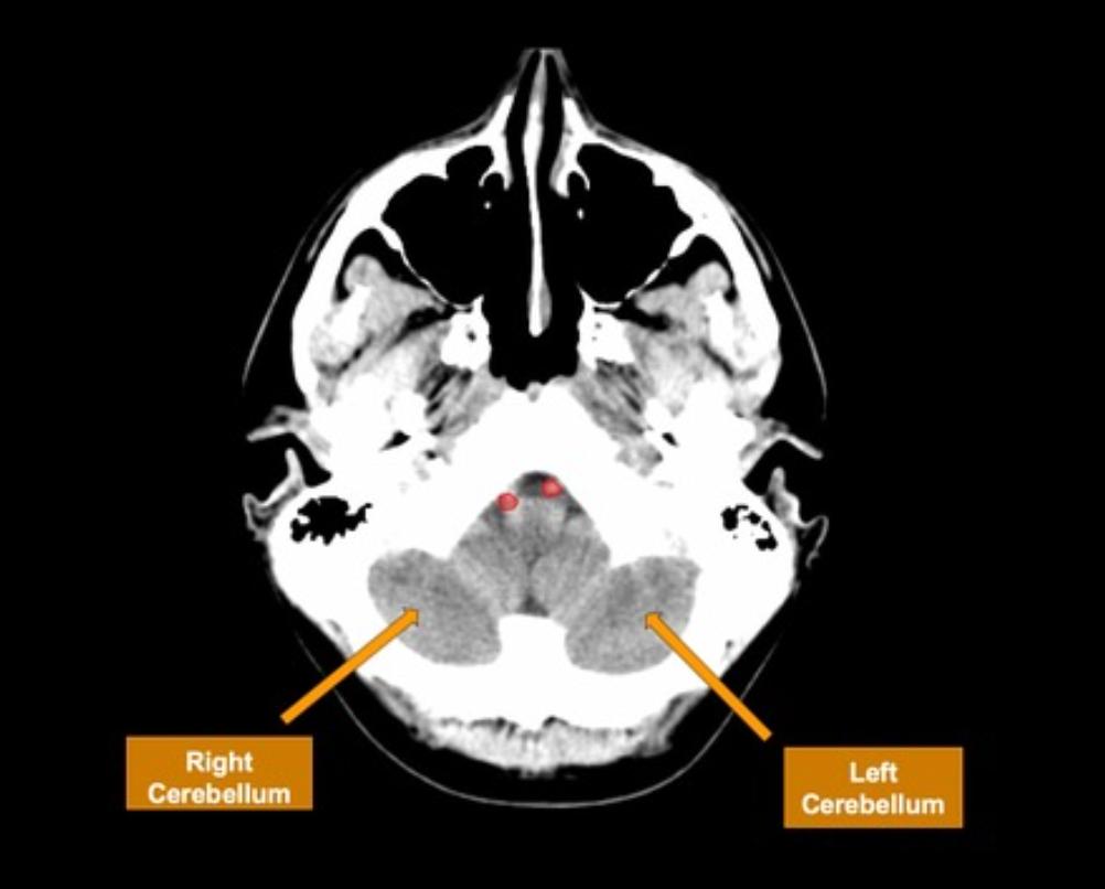
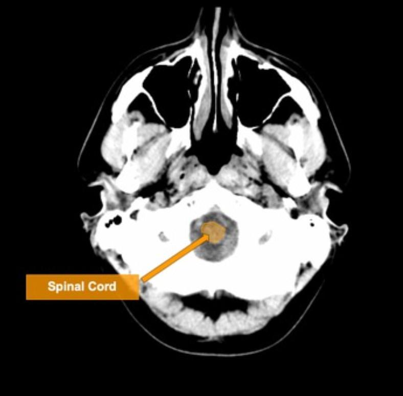
1 foramen magnum[…]
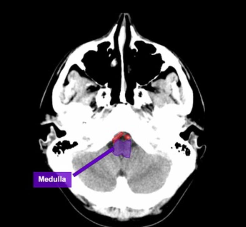
2 medulla oblongata[…]


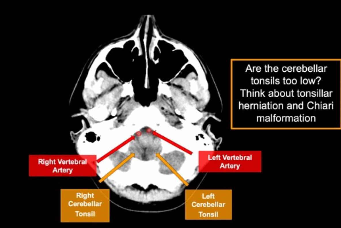
3 vertebral artery[…]
4 cerebellar tonsil[…]


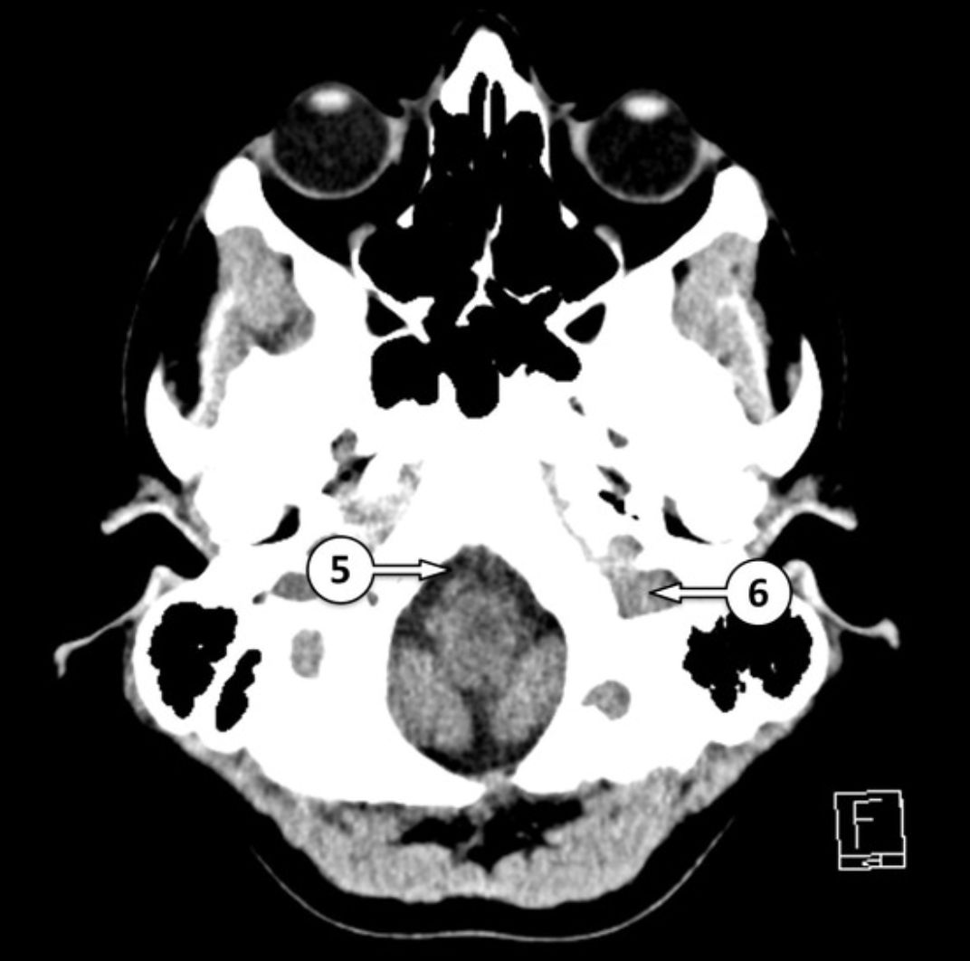
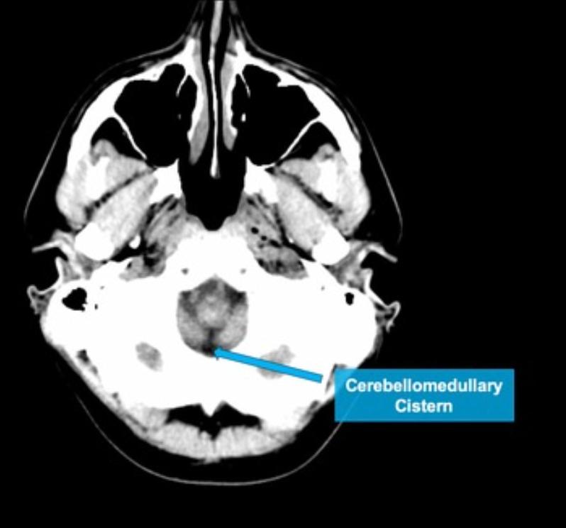
5 premedullary cistern[…]
6 internal jugular vein[…]


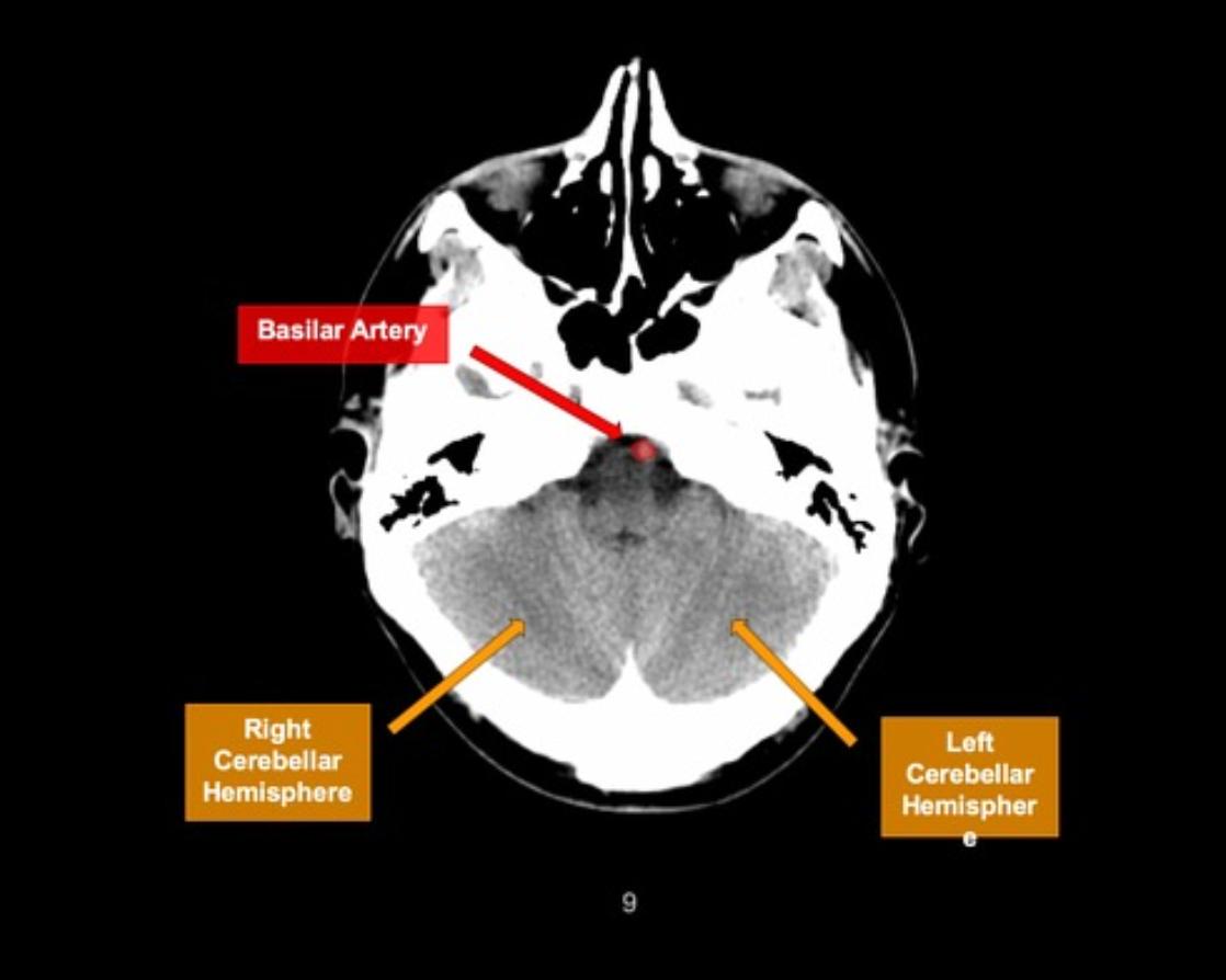
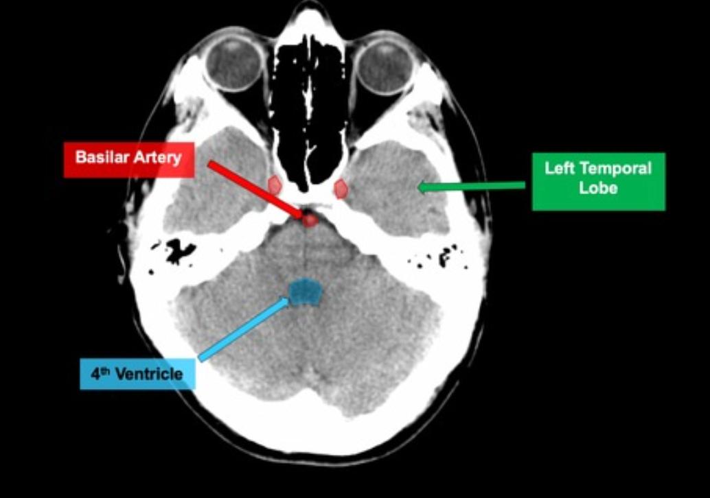
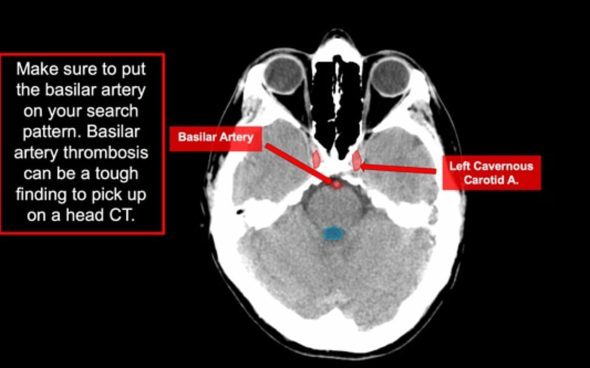
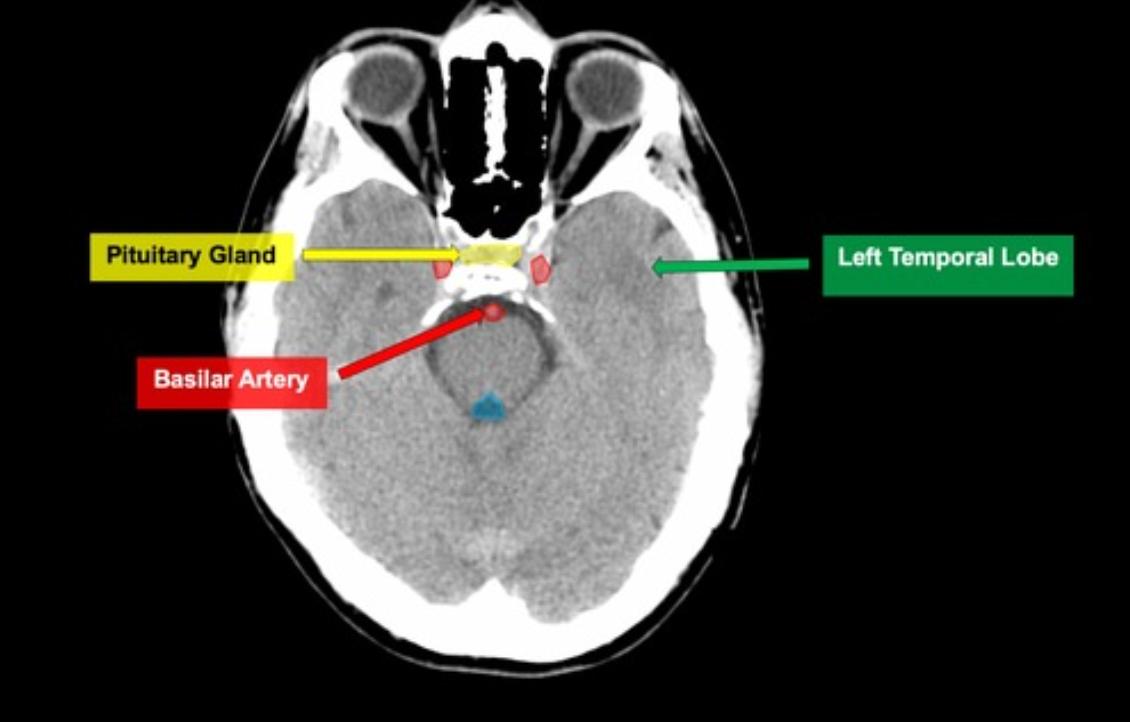
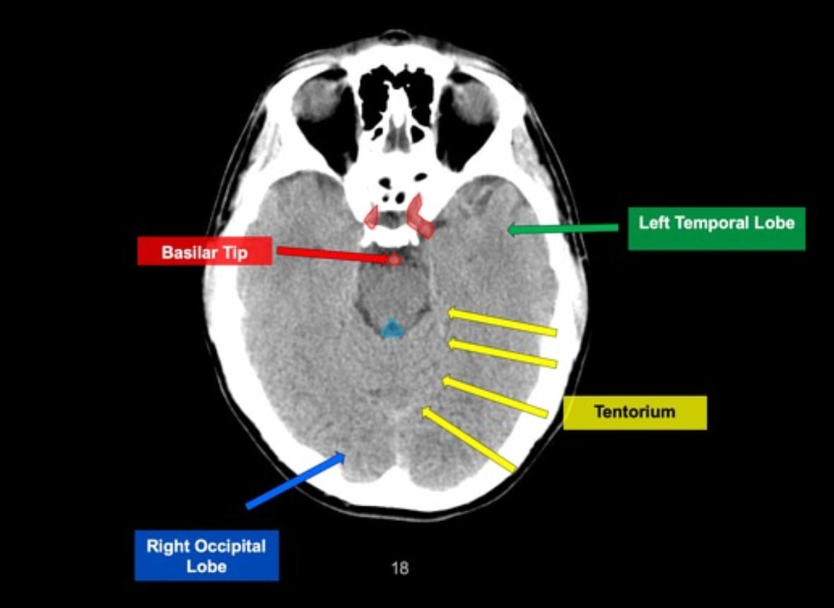
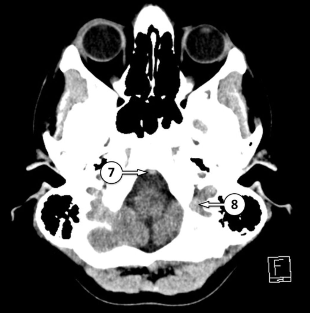
7 basilar artery[…]
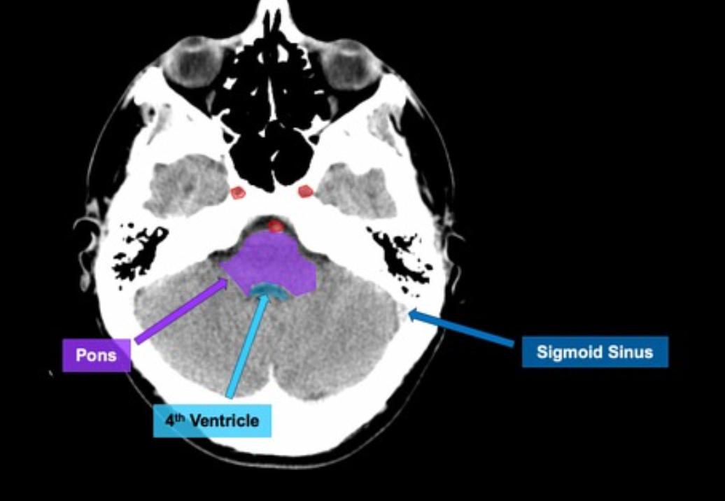
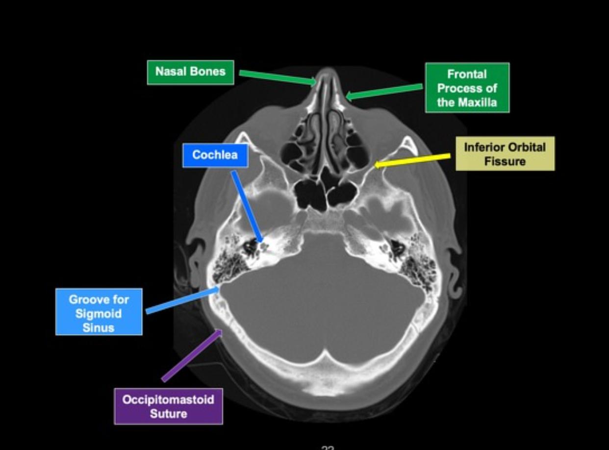
8 sigmoid sinus[…]


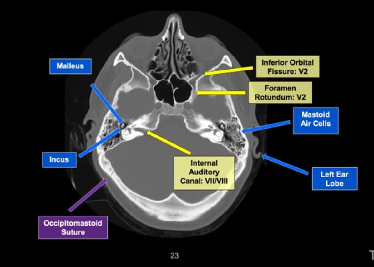
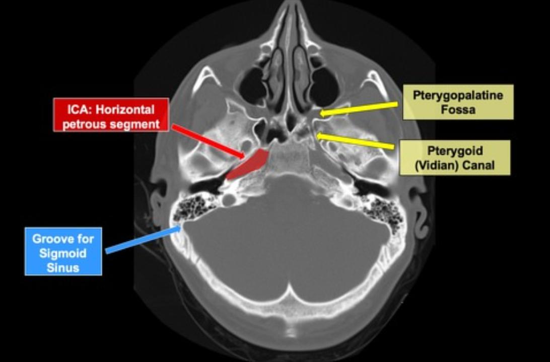
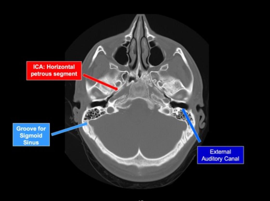
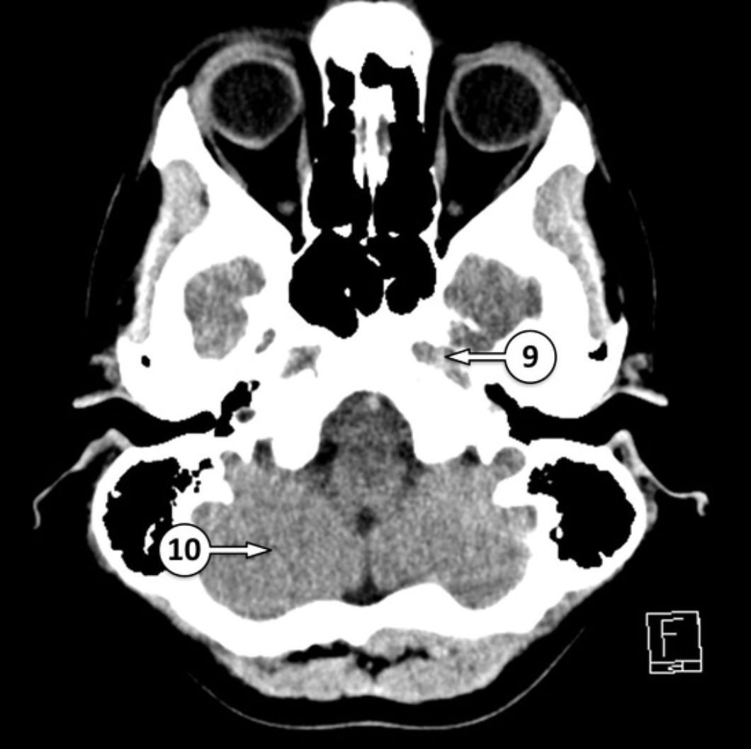
9 petrous ICA[…] in the carotid canal[…]
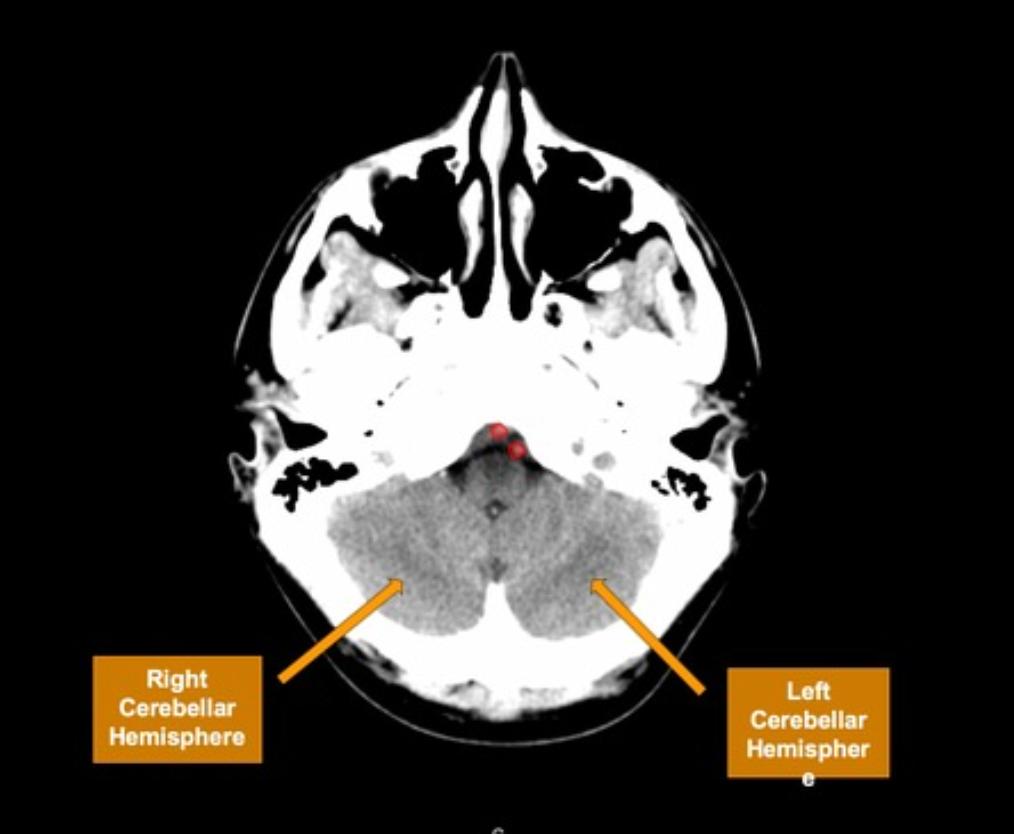
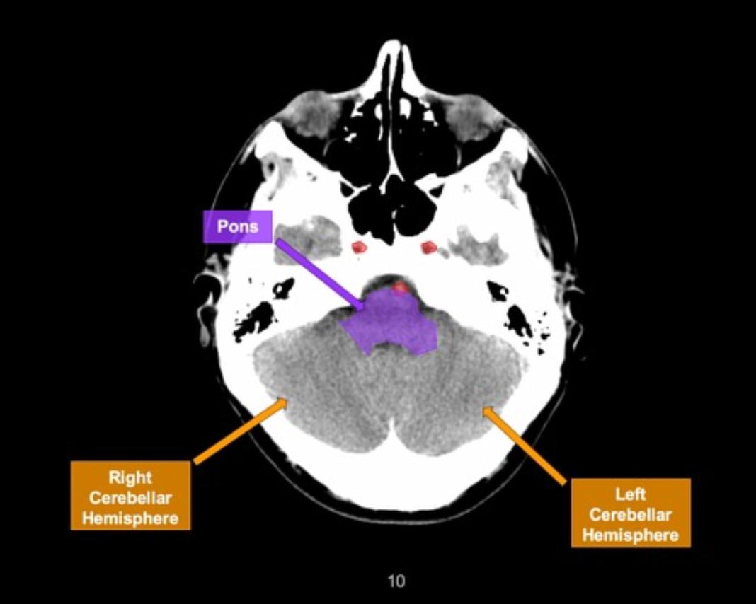
10 cerebellar hemisphere[…]


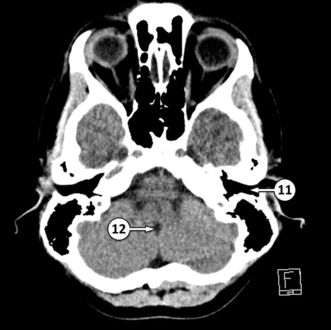
11 external auditory canal[…]
12 foramen of Magendie[…]


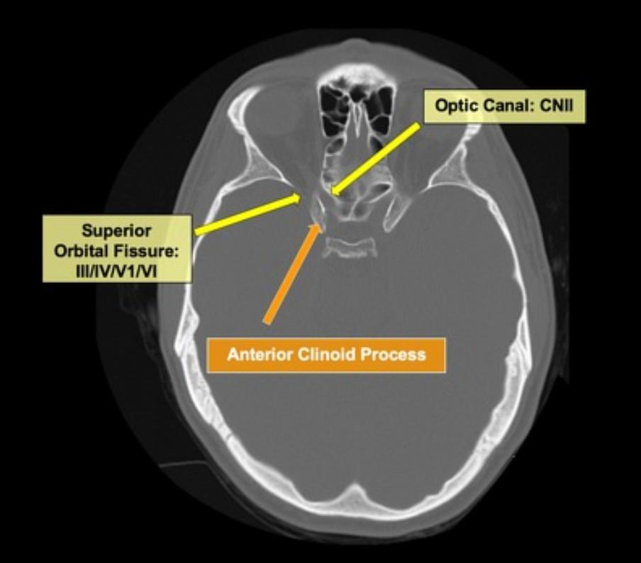
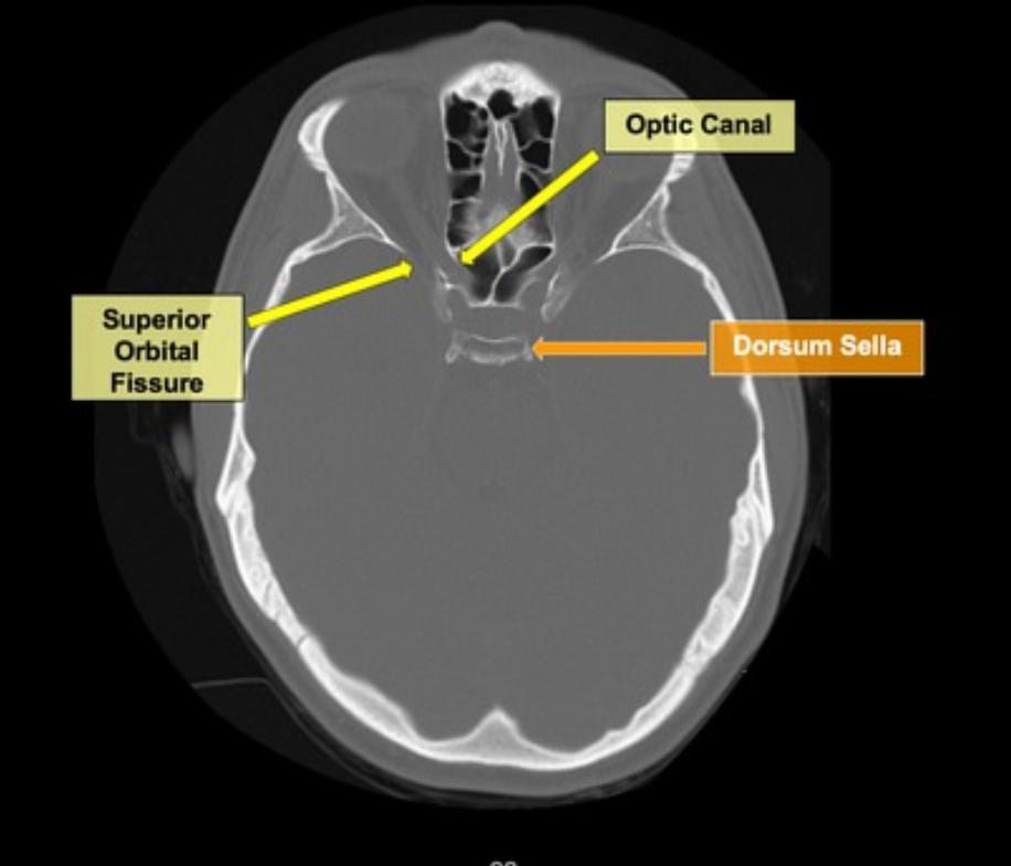
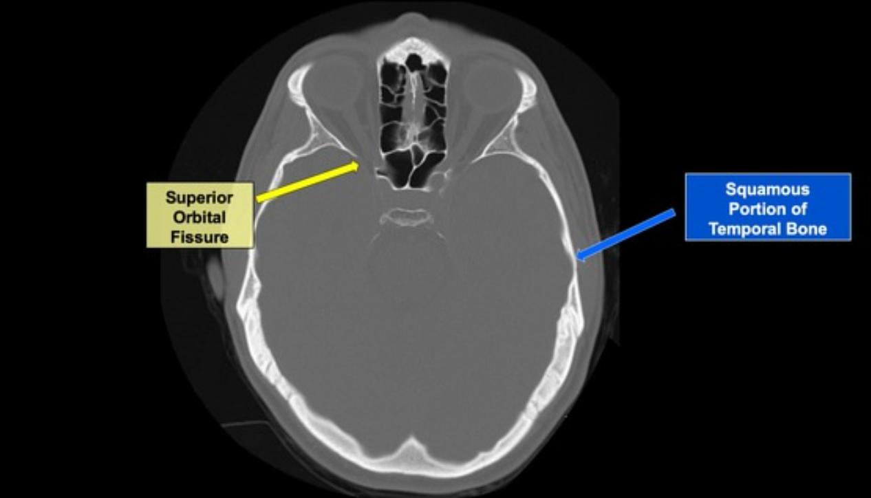
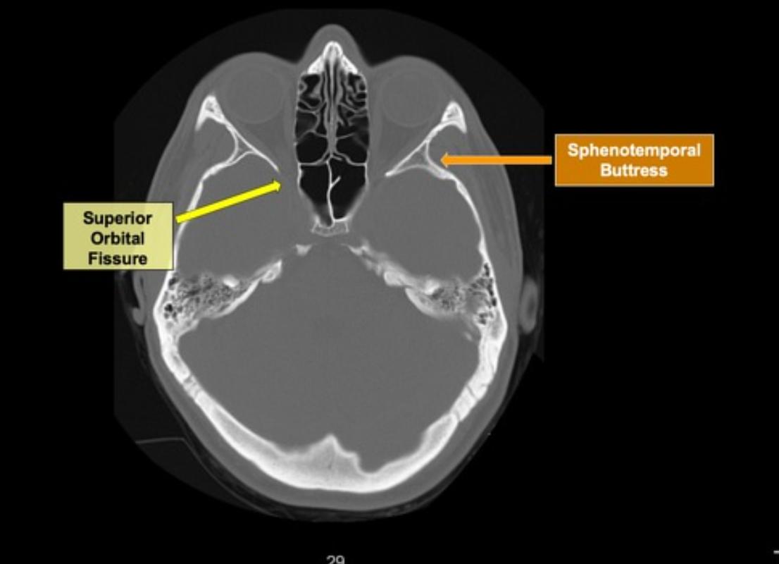
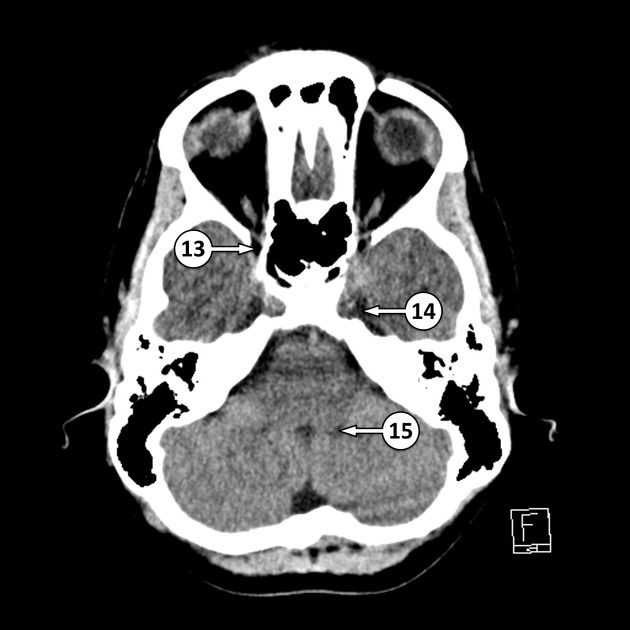
13 superior orbital fissure[…]
14 Meckel's cave[…]
15 foramen of Luschka[…]


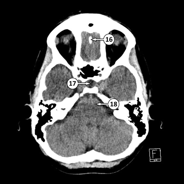
16 crista galli[…]
17 pituitary fossa[…]
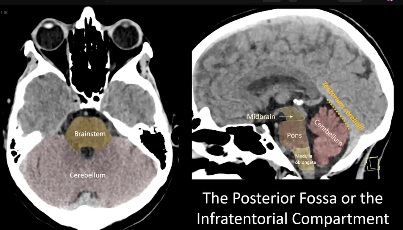
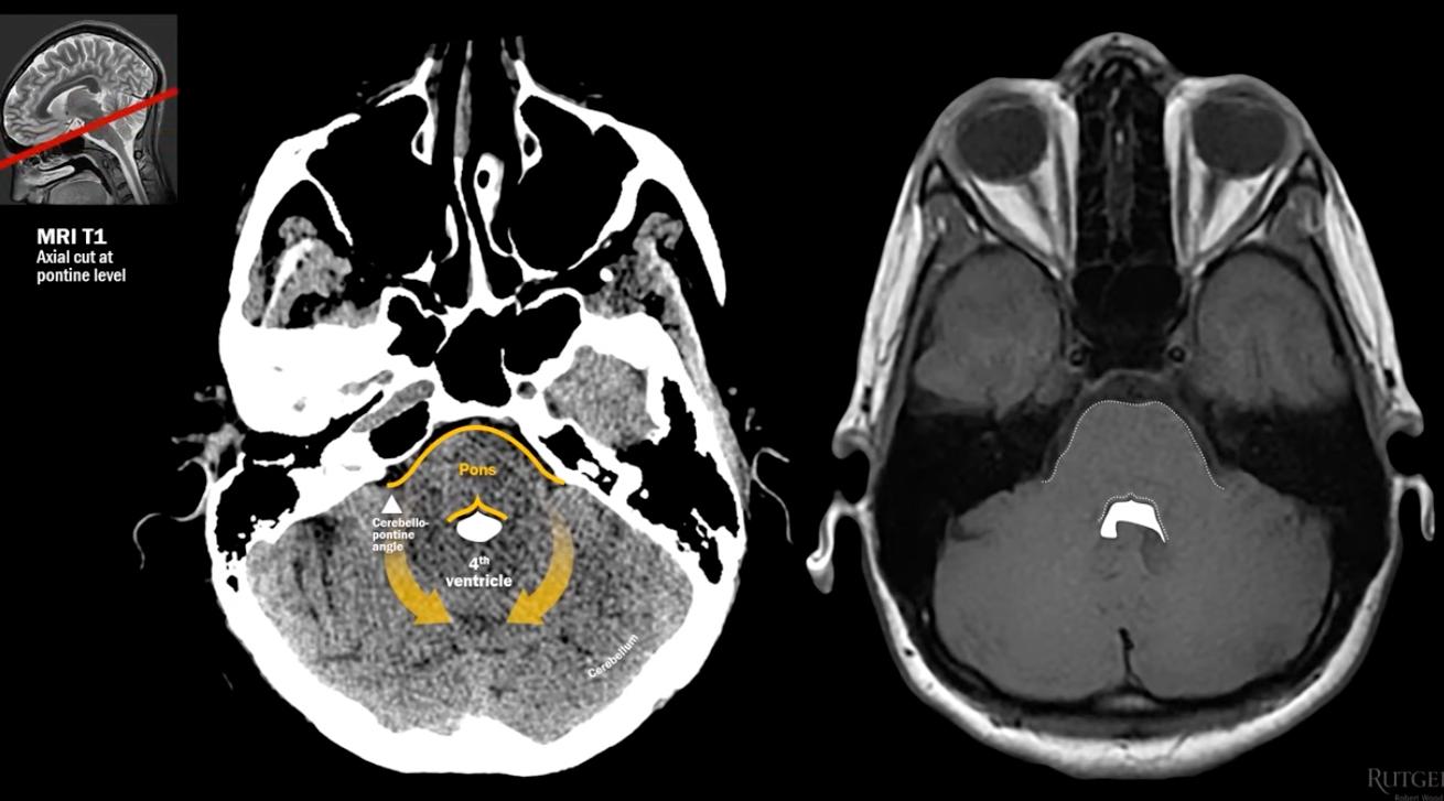
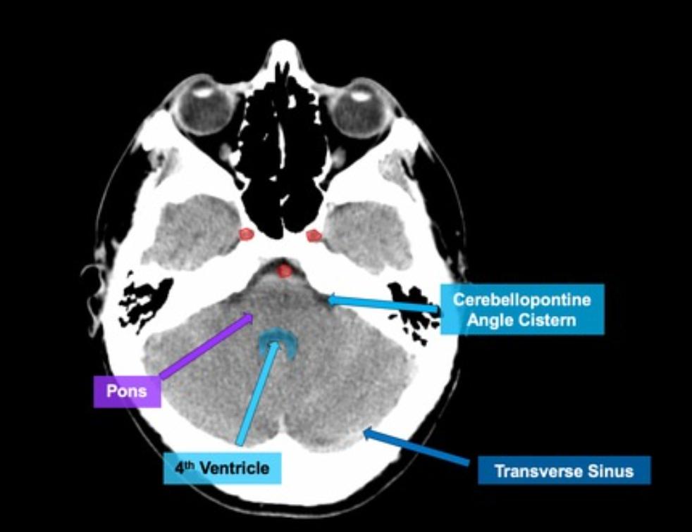
18 pons[…]


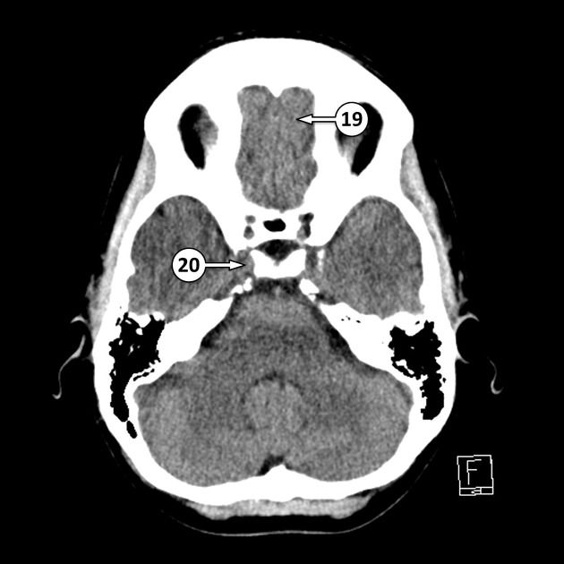
19 gyrus rectus[…]
20 cavernous ICA[…] in the cavernous sinus[…]


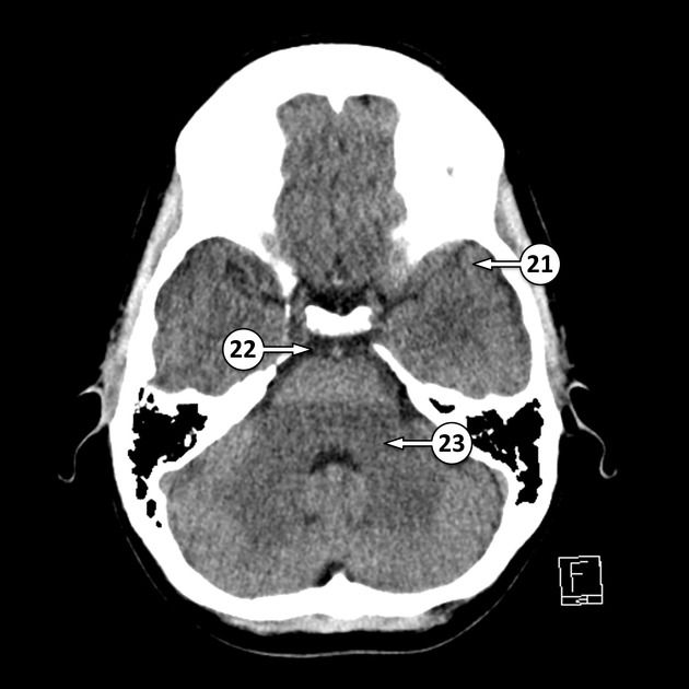
21 left temporal pole[…]
22 prepontine cistern[…]
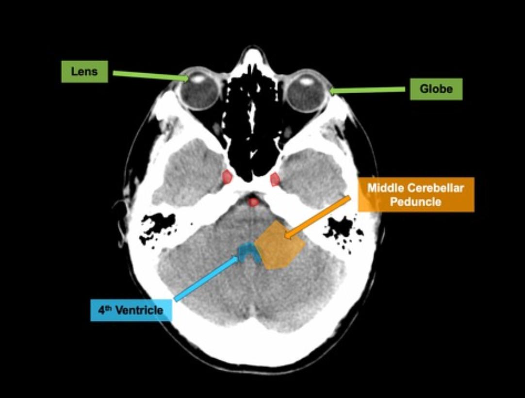
23 middle cerebellar[…] peduncle


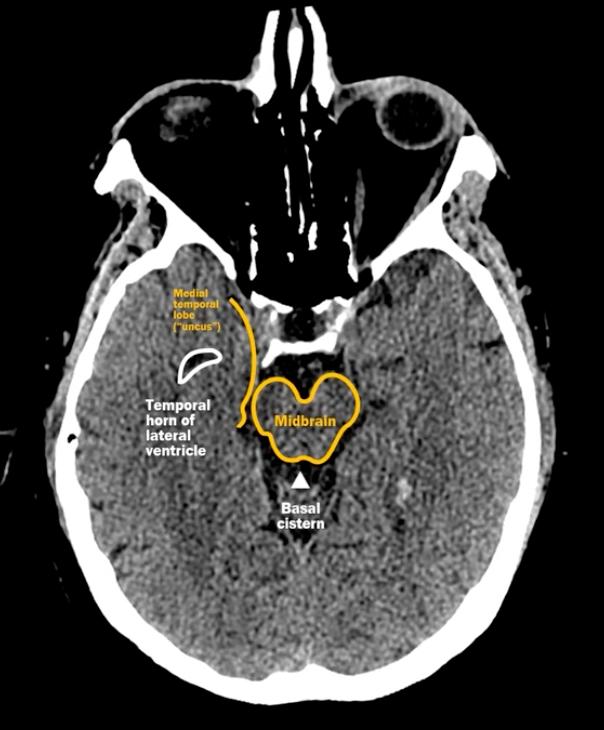
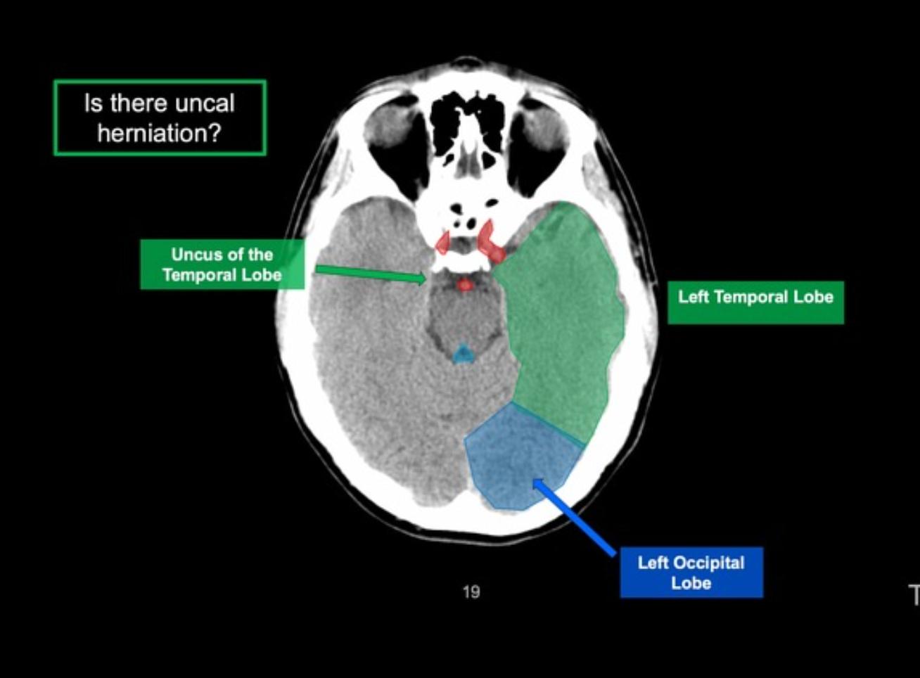
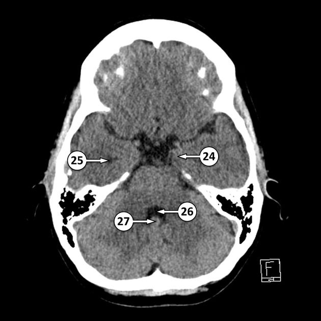
24 uncus[…]
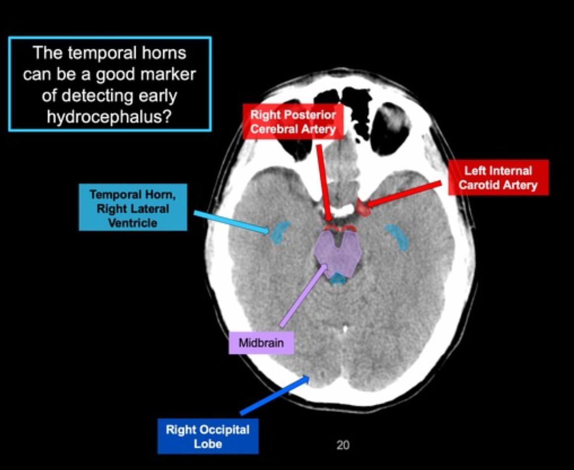
25 temporal horn[…] of the lateral ventricle[…]
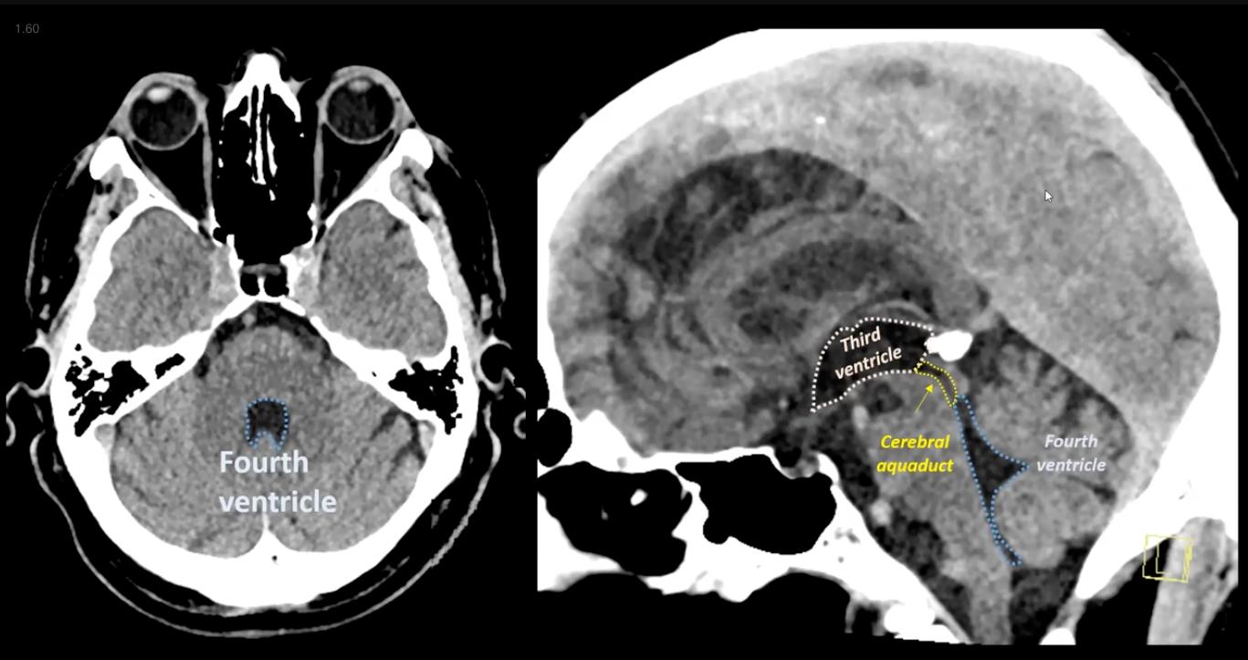
26 4th ventricle[…]
27 nodule of the cerebellum[…]


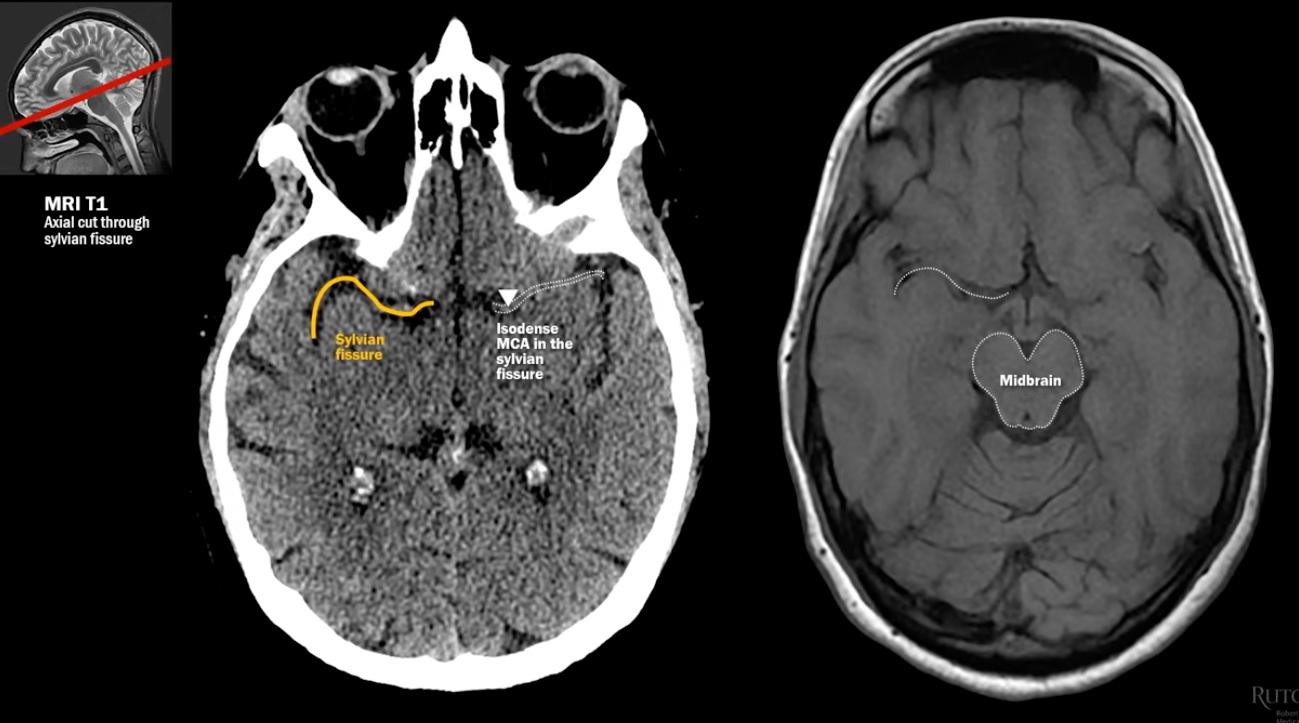
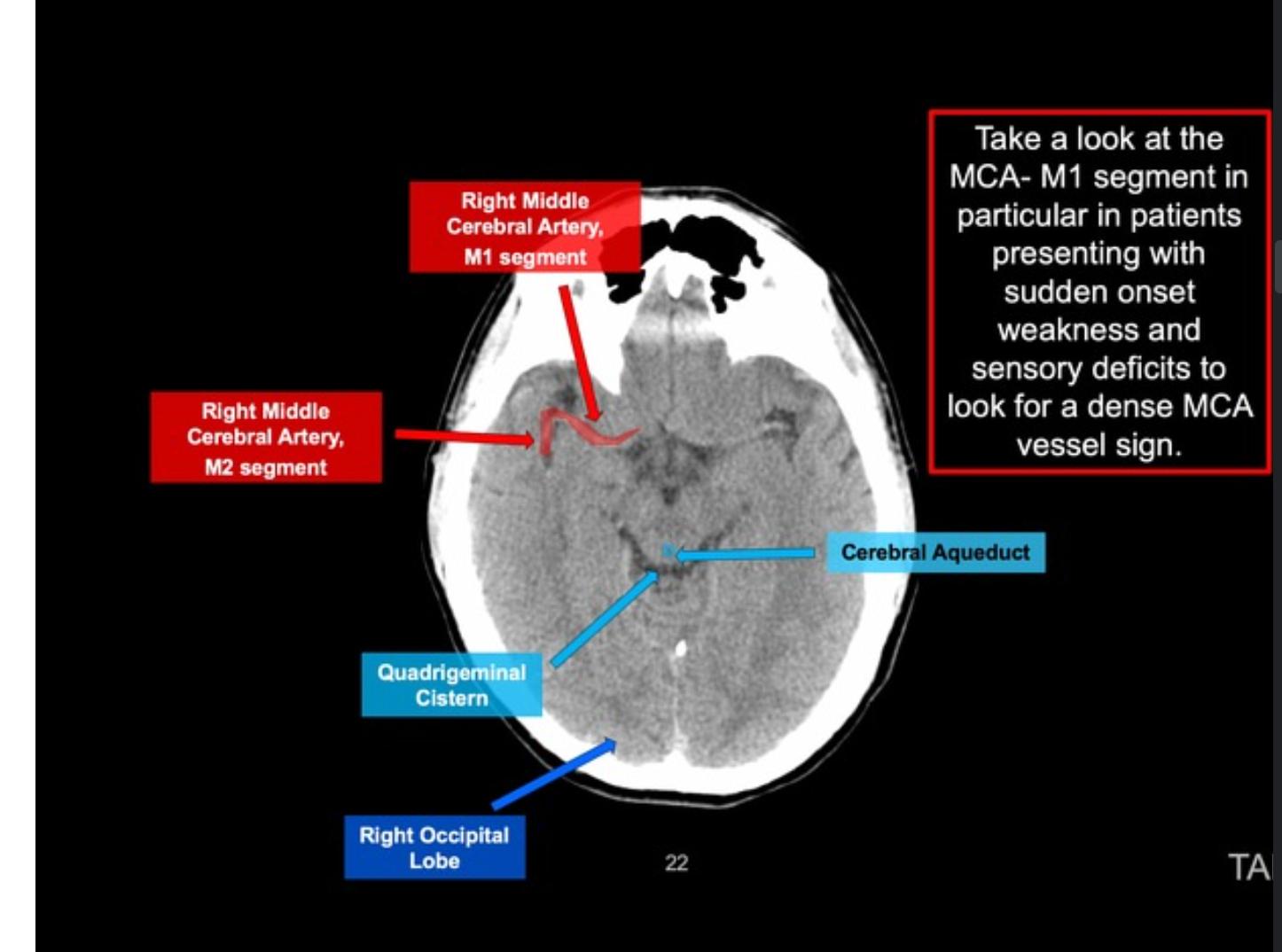
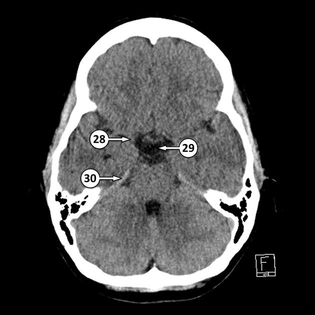
28 MCA M1[…] segment
29 suprasellar[…] cistern
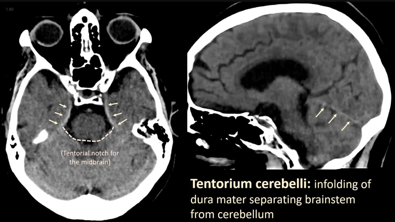
30 tentorium cerebelli[…]


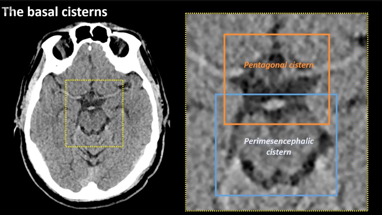
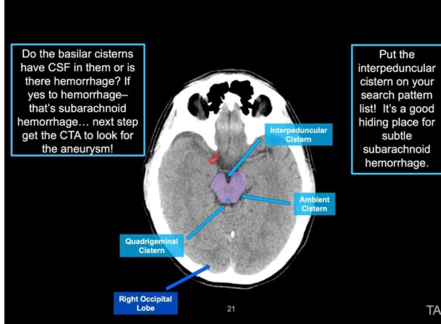
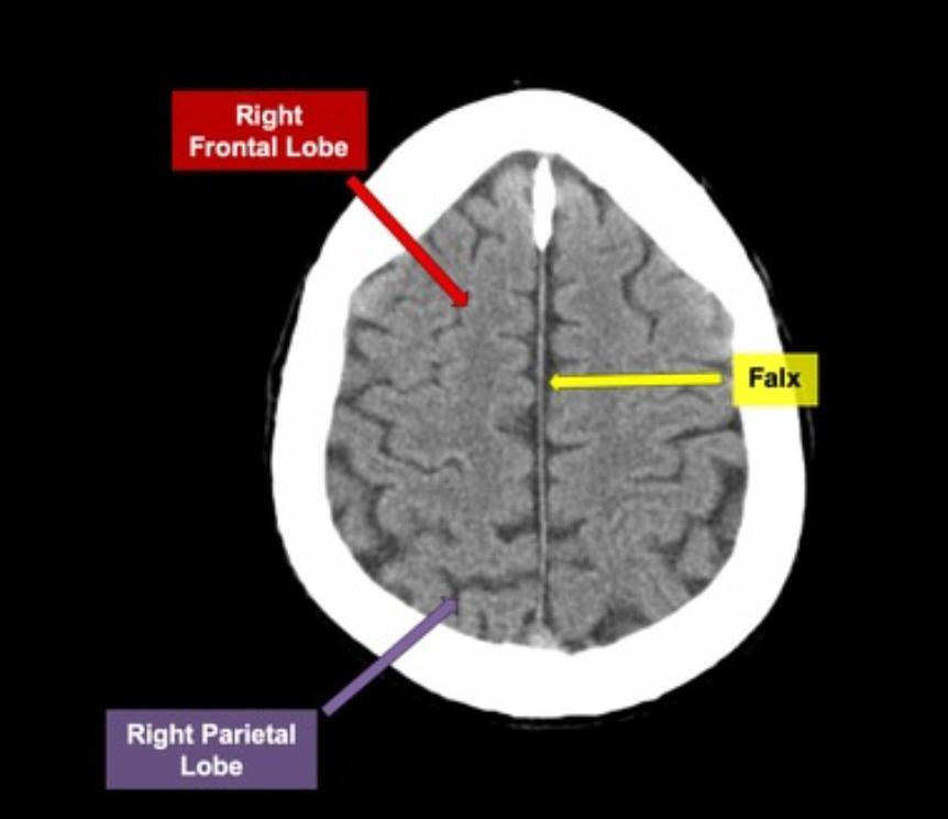
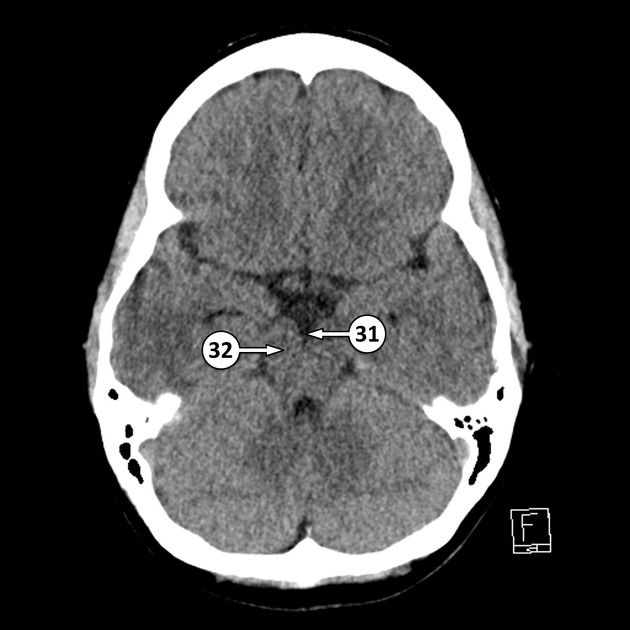
31 interpeduncular[…] cistern
32 cerebral peduncle[…]


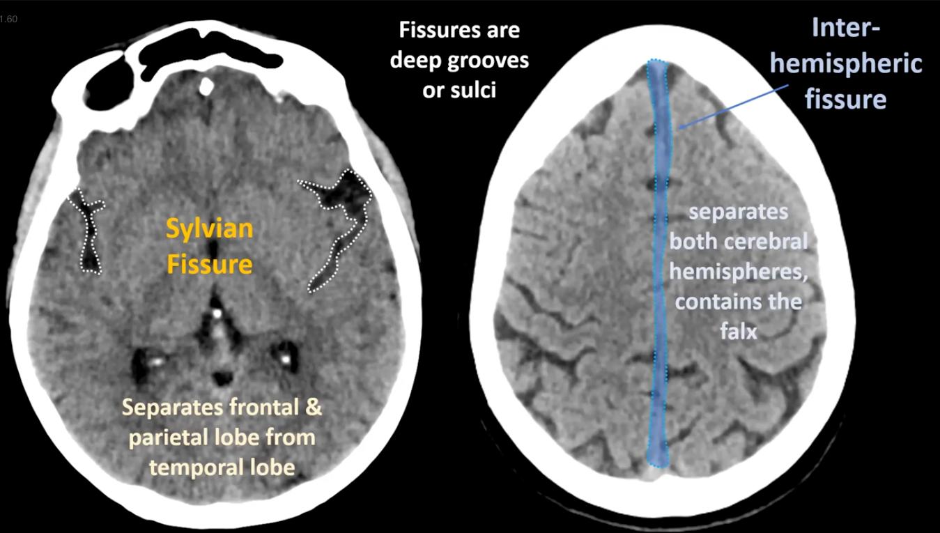
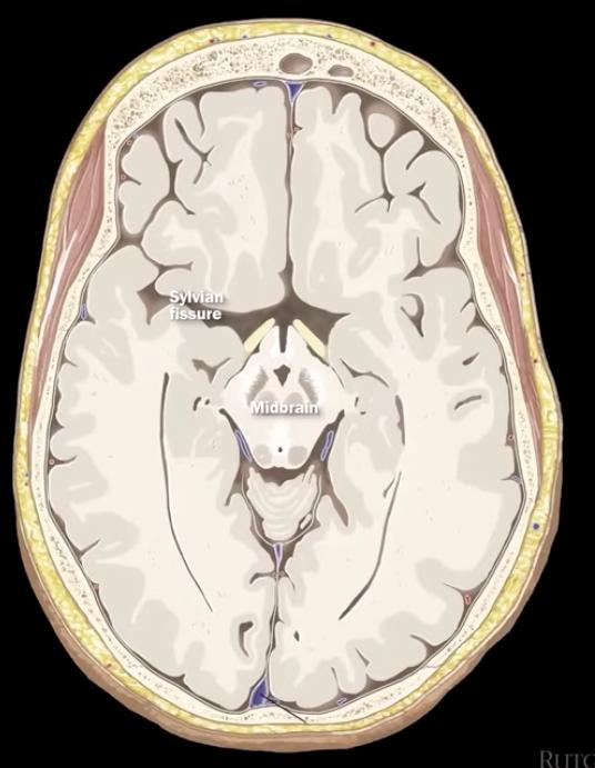
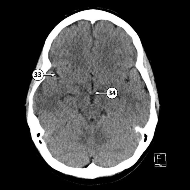
33 Sylvian fissure[…]
34 mammillary bodies[…]


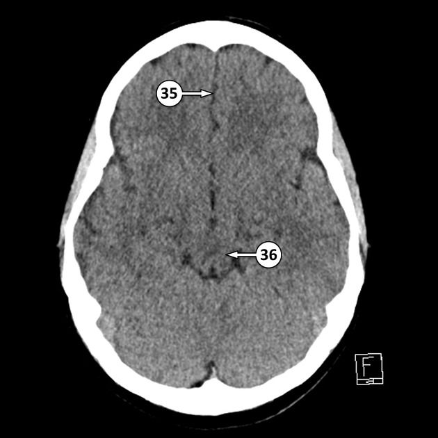
35 median interhemispheric fissure[…]
36 midbrain[…]


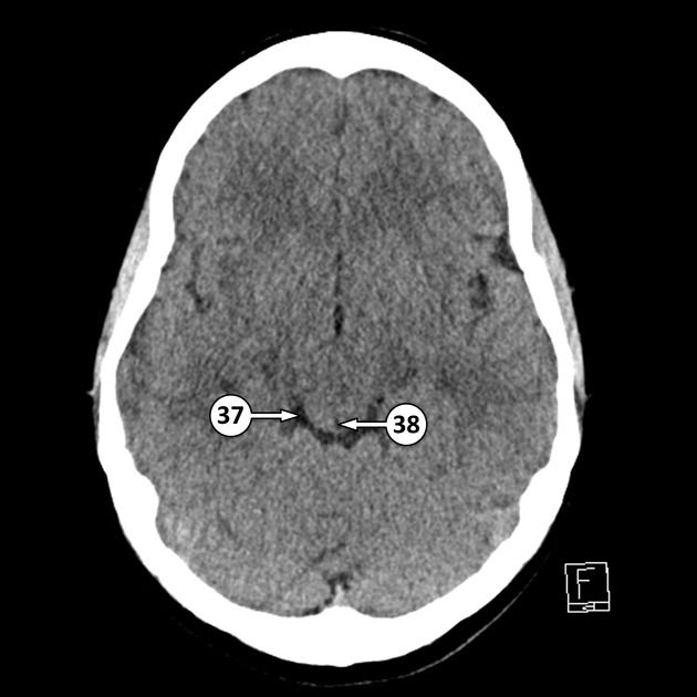
37 ambient cistern[…]
38 cerebral aqueduct (of Sylvius)[…]
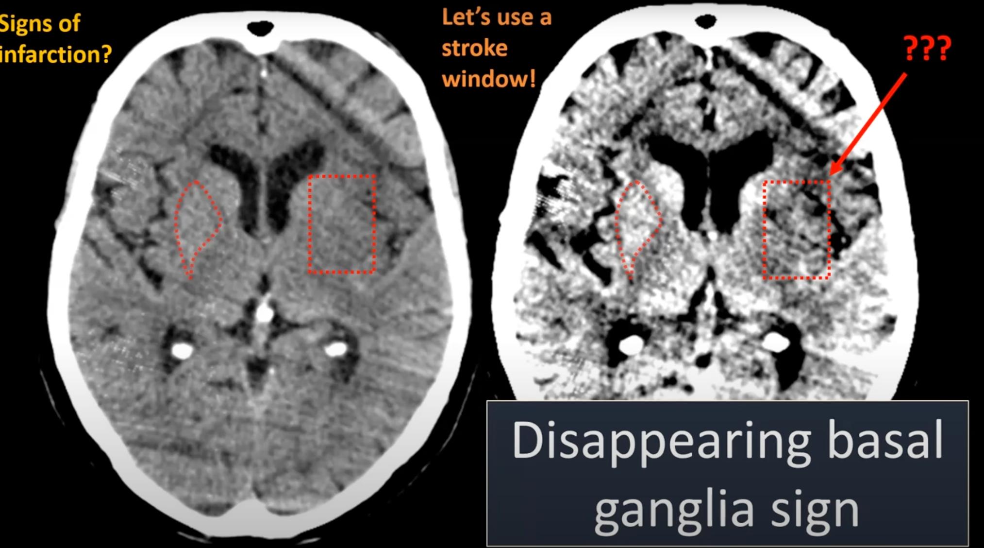
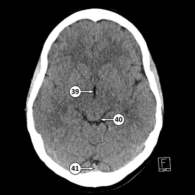
39 third ventricle[…]
40 inferior colliculus[…]
41 confluence of the sinuses[…]

41 anterior limb of the internal capsule[…]
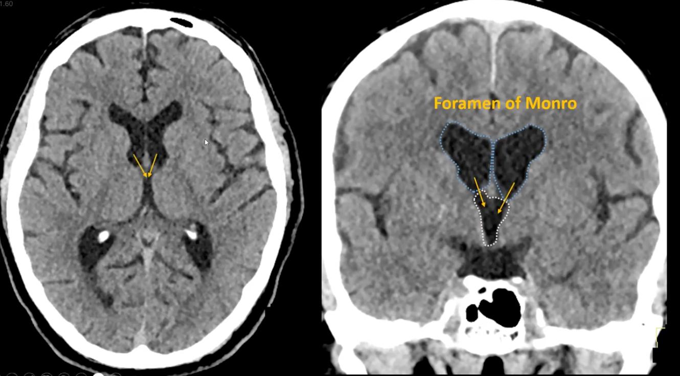
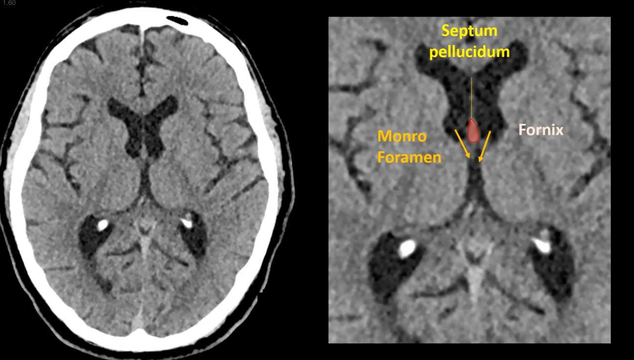
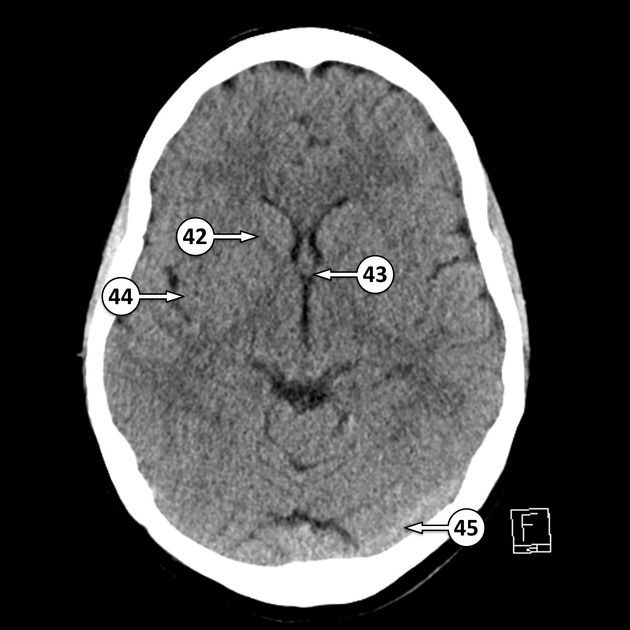
42 interventricular foramen (of Monroe)[…]
43 insular cortex[…]
44 transverse sinus[…]

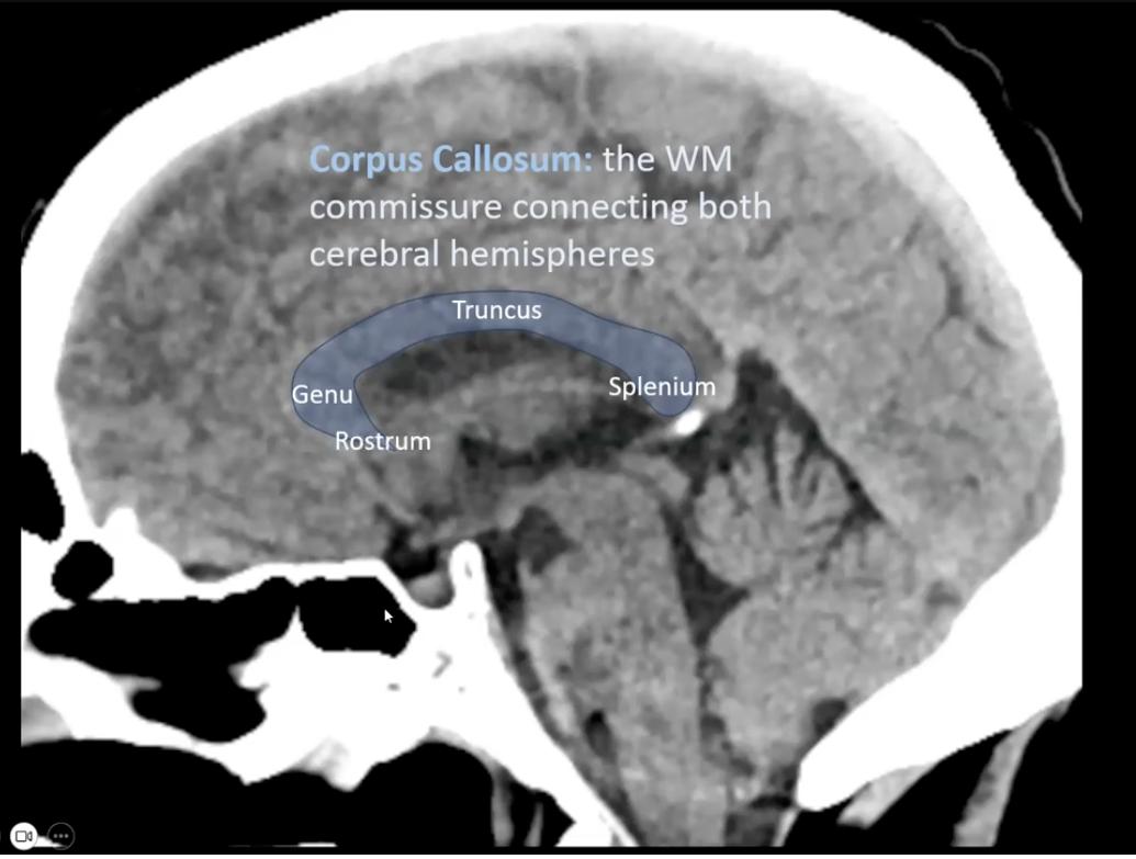
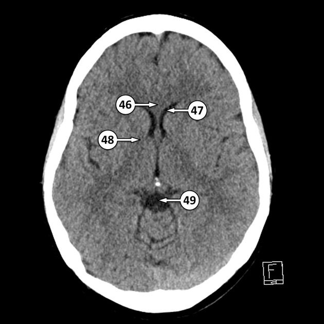
46 genu of the corpus callosum[…]
47 frontal horn of the lateral ventricle[…]
48 genu of the internal capsule[…]
49 quadrigeminal[…] cistern


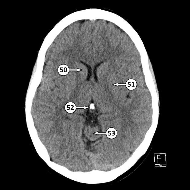
50 head of the caudate nucleus[…]
51 lentiform nucleus[…]
52 pineal gland[…]
53 superior vermis of the cerebellum[…]


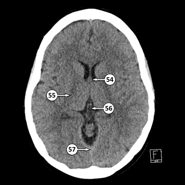
54 column of the fornix[…]
55 posterior limb of the internal capsule[…]
56 internal cerebral vein[…]
57 straight sinus[…]


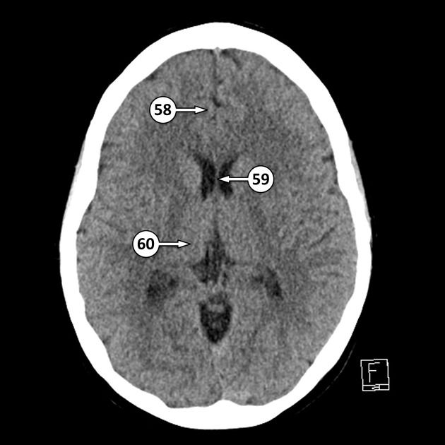
58 cingulate gyrus[…]
59 septum pellucidum[…]
60 thalamus[…]


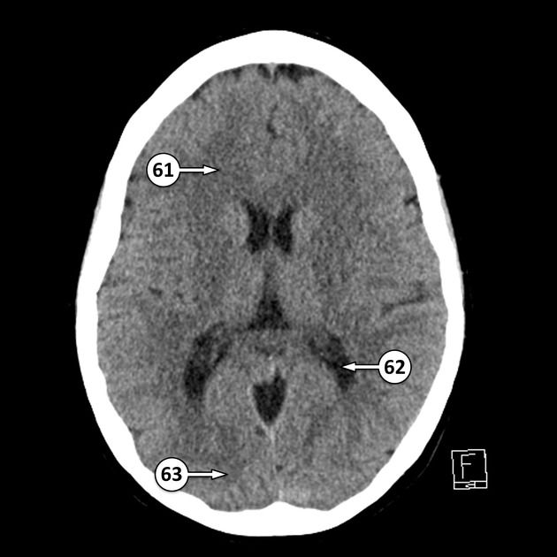
61 forceps minor[…]
62 trigone (atrium) of the lateral ventricle[…]
63 occipital lobe[…]


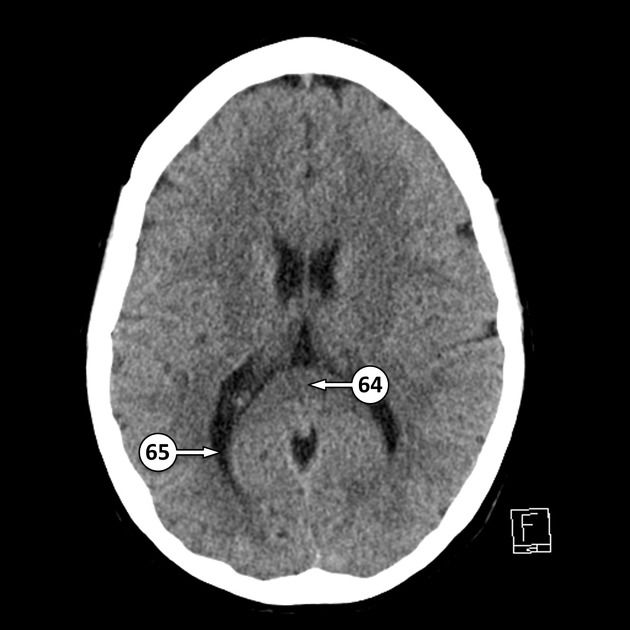
64 splenium of the corpus callosum[…]
65 occipital horn of the lateral ventricle[…]


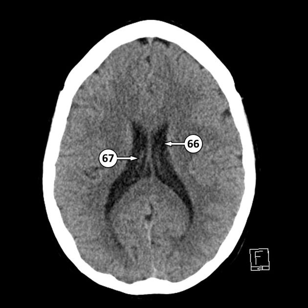
66 body of the lateral ventricle[…]
67 choroid plexus[…]


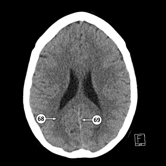
68 forceps major[…]
69 inferior sagittal sinus[…]


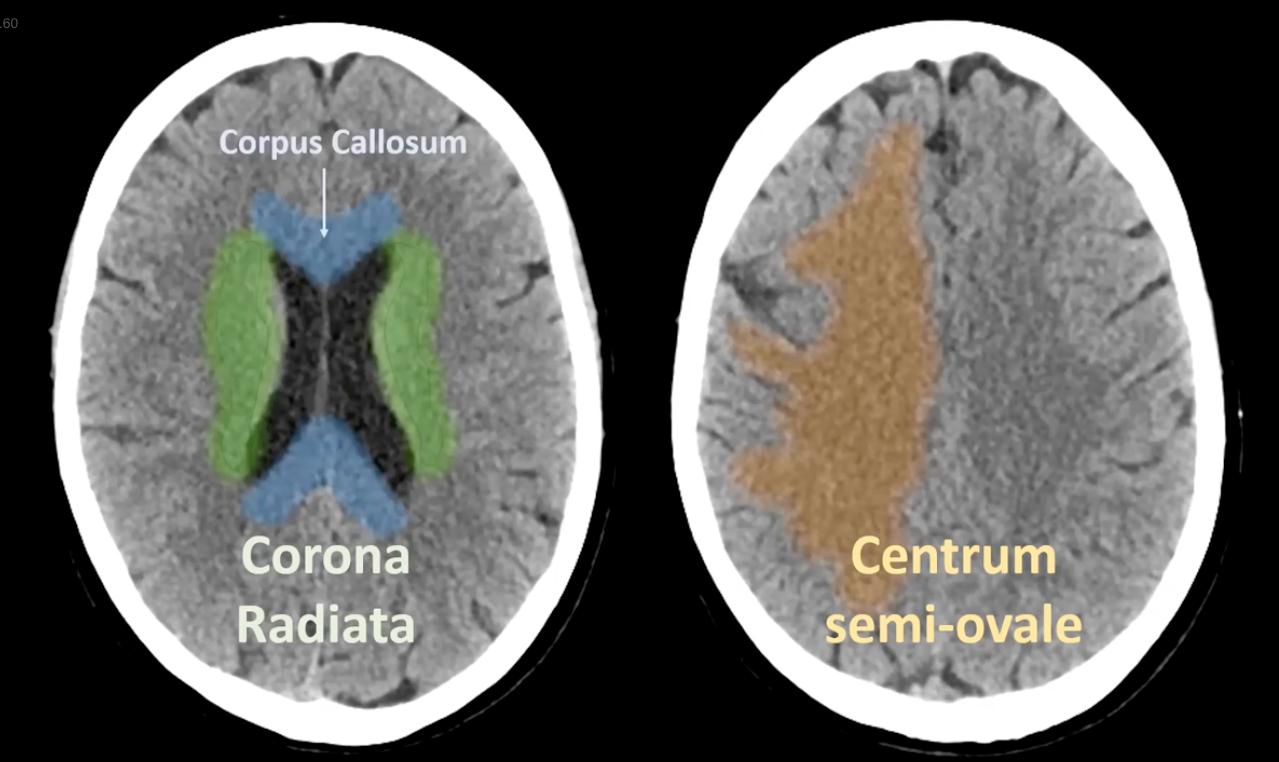
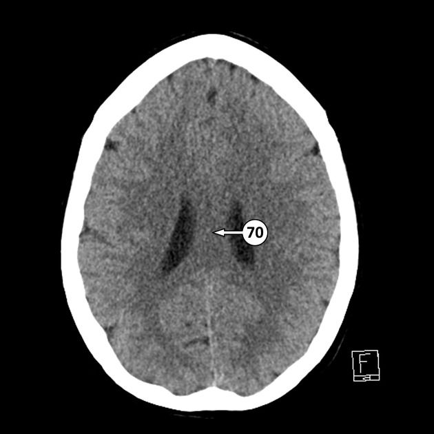
70 body of the corpus callosum[…]


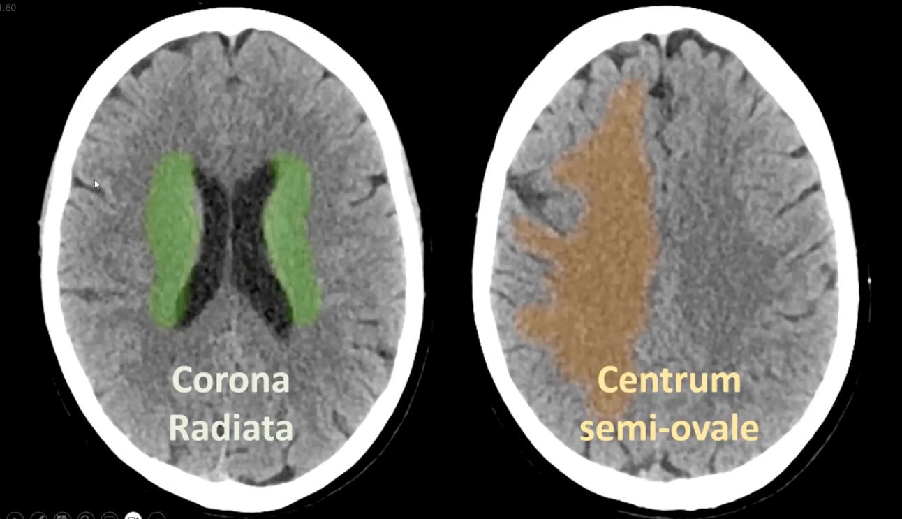
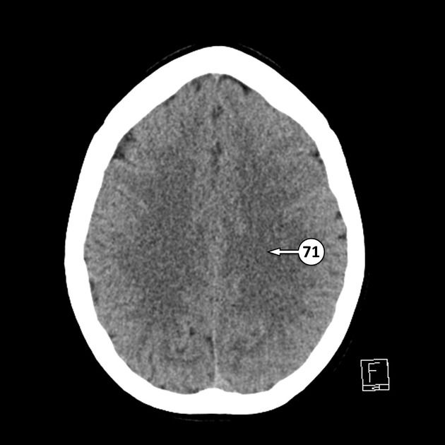
71 centrum semiovale[…]

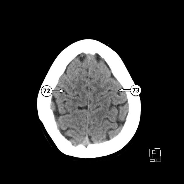
72 central sulcus (of Rolando)[…]
73 hand bump of the pre central gyrus (primary motor cortex)[…]


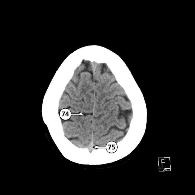
74 marginal sulcus[…]
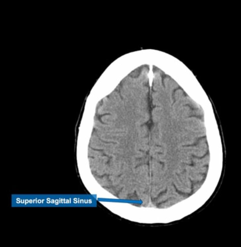
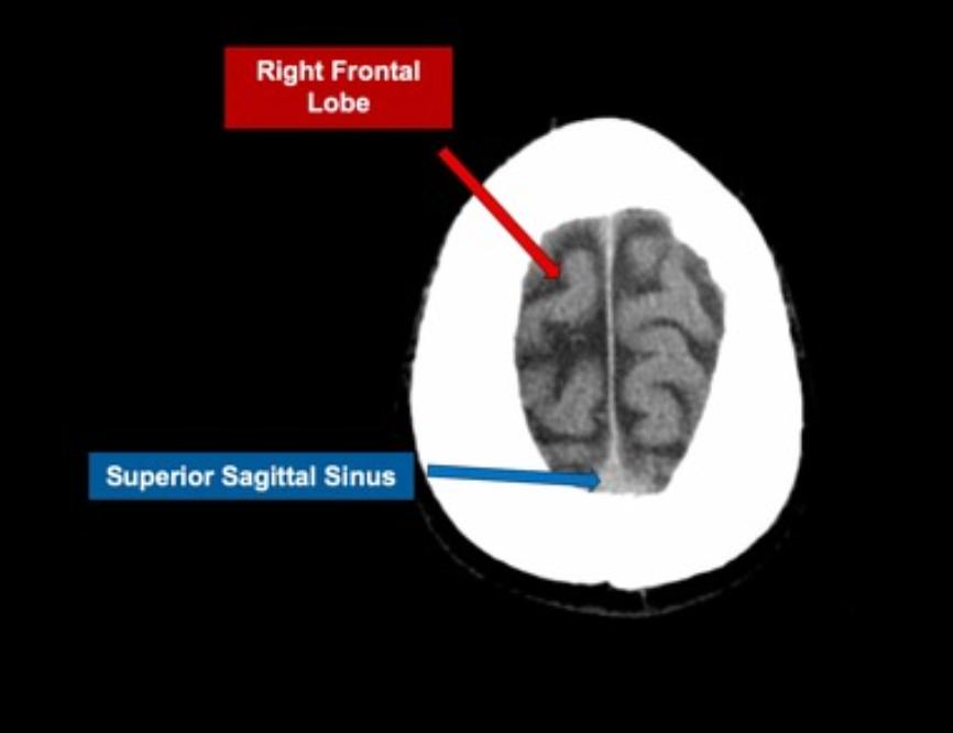
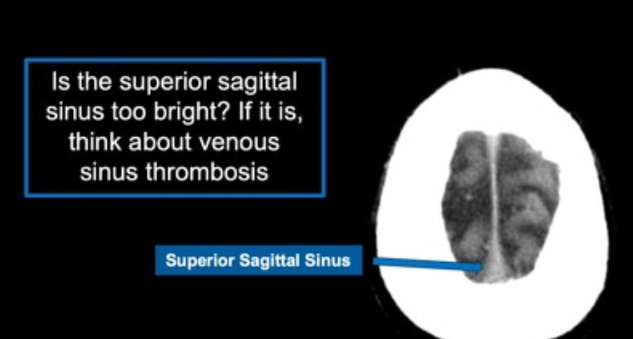
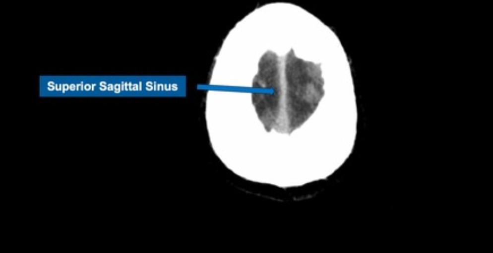
75 superior sagittal sinus[…]


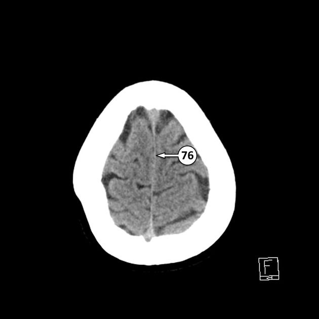
76 falx cerebri[…]