CTA

CTA

Radiology · 23 cards

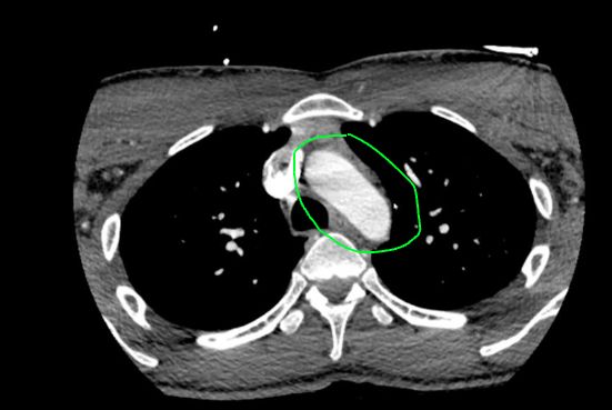
CTA (1)
  • This is the aortic arch[…]

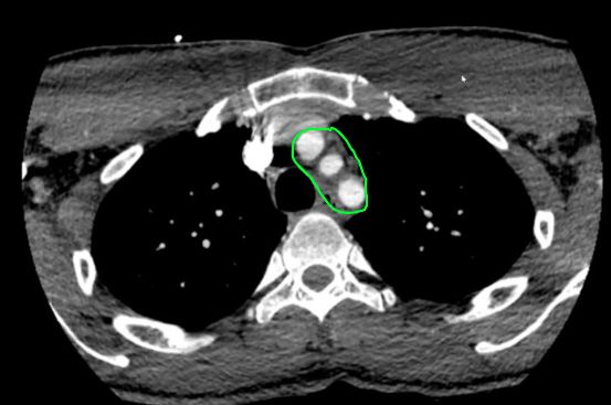
CTA (2)
  • These are the 3 great vessels (R brachiocephalic, L CCA, L Subclavian)[…]

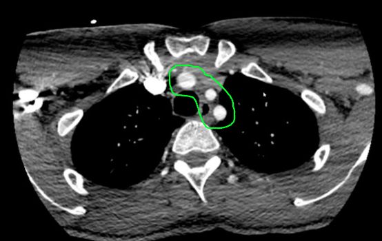
CTA (3)
  • These are the 3 great vessels (R brachiocephalic, L CCA, L Subclavian)[…]

CTA (4)
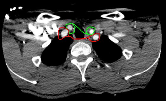
  • green: CCA[…]
  • red: Subclavians[…]

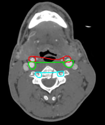
CTA (5)
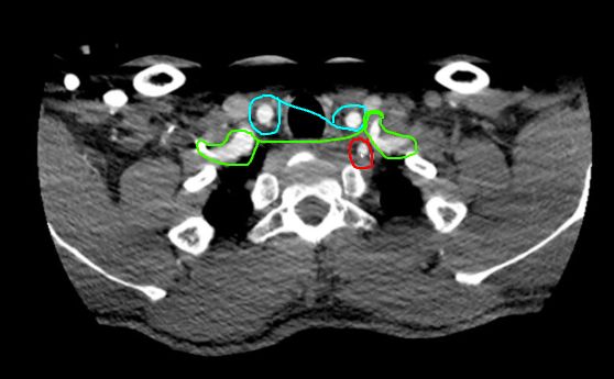
  • this shows the
    • blue: CCA[…]
    • green: Subclavian[…]
    • red: vert[…] coming off the subclavian[…] (green)

CTA (6)
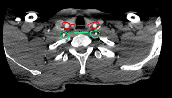
  • this shows the 4 main vessels:
    • red: CCA[…]
    • green: Verts[…]

CTA (7)
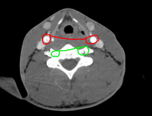
  • this shows the 4 main vessels:
    • red: CCA[…]
    • green: Verts[…]

CTA (8)
  • this shows the:
    • ECAs[…] (red, anterior) and ICAs[…] (green, posterior) at the level of the carotid bifurcation[…]
    • blue: vertebral[…] arteries

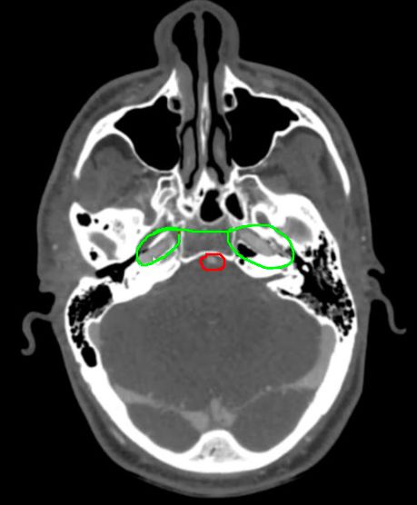
CTA (9)
  • this shows the: 
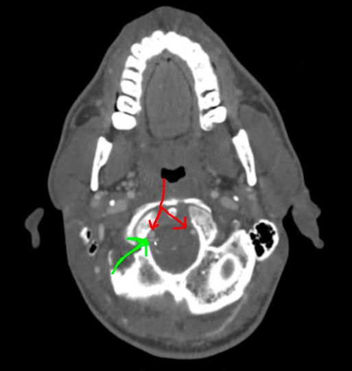
    • green: petrous ICAs (C2 segment)[…]
    • red: basilar[…] artery

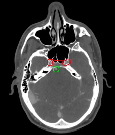
CTA (10)
  • this shows the: 
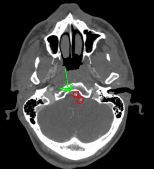
    • red: lasserum ICAs (C3 segment)[…]
    • green: basilar[…] artery

CTA (11)
  • this shows the: 
    • red: cavernous ICA (C4 segment)[…]
    • green: PCA[…] branching off the basilar[…]

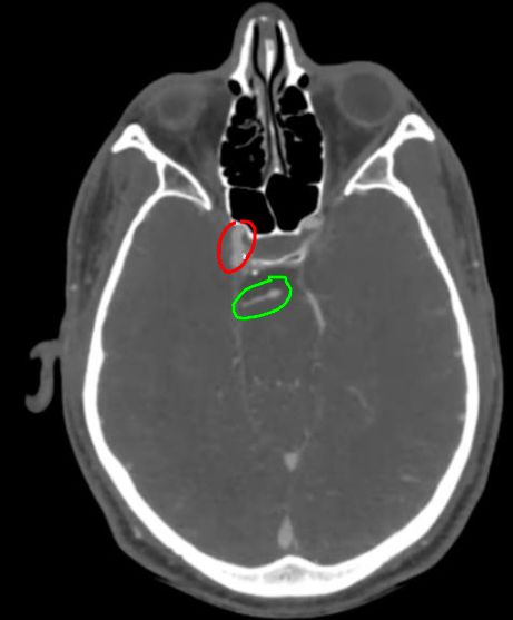
CTA (12)
  • this shows the: 
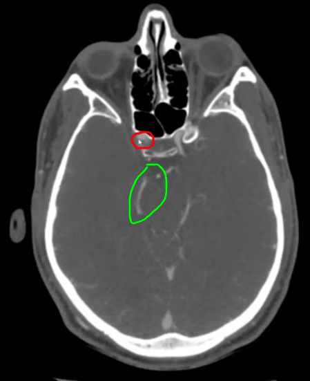
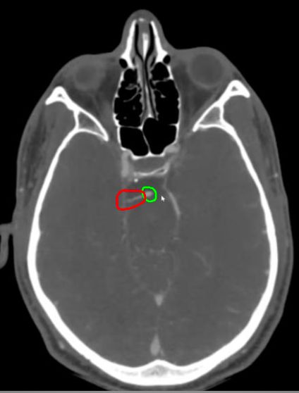
    • red: supraclinoid ICA (C5 segment)[…] becoming the ophthalmic ICA (C6 segment)[…] after giving off the ophthalmic[…] artery briefly before turning around posteriorly
    • green: PCA[…]

CTA (13)
  • this shows the: 
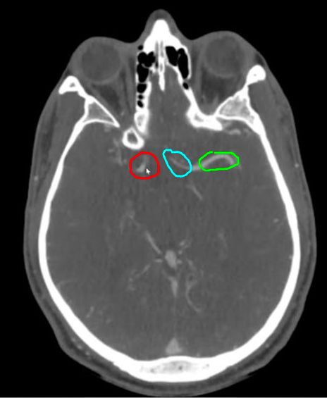
    • red: tICA aka the communicating ICA (C7 segment)[…] bifurcating into the 
      • green: M1 MCA[…]
      • blue: A1 ACA[…]

CTA (14)
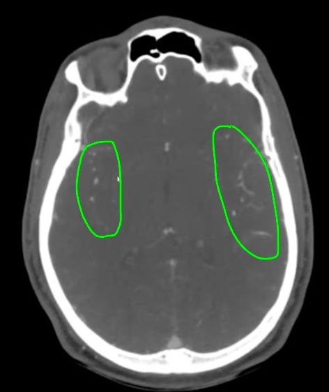
  • these are the MCA[…] branches traveling through the sylvian fissure[…]

CTA (16)
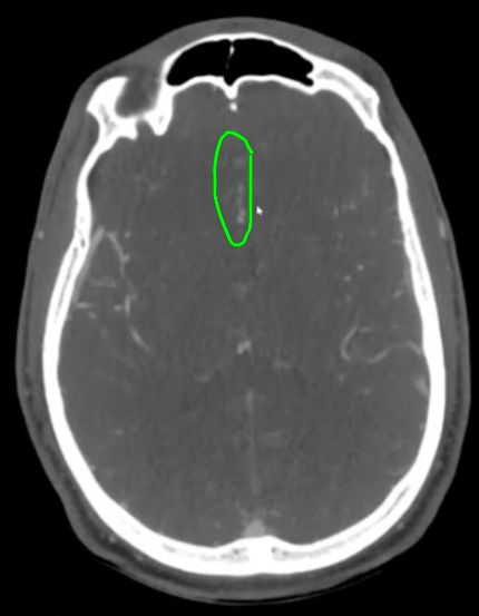
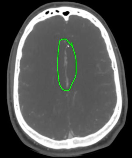
  • these are the branches of the ACA[…] traveling through the falx cerebri[…]

CTA (16)
  • these are the branches of the ACA[…] traveling through the falx cerebri[…]

CTA (17)
  • this first branch off the vert (intracranial segment)[…] (red) is the PICA[…] (green)

CTA (18)
  • this 2nd branch off the vert (intracranial segment)[…] (red) or first branch off the basilar[…] (not in this case) is the AICA[…] (green)

CTA (19)
  • this 2nd to last branch of the basilar[…] artery (green) is the SCA[…] (red)

CTA (20)
  • the basilar[…] artery finally bifurcates into the PCAs[…] (red)

CTA (21)
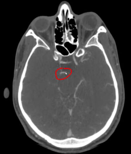
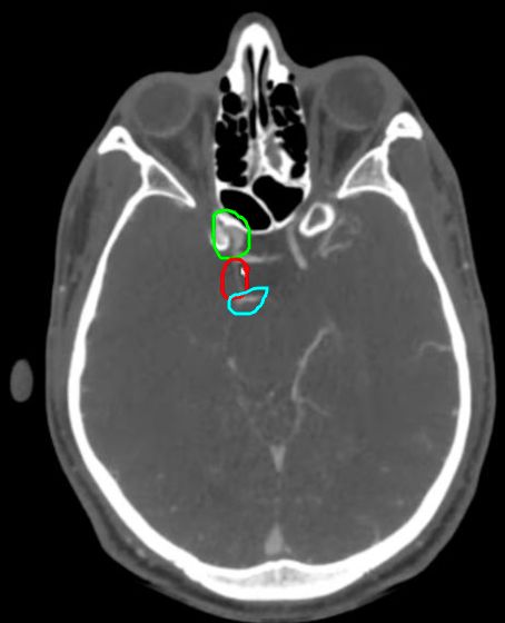
  • the vessel connecting the ICA[…] (green) to the PCA[…] (blue) is the Pcomm[…] (red)

Basilar artery fenestration[…] is the most common congenital anomaly of the basilar[…] artery, resulting from failed fusion of paired longitudinal neural[…] arteries, and may be associated with vertebrobasilar junction aneurysms[…]
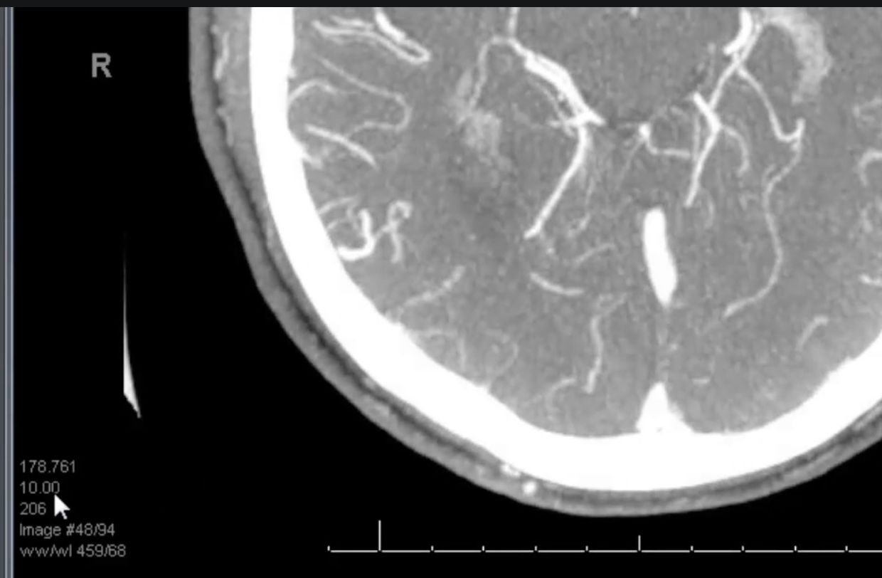
MIP stands for Maximum Intensity Projection[…] reconstructed image (recontructs the 2D w/multiple slices > into 3D slabs) and is very helpful for a 1st-pass view to rule out LVOs before confirming on the source[…] images; slice thickness[…] shown in bottom left (here it's 10mm)