EMG

EMG

Radiology · 24 cards · 1 labeled figure

In AIDP, increased CMAP duration is due to temporal dispersion[…] from nerves losing their insulation, so signals arrive at muscles at different times and the electrical signal gets stretched out. Low temperature[…] just slows everything but doesn’t cause temporal dispersion[…].
  • CMAP[…] is the electrical sum of many muscle fibers summed up
  • Delay at the NMJ[…] affects onset latency[…], not duration (delays start time for the signal) 
EMG - Explain the following terms
  • Motor Nerve
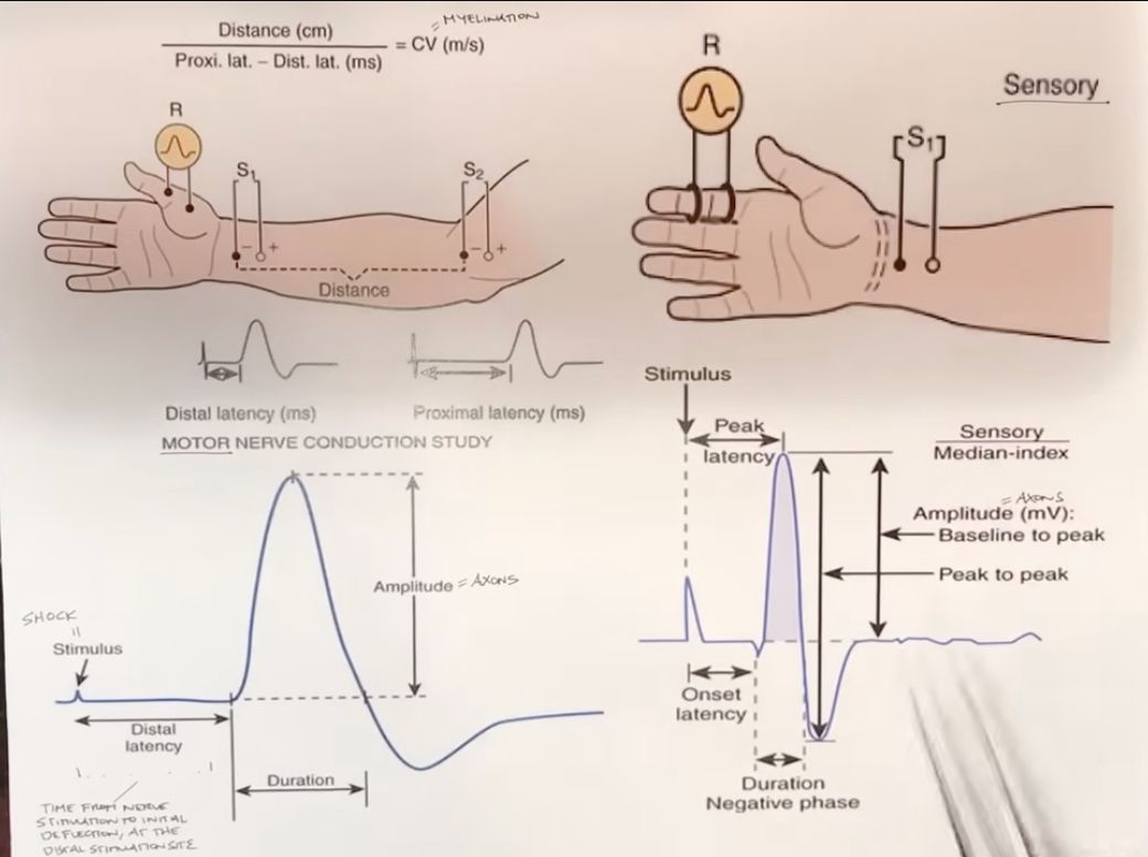
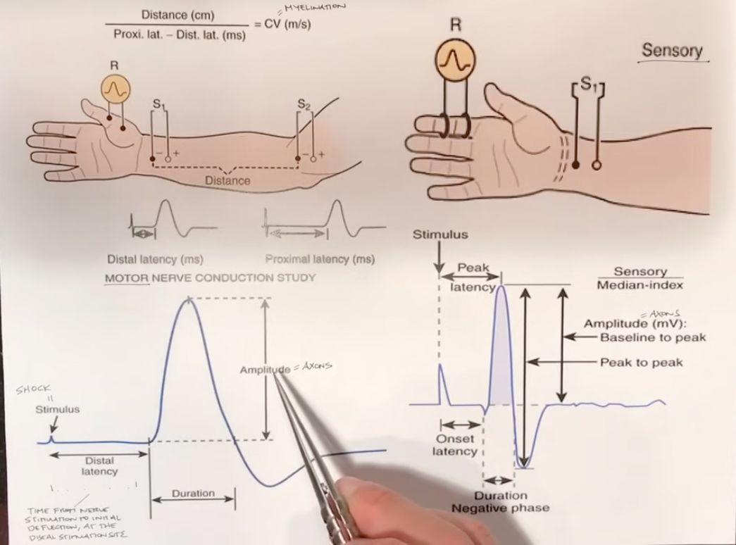
    • distal latency[…]: time from shock/stimulus till electricity initially arrives at the muscle (NMJ)[…] where your recording electrode[…] is located
      • ex) for median[…] motor nerve, this would be the abductor pollicis brevis[…] muscle recording electrode
      • abnormal finding: longer[…] distal latency = demyelination[…] causing conduction to propogate more slowly
    • Amplitude[…]: sums up how many axons[…] are participating in depolarization
      • the more axons[…] that participate, the higher[…] the amplitude
      • abnormal finding: decreased[…] amplitude = axonal[…] loss 
    • Duration[…]: how fast/slow the axons[…] depolarize
      • abnormal finding: increased[…] duration = axonal[…] loss
EMG - Explain the following terms
  • Sensory Nerve
    • unlike motor nerve recordings you only look at 2[…] points because you eliminate the need for measurement at an NMJ[…]
    • Onset Latency[…]: time from shock/stimulus to the sensory electrode pickup (similar to distal latency[…] in cases of motor nerve recording) 
      • this represents how well that nerve conducts so delayed[…] onset latency = demyelination[…]
    • Amplitude[…]: how many intact axons[…] are participating in the study
      • decreased[…] Amplitude = axonal[…] loss
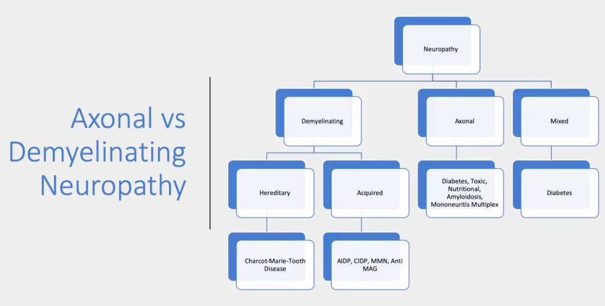
Peripheral Neuropathy Classification
  • Axonal[…]
    • normal[…] latency[…]conduction velocity[…], and F wave/H reflex[…]
    • decreased[…] amplitude[…]
    • no[…] conduction blocks[…] or temporal dispersion[…]
  • Demyelinating[…]
    • prolonged[…] latency[…] and F wave/H reflex[…]
    • normal[…] amplitude[…]
    • decreased[…] conduction velocity[…]
    • +conduction blocks[…]
    • +temporal dispersion[…]
  • Mixed[…]
Peripheral Neuropathy Classification
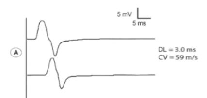
A is normal

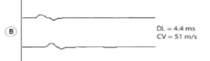
What's wrong with B?

  • preserved latency[…] and relatively preserved conduction velocity[…]
  • decreased amplitude[…]
  • this is characteristic of axonal[axonal or demyelinating?] neuropathies
Peripheral Neuropathy Classification
A is normal

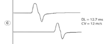
What's wrong with C?

  • preserved amplitude[…]
  • increased latency[…]
  • decreased conduction velocity[…]
  • this is characteristic of demyelinating[axonal or demyelinating?] neuropathies
Peripheral Neuropathy Classification
A is normal

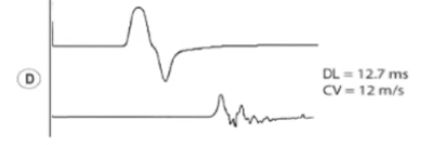
What's wrong with D?

  • decreased amplitude[…]
    • this shows conduction block[…]: reduction of CMAP[…] amplitude[…] and/or area >20[…]%
  • increased duration[…] and distal latency[…] 
      • this also shows temporal dispersion[…]: increase in CMAP[…] duration[…] >15[…]%
    • this is characteristic of acquired demyelinating[axonal or demyelinating?] neuropathies
    Peripheral Neuropathy Classification
    • Axonal Neuropathy
      • decreased amplitude[…] + conduction velocity[…] (never below 75[…]% of LLN)
        • they may lose the faster conducting fibers but they'll still preserve the slower[…] conducting fibers
      • + normal to increased distal latency[…] (never above 130[…]% of ULN)
    • Demyelinating Neuropathy
      • decreased conduction velocity[…] (<75[…]% LLN) + markedly increased distal latency[…] (>130[…]% ULN)
        • because the nerves lose myelin[…] on the faster AND slower nerves
      • + decreased amplitude[…] if conduction block[…] is present or secondary axonal loss[…] is present
        • difference between different nerve fibers can also result in temporal dispersion[…] resulting in loss of amplitude (different nerves conduct differently)
    Diabetic Neuropathy
    • predominantly axonal[axonal or demyelinating?]; can be mixed[…]
    • symmetric[asymmetric or symmetric?]length[…]-dependent pattern
    • EMG: neurogenic[…] MUAPs[…] in length[…]-dependent patterns
      • Sensory: involvement of sural[…] first > ulnar[…] > median[…]radial[…]
      • Motor: peroneal[…] first > tibial[…]ulnar[…] > median[…]
    • NCS: axonal[…] pattern
      • reduced amplitude[…]
      • preserved latencies[…]
      • length[…]-dependent pattern (distal muscles first)
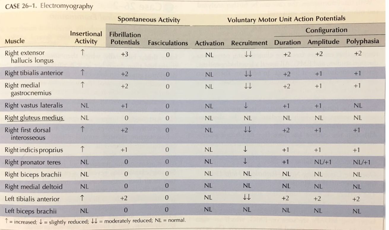
    EMG Case Example - Interpret This

    • sensory EMG
      • sural[…] sensory nerve has NR[…]
      • ulnar[…] and median[…] sensory nerves have reduced amplitudes[…], but preserved latency[…] and preserved conduction velocity[…]
    • motor EMG
      • peroneal[…] motor nerve has reduced amplitudes[…], relatively preserved latency[…]
      • tibial[…] nerve has reduced amplitude[…]
      • median[…] then ulnar[…] motor nerves: normal amplitude[…]
    • NCS
      • there is increased spontaneous activity[…] and increased voluntary MUAPs[…] in a lenght-dependent[…] pattern 
        • more in distal[…] muscles like the right extensor hallicus longus[…] in comparison to proximal[…] muscles like the right tibialis anterior[…] or right medial gastrocnemius[…] where they're a bit less or the right vastus lateralis[…] and right gluteus medius[…] which have no spontaneous activity or voluntary MUAPs
    • this is characteristic of axonal[…] neuropathy in a length[…]-dependent pattern c/f diabetic[…] neuropathy
    GBS and CIDP  
    • predominantly demyelinating[…] neuropathy
    • EFNS[…] Criteria is used to diagnose CIDP
    • NCS Changes
      • increased latencies[…]
      • decreased conduction velocity[…]
      • relatively preserved amplitude[…]
      • presence of conduction blocks[…] and temporal dispersion[…] indicates acquired etiology
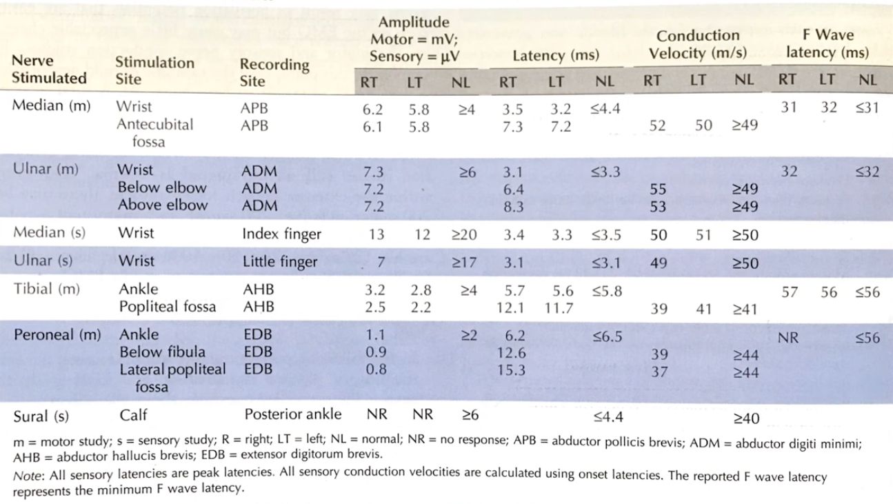
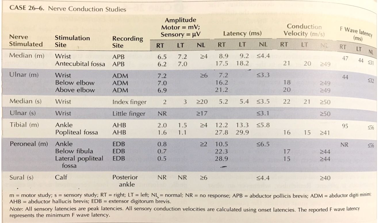
    EMG NCS Case Example - Interpret This

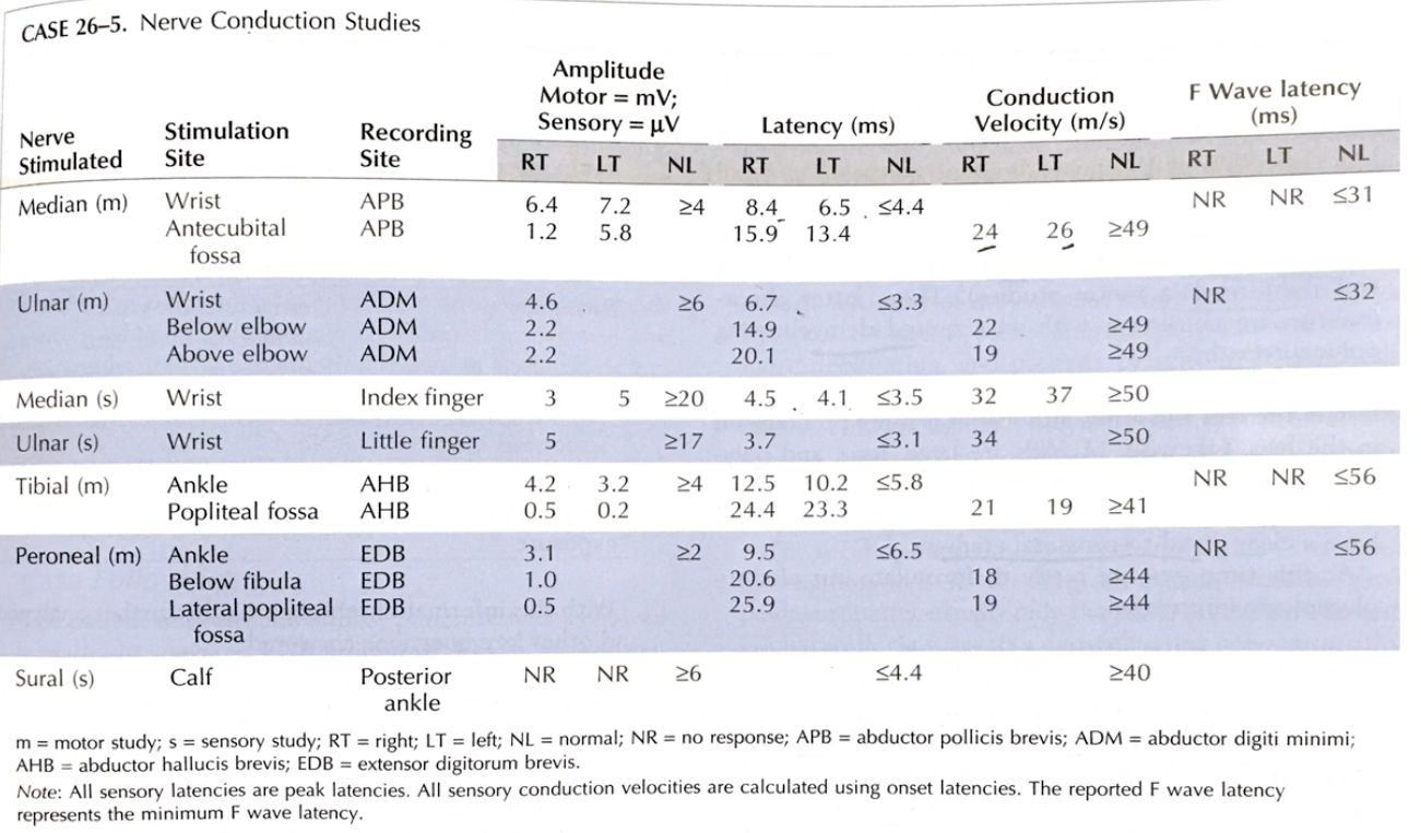
    • there is a conduction block[…] in the median[…]ulnar[…]tibial[…], and peroneal[…] nerves as evidenced by the decrease[…] in amplitude[…] from distal[…] to proximal[…]
    • latency[…] is prolonged[…]
    • conduction velocity[…] is reduced[…]
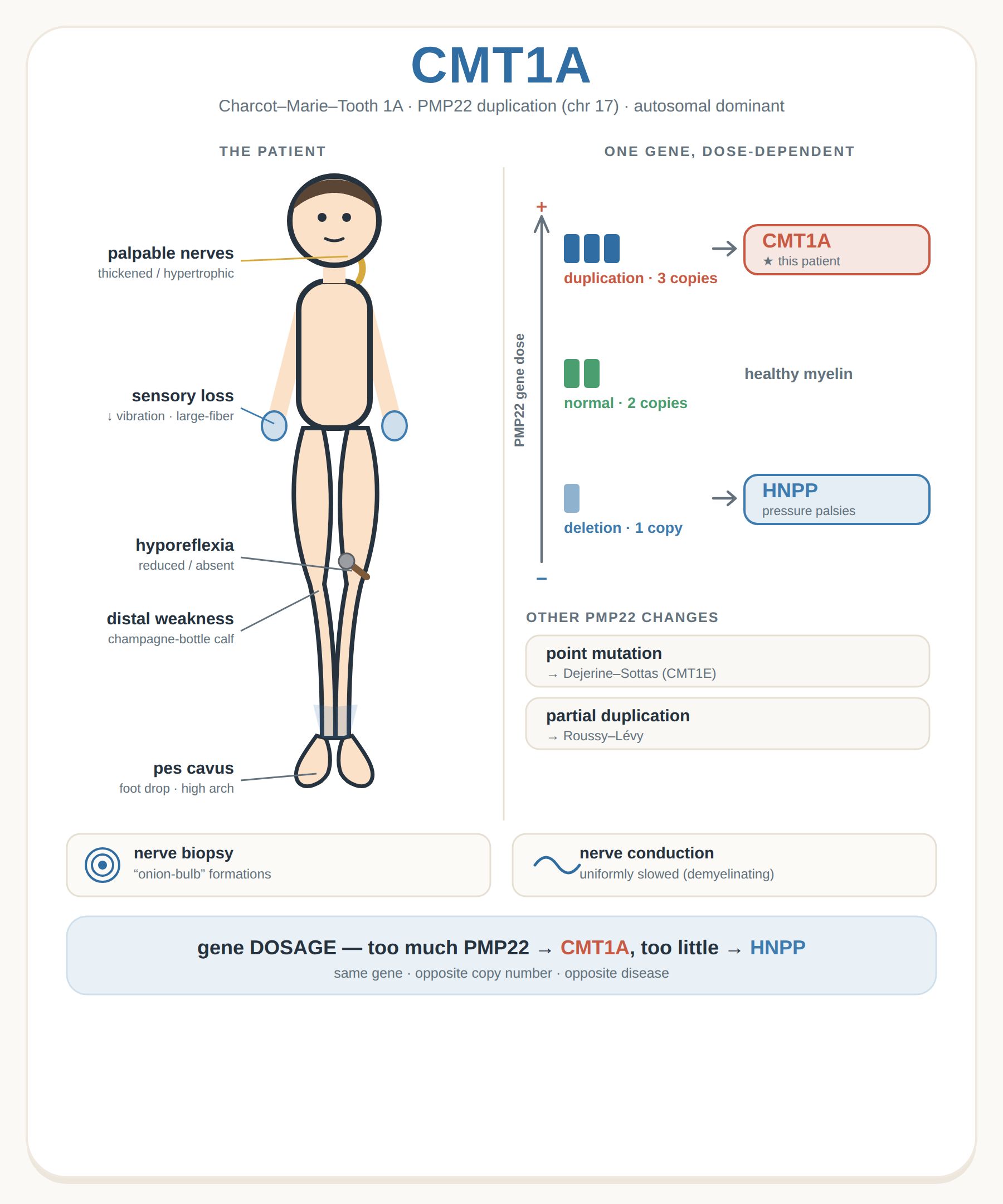
    Charcot Marie Tooth Disease (CMT)
    • this is an inherited demyelinating[…] neuropathy except a few axonal[…] forms such as CMT2
    • this will present with uniform demyelination[…] unlike acquired[…] demyelinating neuropathies which have a patchy[…] pattern
    • NCS
      • increased latencies[…]
      • decreased conduction velocity[…]
      • relatively preserved amplitudes[…]
      • since it is hereditary: you will typically NOT see conduction blocks[…] and temporal dispersion[…]
    EMG NCS Case Example - Interpret This

    • prolonged latencies[…]
    • decreased conduction velocities[…]
    • relatively preserved amplitudes[…]
    • this supports a demyelinating[…] neuropathy
      • there are no conduction blocks[…] (decrease in amplitude>20[…]%) which tells us that this is NOT an acquired[…] subtype of this neuropathy
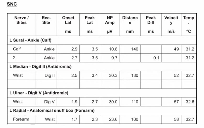
    When Interpreting EMG sensory component

    • SNAP (sensory nerve action potential)[…] shown first
      • usually in the LE it's tested in the sural[…] nerve
      • usually in the UE it's tested in the median[…]ulnar[…], and radial[…] nerves
    • first you look at peak latency[…] which is when the stimulus[…] is first given to the point the amplitude[…] peaks[…] called the peak latency[…]
    • next you look at the amplitude[…] itself
    • finally you check the conduction velocity[…]distance[…]/onset latency[…]; note that this latency is the time from stimulus to response first being felt[…] and not the same as the peak latency[…] which we look at for its own stand-alone value
    • note: the temperature[…] of the skin can affect these recordings
    When Interpreting EMG motor component

    • CMAP (compound muscle action potential)[…] shown first
      • typically in the LE this is the peroneal[…] and tibial[…] motor nerves
      • typically in the UE this is the median[…] and ulnar[…] motor nerves
    • first you look at distal latency[…] which is when the stimulus[…] is first given to the point the G1[…] recording is felt
      • note this is similar in concept but different in name compared to the onset latency[…] with SNAPs
    • second look at the amplitude[…] and ensure it does not drop from distal[…] to proximal[…] by more than 20[…]% (which is called conduction block[…])
    When Interpreting EMG needling component

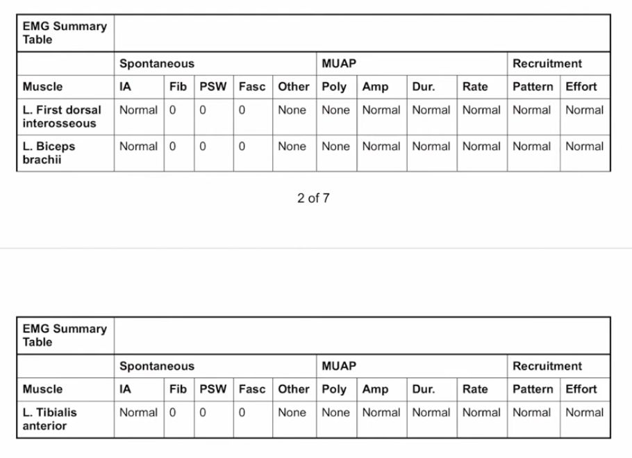
    • First you look at the spontaneous[…] activity of the muscle[…] which is the activity of the muscle[…] while at rest[…]
      • IA[…]insertional activity[…]
      • then keep the needle in for few secs: abnormal findings include presence of Fibrillations[…]Positive Sharp Waves (PSW)[…]Fasciculations[…], or Other[…] findings like myokymia
    • Second you ask the patient to activate[…] the muscle and look at the morphology[…] of the MUAP[…]
      • abnormal findings include that it is polyphasic[…], whether the amplitude[…] is small or larger, the duration[…], the firing rate[…]
    • Third you look for Recruitment[…] 
      • in neuropathy you will see this decreased[…] bc the nerve[…] is injured and cannot recruit muscle motor[…] units so it has to fire more[…] (increased rate) 
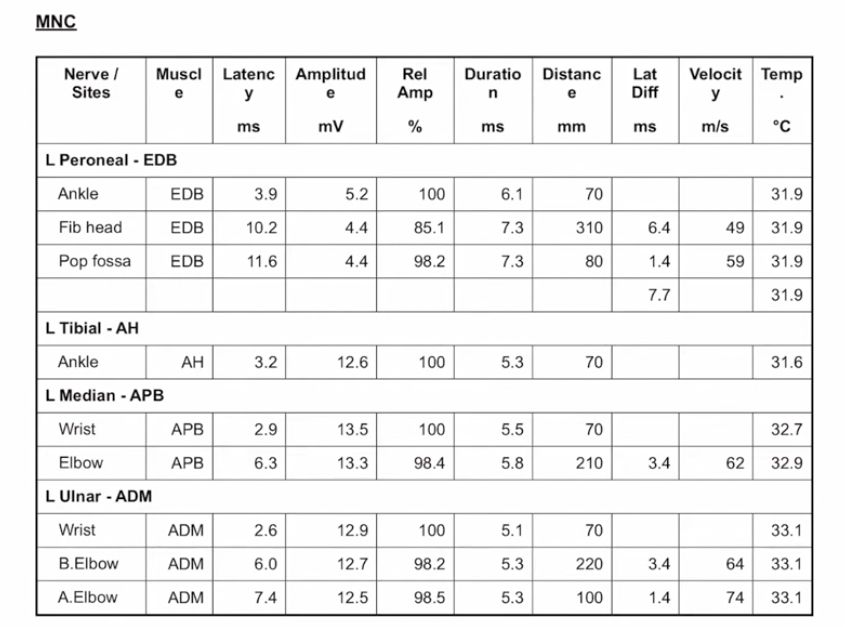
    Easily remember ENG/NCS Abnormal Findings
    • Onset/Peak Latency[…] (SNAP) and Distal Latency[…] (MUAP)
      • SNAP UE: Ulnar[…] & Radial[…] & Median[…] >2-3.5[…]
      • MUAP UEUlnar (FDI)[…]Ulnar (ADM)[…]Median[…] >3-4.5[…]
      • SNAP LE: Sural[…] & Superficial Peroneal[…] >4-4.5[…]
      • MUAP LE: Peroneal (EDB)[…]Peroneal (TA)[…]Tibial[…]Femoral[…] >4-6.5[…]
    • Amplitude[…]
      • SNAP UE: Ulnar[…] & Radial[…] & Median[…]<10-15[…]
      • MUAP UEUlnar (FDI)[…]Ulnar (ADM)[…]Median[…]<6-7[…]
      • SNAP LESural[…]Superficial Peroneal[…]<5[…]
      • MUAP LE: Peroneal (EDB)[…]Peroneal (TA)[…]Tibial[…]Femoral[…]<2.5-4[…]
    • CV[…]
      • SNAP UE: Median[…] & Ulnar[…] & Radial[…] <50[…]
      • MUAP UE: Ulnar (FDI)[…]Ulnar (ADM)[…]Median[…]<50[…]
      • SNAP LE: Sural[…] & Superficial Peroneal[…] <40[…]
      • MUAP LE: Peroneal (EDB)[…]Peroneal (TA)[…]Tibial[…]Femoral[…]<40[…]

    extensor digitorum brevis (EDB)
    tibialis anterior (TA)
    First Dorsal Interossei (FDI)
    Abductor Digiti Minimi (ADM)