Vascular / Heme

Vascular / Heme

Anatomy · 19 cards

Circle of Willis Anatomy Overview
  • The Circle of Willis is formed anteriorly by the anterior communicating artery (AComm)[…] connecting the two ACAs, and posteriorly by the posterior communicating arteries (PComm)[…] connecting each ICA to the corresponding PCA.
  • Anterior circulation = ICA → MCA + ACA (supplies ~80% of cerebrum).
  • Posterior circulation = 2 vertebral arteries[…] → 1 basilar artery → 2 PCAs (supplies brainstem, cerebellum, occipital lobes, and medial temporal).
  • Fetal PCA[…] variant: PCA arises from the ICA rather than the vertebrobasilar system — important for embolic stroke source determination.
ACA Stroke :: Localization

ACA supplies the medial frontal and parietal cortices.

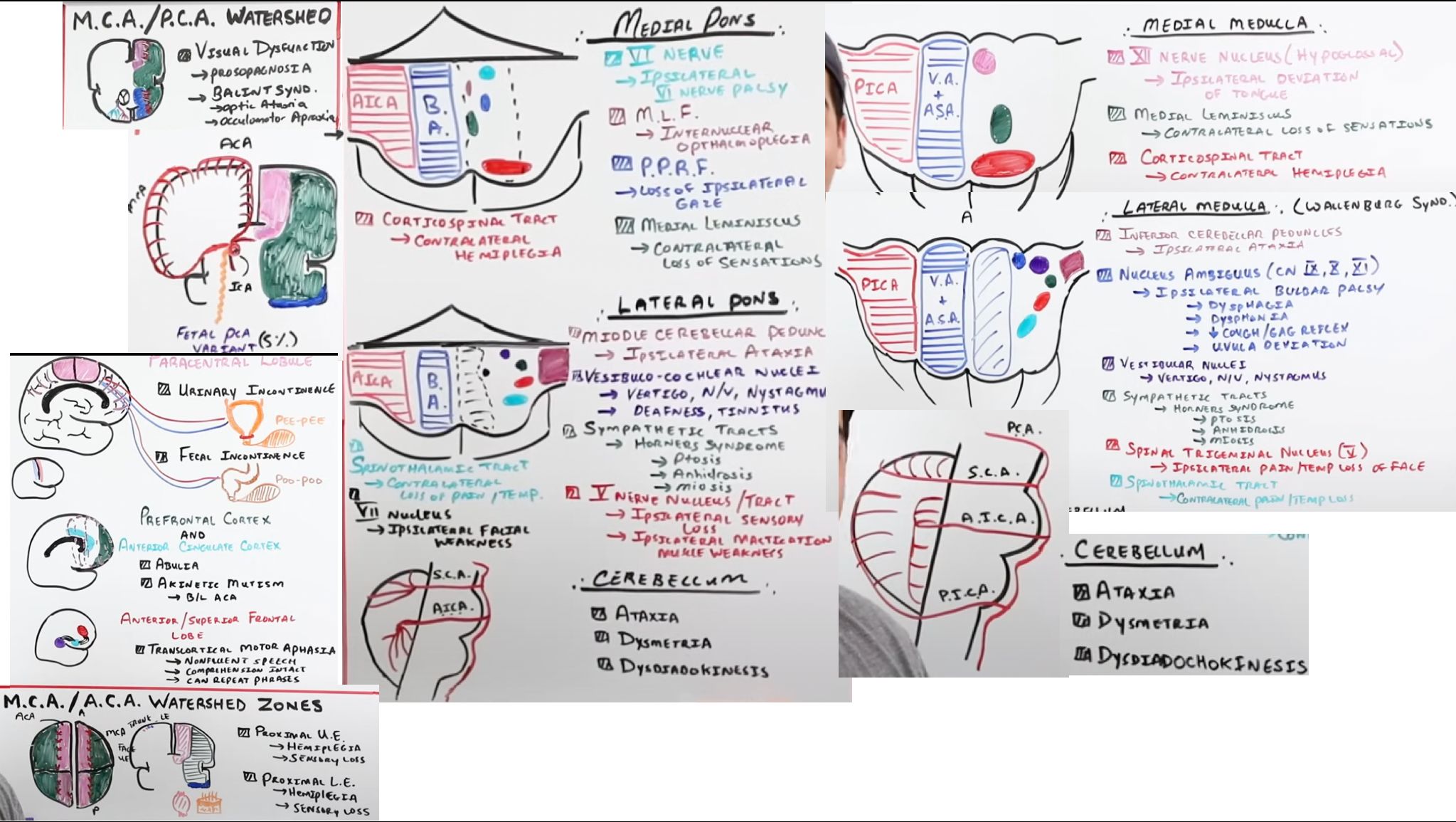
Knockout of the paracentral lobule[…] (motor/sensory leg area) → contralateral lower-extremity weakness/sensory loss, plus urinary and bowel incontinence[…].

Knockout of the prefrontal cortex (PFC)[…] or anterior cingulate cortex (ACC) → abulia[…] (apathy/lack of motivation).

Bilateral[…] ACA territory infarcts (e.g., from an AComm aneurysm rupture or single A1 supplying both) → akinetic mutism — much worse than unilateral.
MCA Stroke :: Dominant and Nondominant Localization

MCA supplies the lateral cortex (face/arm area), Broca's, Wernicke's, and most of the basal ganglia via lenticulostriate branches.

Dominant (usually LEFT) MCA stroke:
- Broca's aphasia[…] (superior division) — non-fluent, expressive.
- Wernicke's aphasia[…] (inferior division) — fluent, receptive.
- Gerstmann syndrome[…] from inferior parietal lobule (agraphia, acalculia, finger agnosia, L-R disorientation).

Nondominant (usually RIGHT) MCA stroke:
- Apraxias[…]: ideomotor (can't perform motor command), eyelid apraxia (can't open eyes despite intact motor), ideational apraxia (can't sequence tool use).
- Hemineglect[…] (visual or sensory) of the contralateral side.
- Anosognosia (denial of deficit).

Either side: contralateral face/arm weakness > leg (face/arm somatotopy on the lateral cortex).
PCA Stroke :: Syndromes

PCA supplies the occipital lobe, medial temporal lobe (hippocampus), thalamus, and midbrain.

Cortical PCA stroke: contralateral homonymous hemianopia with macular sparing[…] (collateral MCA supply to the macular cortex).

Bilateral PCA stroke: cortical blindness (Anton syndrome)[…] — patients deny their blindness.

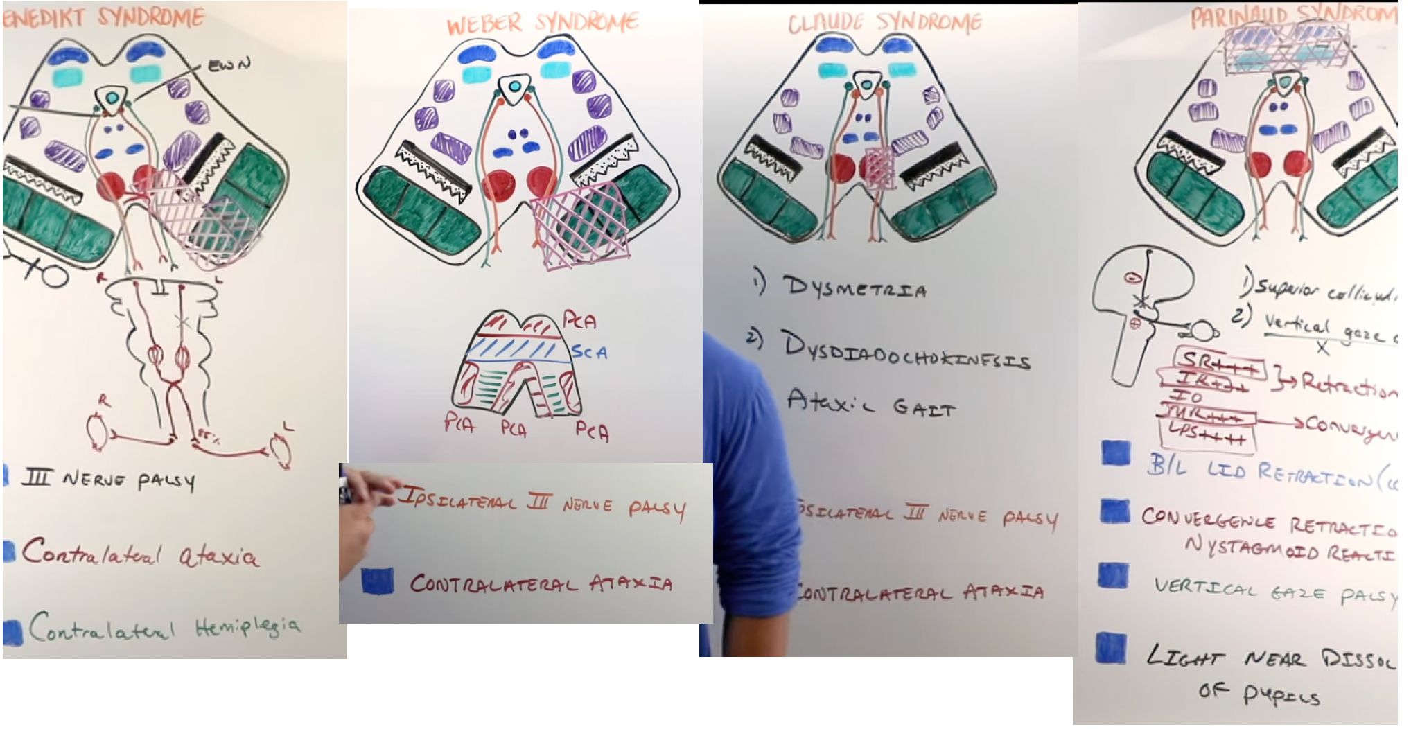
Thalamic/midbrain PCA stroke: brainstem syndromes — Weber[…] (CN III + contralateral hemiparesis), Claude[…] (CN III + contralateral ataxia), and Benedikt[…] (CN III + red nucleus tremor + sensory loss).
Watershed Strokes :: Localization

Watershed (border-zone) strokes occur at the boundaries between major arterial territories, typically from hypotension/hypoperfusion[…] rather than embolic occlusion.

ACA/MCA watershed[…]: "man in a barrel" syndrome — proximal weakness and sensory loss in both upper extremities (homunculus shoulders sit at this watershed).

MCA/PCA watershed[…] (parieto-occipital): prosopagnosia[…] (face blindness) and Balint syndrome[…] (simultanagnosia, oculomotor apraxia, optic ataxia).
Posterior Circulation :: Brainstem Blood Supply

Two vertebral arteries[…] ascend along the anterior medulla; at the pontomedullary junction[…] they fuse to form the basilar artery[…], which courses along the anterior pons and midbrain before bifurcating into the two PCAs.

Vertebral branches: ASA[…] (anterior spinal artery), PICA[…] (posterior inferior cerebellar artery → inferior cerebellum + lateral medulla), PSA.

Basilar branches: paramedian, short circumferential, and long circumferential[…] branches; AICA[…] (anterior inferior cerebellar artery → lateral pons + anterior inferior cerebellum); SCA[…] (superior cerebellar artery → superior cerebellum + lateral midbrain).
Posterior Circulation :: Medial Pontine Syndrome

Occlusion of paramedian basilar branches[…] → medial pontine syndrome:

- At CN VI nucleus[…] → ipsilateral horizontal gaze palsy.
- At MLF[…] → INO (impaired adduction with abducting nystagmus of the other eye).
- At PPRF[…] → contralateral horizontal gaze palsy.
- At medial lemniscus[…] → contralateral loss of vibration/proprioception/discriminative touch.
- At CST[…] → contralateral hemiparesis.

Classic combination: Millard-Gubler syndrome[…] (ventral pontine) = ipsilateral CN VI + CN VII palsy + contralateral hemiparesis.
Posterior Circulation: AICA and PICA Syndromes

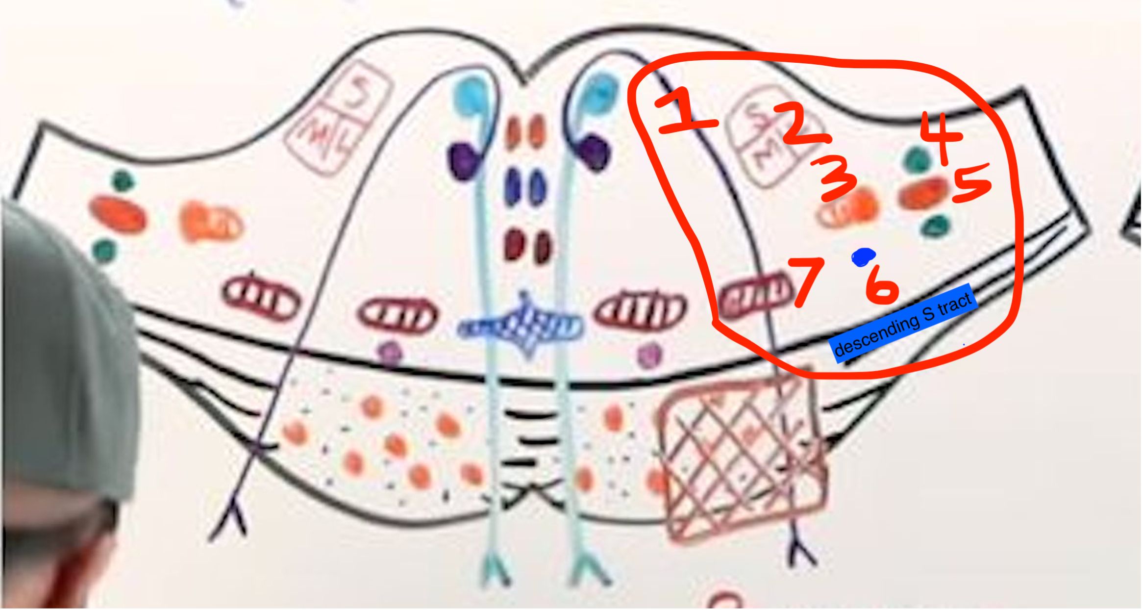
  • AICA[…] occlusionLateral Pontine[…] Syndrome:
    1. CN VII Motor[…] fibers → ipsilateral facial[…] weakness (LMN pattern)
    2. CN VIII Sup/M/Lat Vestibular Nuclei[…] → ipsilateral vertigo[…]/n/v[…]/ataxia[…]
    3. Spinal[…] nucleus & tract of CN V[…] ipsi[…]lateral facial pain/T loss
    4. Dorsal/Ventral Cochlear Nuclei[…] → ipsilateral hearing[…] loss 
    5. Middle Cerebellar Peduncle[…] → ipsilateral ataxia[…]
    6. S[…] tract → ipsilateral Horner[…] syndrome
    7. Spinothalamic Tract / Spinal Lemniscus[…] → contra[…]lateral body pain/T loss
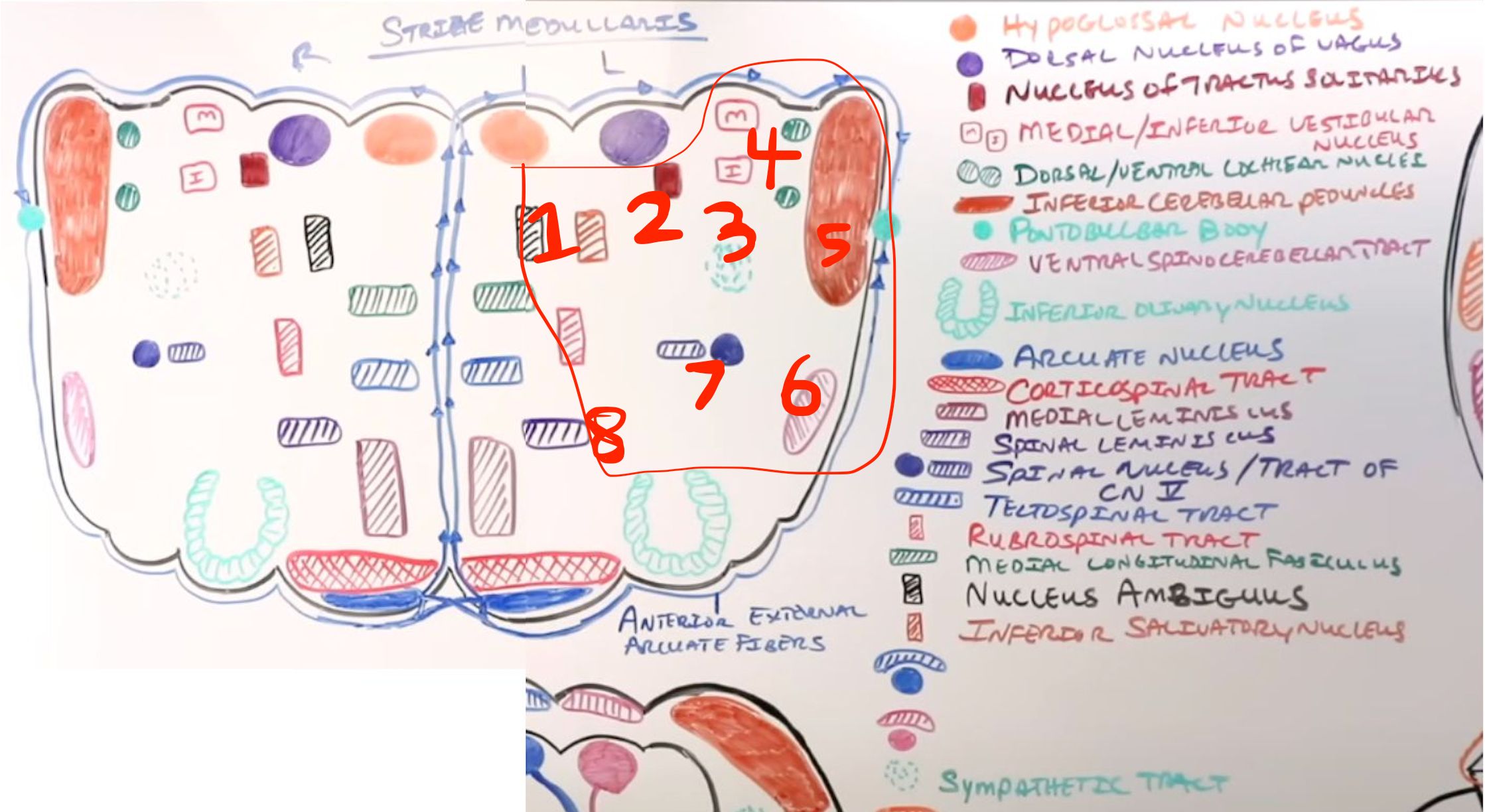
  • PICA[…] occlusion → Lateral Medullary (Wallenberg)[…] syndrome
    • similar crossed pattern as AICA but major difference is involvement of nuclei that canuse dysphagia/dysphonia (nucleus ambiguus)[…] instead of facial weakness/more hearing loss

Midbrain Stroke Syndromes (PCA Territory)

Vascular supply: PCA[…] → tectum + ~75% of anterior tegmentum; PComm[…] → ~25% of anterior tegmentum; SCA[…] → midline portion.

Weber syndrome[…]: at CN III + crus cerebri → ipsilateral CN III palsy (down/out eye, dilated pupil, ptosis) + contralateral hemiparesis (CST/CBT involvement).

Claude syndrome[…]: at CN III + red nucleus + SCP → ipsilateral CN III palsy + contralateral ataxia (loss of cerebellar output via SCP).

Benedikt syndrome[…]: at CN III + red nucleus + medial lemniscus → ipsilateral CN III palsy + contralateral ataxia + contralateral sensory loss (DCML).

Bilateral occipital infarct (top of basilar) → cortical blindness[…].
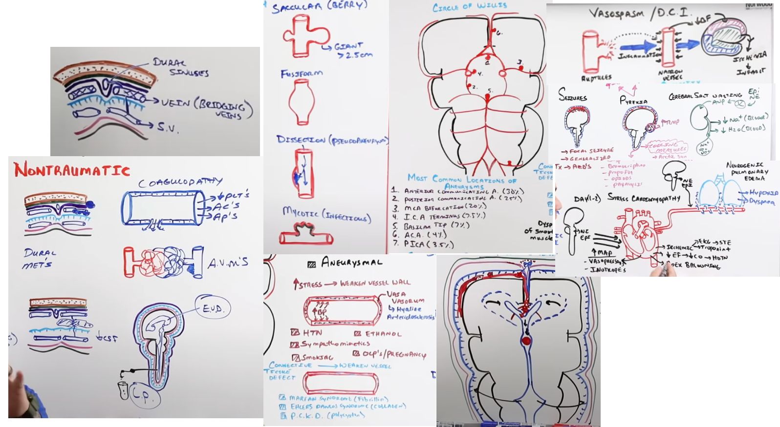
Meninges :: Anatomy

Layers (superficial to deep): dura, arachnoid, pia[…].

Dura mater[…] has an outer periosteal[…] layer and an inner meningeal[…] layer; the two separate to form venous sinuses[…]. Inward dural folds include the falx cerebri[…] (separates hemispheres) and tentorium cerebelli[…] (separates supra- from infratentorial structures).

Arachnoid mater[…] has trabeculae and arachnoid granulations (villi)[…] that drain CSF into the dural sinuses.

Pia mater[…] ("soft mother") is adherent to the brain surface and follows every sulcus/gyrus.

The subarachnoid space[…] contains CSF and the cerebral arteries — site of SAH.
Intracerebral Hemorrhage (ICH)

ICH is parenchymal[…] bleeding into brain tissue.

Common arterial sources by location: lenticulostriate[…] arteries (basal ganglia/putamen), thalamogeniculate[…] arteries (thalamus), paramedian pontine[…] branches (pons), and SCA/AICA/PICA (cerebellum).

Major causes: hypertension[…] (most common — deep ICH), cerebral amyloid angiopathy (CAA)[…] (lobar ICH in elderly), coagulopathy, hemorrhagic transformation of ischemic stroke, malignancy metastases, CVST[…], AVM, and mycotic aneurysms.

Brain mets that classically hemorrhage: melanoma, papillary thyroid, small-cell lung, renal cell, and choriocarcinoma[…].

Contraindicated procedure: lumbar puncture[…] (risk of herniation if mass effect).
Subarachnoid Hemorrhage

  • SAH most commonly results from rupture of a saccular (berry)[…] aneurysm; other aneurysm types include fusiform, dissection, and mycotic.
  • Most common berry aneurysm sites (7): AComm > PComm > MCA bifurcation > ICA terminus > Basilar tip > ACA > PICA[…] 
  • Risk factors: hypertension[…], sympathomimetics, smoking, EtOH, OCPs.
  • Connective tissue diseases associated with aneurysms and implicated protein (3): Marfan (fibrillin), Ehlers-Danlos (collagen), and ADPKD (polycystin)[…].
  • Other causes: fibromuscular dysplasia[…] and septic emboli from infective endocarditis.
SAH :: Diagnosis, Grading, and Complications

Diagnosis: CT head shows blood in sulci or basal cisterns[…]. If CT is negative within 6 hours and suspicion remains, perform LP for xanthochromia and opening pressure[…].

Grading scales: Hunt-Hess[…] score grades mortality risk; modified Fisher[…] score grades vasospasm risk based on clot thickness and IVH presence.

Complications:
- Obstructive hydrocephalus[…] from IVH; later, communicating hydrocephalus[…] from arachnoid granulation impairment.
- Rebleeding[…] (highest in first 24 hrs).
- Vasospasm[…] typically days 4-14[…] → delayed cerebral ischemia.
- Hypothalamic pyrexia, catecholamine surge → Takotsubo[…] cardiomyopathy, neurogenic pulmonary edema.
- ANP release blocks ALD and ADH → hyponatremia and hypovolemia[…] (cerebral salt wasting).
SAH :: Treatment and ICU Management

Definitive treatment: secure the aneurysm by endovascular coiling[…] or surgical clipping[…] (or flow diversion for complex aneurysms).

Rebleed prophylaxis (before securing): aminocaproic acid or TXA[…] (antifibrinolytics).

Vasospasm prevention: nimodipine[…] for all SAH patients × 21 days.

Vasospasm/DCI treatment: induced hypertension[…] with pressors; endovascular angioplasty/intraarterial vasodilators if refractory.

Hydrocephalus: CSF diversion via EVD[…], possible eventual VP shunt.

Fever (pyrexia) management: Arctic Sun[…] surface cooling, Zoll[…] intravascular catheter, or cold normal saline infusions.

Shivering: magnesium, buspirone, bromocriptine[…]; escalate to sedation, opioids, or paralytics if needed.
Subdural Hemorrhage (SDH)

SDH = rupture of bridging veins[…] between the cerebral cortex and dural sinuses.

Imaging: crescent-shaped[…] blood collection that CROSSES suture lines but is bounded by dural reflections[…] (falx, tentorium).

Causes: trauma (especially in elderly with cerebral atrophy or EtOH), infection, coagulopathy, AVM, dural metastases, and intracranial hypotension (SIH)[…].

Mimic: subdural hygroma[…] (CSF collection from arachnoid tear, often after CSF leak repair).

Stages (time course):
- Acute = fresh blood, hyperdense[…] on CT (days).
- Subacute = clotted blood + exudate, isodense (weeks) — easily missed on CT.
- Chronic = clotted blood + exudate + neocapillaries[…], hypodense (weeks-months) — prone to rebleed.
Epidural Hemorrhage (EDH)

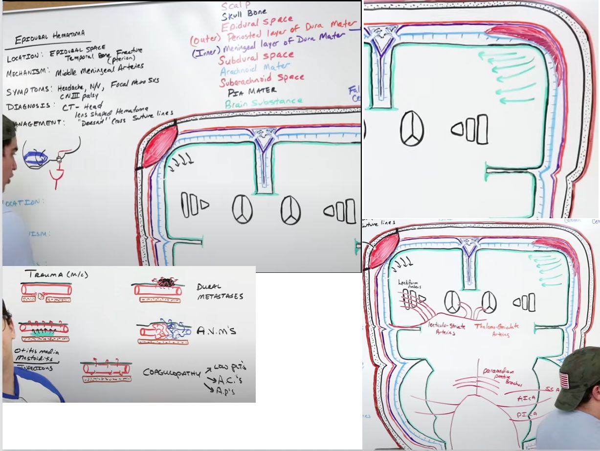
EDH most commonly results from laceration of the middle meningeal artery[…] at the pterion[…] (thinnest part of the skull, where temporal/parietal/frontal/sphenoid bones meet).

Can also be venous (sinus injury) or spinal in origin.

Imaging: biconvex (lentiform)[…] blood collection that is LIMITED BY sutures[…] (dura is fused to sutures) but can cross dural reflections[…].

Classic clinical course: head trauma → brief LOC → lucid interval[…] → rapid decline as the hematoma expands.

Causes besides trauma: infection, coagulopathy, AVM, and metastases.

Treatment: urgent surgical craniotomy[…] for evacuation in symptomatic or large hematomas.
Herniation Syndromes :: General Signs and Subfalcine

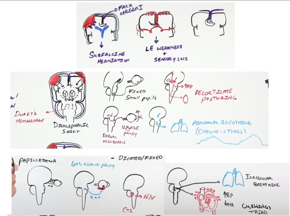
General signs of elevated ICP with herniation:
- Optic nerve compression → papilledema[…].
- CN VI[…] compression (long intracranial course) → palsy with impaired abduction (false localizing sign).
- Chemoreceptor trigger zone near area postrema[…] → nausea/vomiting.
- Brainstem CV/respiratory compression → Cushing reflex[…] (HTN, bradycardia, irregular respirations).

Subfalcine herniation[…]: the cingulate gyrus herniates under the falx cerebri[…].
- Can compress the contralateral ACA[…] → ACA territory infarct.
Herniation :: Transtentorial (Downward Central)

Downward (central) transtentorial herniation[…]: BILATERAL herniation through the tentorial notch.

Features:
- Shearing of pontine perforating arteries[…]Duret hemorrhages[…] (often fatal).
- Sympathetic tract compression → fixed, small (pinpoint)[…] pupils.
- Dorsal midbrain compression → upgaze palsy (Parinaud-like)[…].
- Loss of cortical inhibition of the red nucleus → BUE flexion = decorticate posturing[…].
- Loss of cortical and red nucleus input → BUE/BLE extension = decerebrate posturing[…] (worse).
- Cheyne-Stokes[…] respirations.
Herniation :: Uncal (Transtentorial)

Uncal herniation[…]: UNILATERAL — the uncus of the medial temporal lobe herniates under the tentorium cerebelli.

Features:
- CN III[…] compression → ipsilateral pupil dilation FIRST (parasympathetic fibers run superficially), then down-and-out eye with ptosis (motor fibers later).
- Cerebral peduncle[…] compression → contralateral hemiparesis.
- PCA[…] compression → contralateral homonymous hemianopia.
- Kernohan's notch phenomenon[…]: contralateral peduncle pushed against the tentorial edge → ipsilateral (false localizing) hemiparesis with contralateral CN III palsy.

Tonsillar herniation[…]: cerebellar tonsils herniate through the foramen magnum → compression of the medulla → respiratory arrest and death.