CTP

CTP

Radiology · 19 cards

CTP Basics
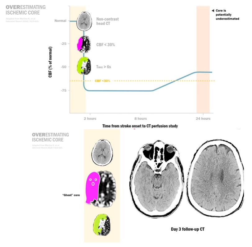
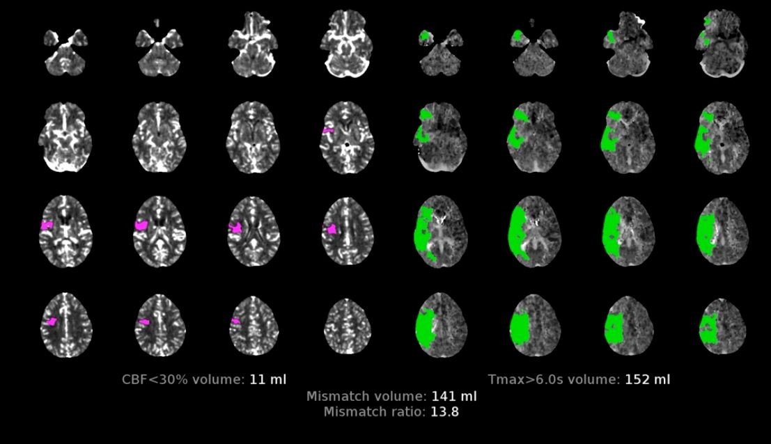
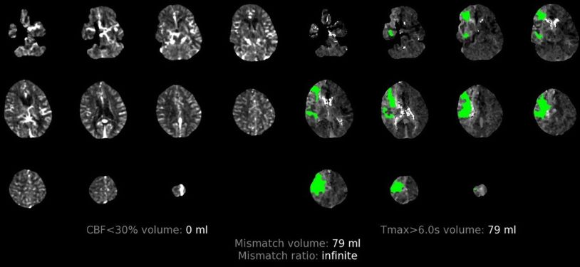
  • CBF<30[…]%: this is defined as the volume of brain tissue with calculated CBF[…] less than 30[…]% compared to the contralateral[…] hemisphere, purple[…] color, corresponding to infarct core[…] = area of irreversible[…] tissue damage
  • Tmax>6[…]s: this is defined as the volume of brain tissue with greater than 6[…]s delay in the arrival of dye bolus, green[…] color, corresponding to the infarct penumbra[…] = area of reversible[…] tissue damage

CTP - Rationale for Use
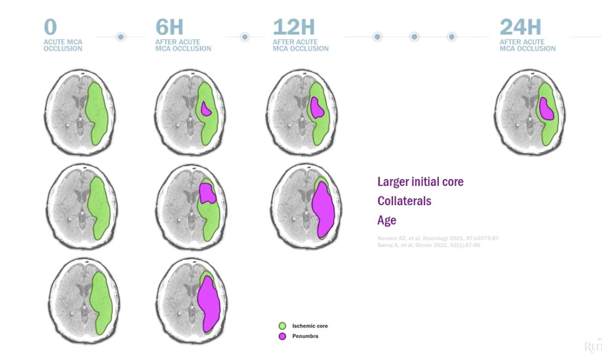
  • The rate of progression of ischemic penumbra to core is highly variable and depends on:
    • size[…] of initial core
    • collaterals[…]
    • age[…]
CTP Parameters
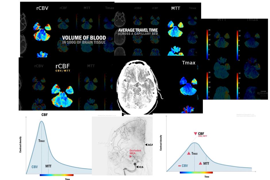
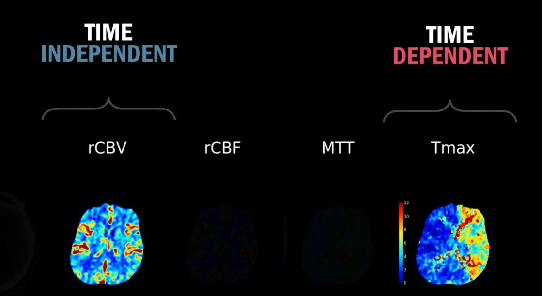
  • rCBV[…]: volume[…] of blood flowing in 100g of brain tissue - an indirect measure of autoregulation[…] and collateralization[…] (the amount of blood that can be delivered to brain tissue)
    • this does not[…] change in LVO because collateralization[…] ensures blood reaches the final target through alternate route 
  • MTT (mean transit time)[…]: average time the contrast spends inside the BV[…]s as measured by the time it takes for blood to travel from intracranial arteries to veins across a capillary bed[…] -- 4[…]-5[…]s is wnl and this increases[…] in LVO because blood reaches the final target through alternate collateral route
  • rCBF[…]: basically the rCBV[…] / 100g brain tissue per minute (aka the time it takes to deliver that volume) = rCBV[…]/MTT[…]
    • it predicts the core[…] infarction relative to the healthy contralateral hemisphere
    • this decreases[…] in LVO because blood reaches the final target through alternate collateral route > increasing the MTT/Tmax which would reduce[…] the volume delivered over a period of time
  • Tmax[…]: predicts the time from start of scan to time to maximum contrast[…] bolus in the brain (delay correlates with penumbra[…])
    • this increases[…] in LVO because blood reaches the final target through alternate collateral route
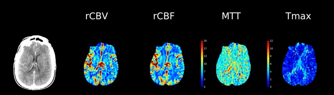
  • Color Schema
    • low values are in blue[…]
    • intermediate values are in green[…]-yellow[…] 
    • high values are in red[…] 
    • colors vary more at baseline with rCBV[…] sequence because different brain tissue types receive different blood flow at rest
    • colors do not vary at baseline with Tmax[…] making it helpful to use to differentiate colors with even smalle
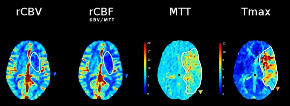
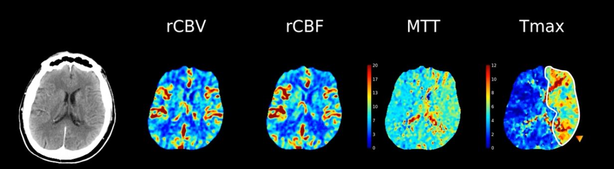
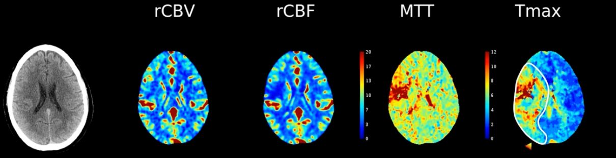
Use the color schema alone to interpret the different values on this CTP in the encircled territories of a patient with L MCA ischemic stroke

  • rCBV: darker blue = lower volume delivered[…]
  • rCBF = rCBV/MTT: darker blue = lower blood flow/time[…]
  • MTT: green/yellow = increased transit time[…]
  • TMax: red/yellow/green = increased TMax[…]
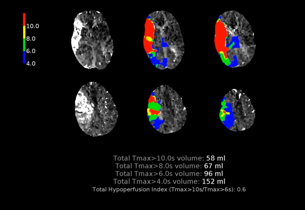
Describe the value of each Tmax threshold in predicting conversion of ischemic penumbra > to core

  • Tmax>6[…]s is typically how we define penumbra
  • Tmax>10[…]s is tissue at immediate risk of infarction
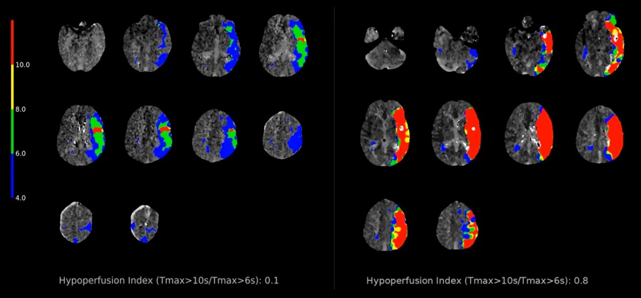
  • Total Hypoperfusion Index[…] also known as Hypoperfusion Intensity Ratio (HIR)[…] is Tmax>10s / Tmax>6s[…]
    • HIR[…]>[…]0.34[…] (per the DEFUSE 3[…] Trial) is a predictor of poor collateral flow and infarct growth
      • higher the value = higher[…] risk of ischemic core growth
Which of these patients have higher risk of ischemic penumbra progressing to core?

Patient on R (HIR>>>0.34)[…]
How can you use CTH + CTP to predict the LKN of a stroke?
  • acutely (say <4-6 hr): CTH wnl, CTP large mismatch[…]
  • intermediately (say ~8-10hr): CTH shows hypodensity, CTP smaller mismatch[…]
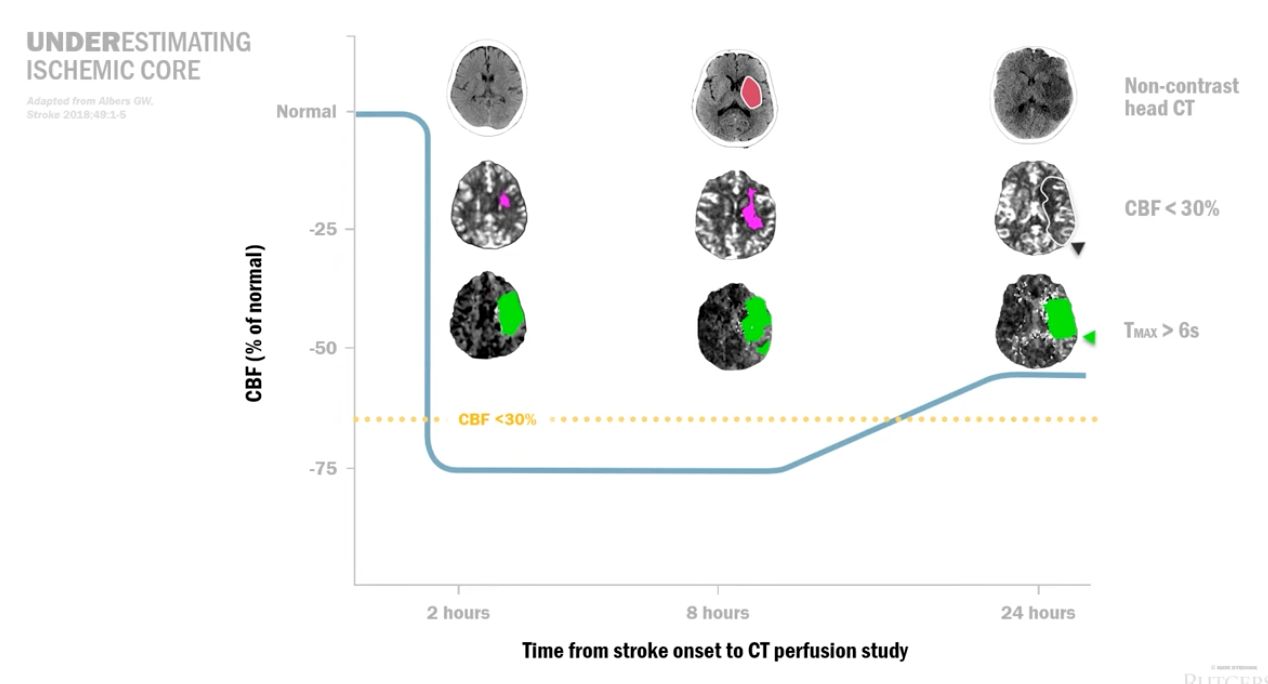
  • chronically (often >24hr): CTH shows further hypodensity, CTP CBF<30% may be absent bc leptomeningeal collaterals are recruited (but too late) while Tmax shows significantly delayed flow >6s[…]
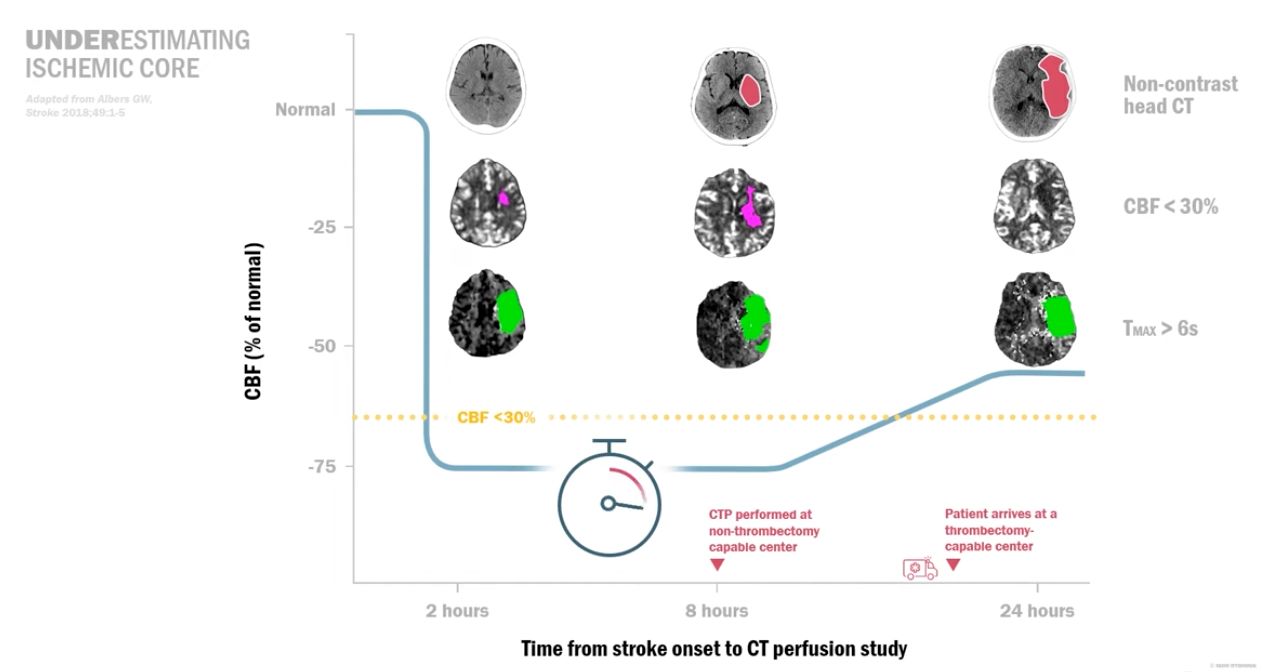
Why do we often need to repeat CTH/CTA/CTP when patients with stroke are transferred from non-EVT capable center to EVT-capable center?
  • CTP are time dependent & only reflect hemodynamics at the time it was performed -- transfer delays can lead to completion of stroke[…]

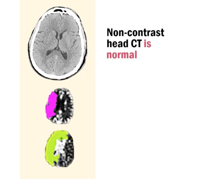
Say CTH wnl but CTP shows large penumbra & large core. What does this tell you about the stroke?

  • patient was within very early time window (<1-2 hours) so CTP overestimated core because it was picking up hypoperfused (but not ischemic) tissue[…]
  • this is also called "ghost[…]" core (because it does not truly exist)

Describe when rCBF may mis-predict ischemic core:
  • it may under[…]estimate core if CTP is performed too late[early or late?] - because surviving penumbra will rely on leptomeningeal collaterals to supply via alternative pathways to keep tissue alive 
  • it may over[…]estimate core if CTP is performed too early[early or late?] - because it may often confuse benign, sluggish blood flow with irreversible tissue death during the hyperacute phase
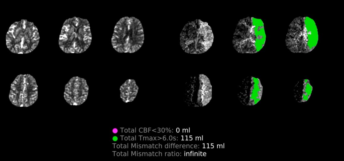
64M, NIHSS 7, LKN 11hr, CTH/CTA wnl, CTP:

rCBV normal and no core (rCBF<30%)
MTT normal to barely increased (some yellow dots)
Tmax>6s: large penumbra (Tmax 115cc) w/red-yellow
  • this is likely a chronically stenosed/occluded L ICA[…] w/ good collaterals thus over-estimated penumbra > shows that high rCBV[…] = good collaterals[…] and time independent[dependent or independent] unlike Tmax which is time dependent[dependent or independent]
62M, LKN 13hr, NIHSS 3, CTH wnl. Interpret the CTP:

  • small core 11cc, large Tmax 152 w/large mismatch 141/13.8 ratio; MTT delayed (red) but rCBV/rCBF wnl so likely chronic R ICA occlusion[…]
CTP
  • Tmax may overestimate penumbra in cases of chronic occlusion[…]
CTP - Determining Quality of Study
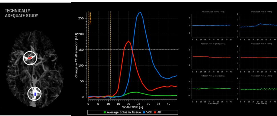
  • the study selects 2[…] points to be technically adequate
    • arterial input function (AIF)[…] (often MCA[…])
    • venous output function (VOF)[…] (often Straight Sinus[…])
  • A technically adequate study should meet the following parameters:
    • AIF[…] change in CT attenuation/density >150[…] HU (look at the y[…]-axis for this)
    • Look for very little side-to-side[…] movement as shown by the side by side rotation[…] and translation[…] X-axes graphs
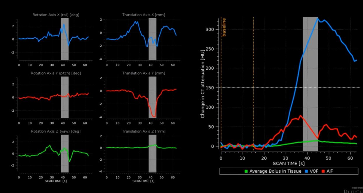
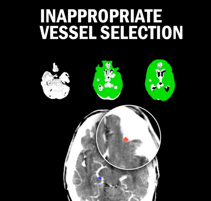
Why is this CTP not a good quality study?

  • (1): lot of motion on rotation/translation X-axes[…]
  • (2): AIF not appropriately selected as it does not cross 150 HU[…]
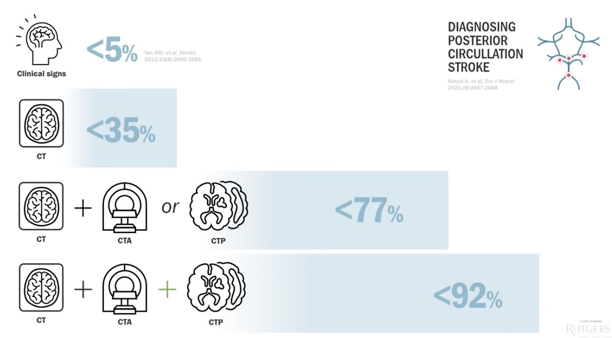
Sensitivity of CTP in tandem with other imaging for catching posterior circulation stroke:
  • no imaging (just clinical s/s alone): <5[…]% sensitivity
  • CTH alone: <35[…]% sensitivity
  • CTH+CTA or CTP alone: <77[…]% sensitivity
  • CTH+CTA+CTP: <92[…]% sensitivity
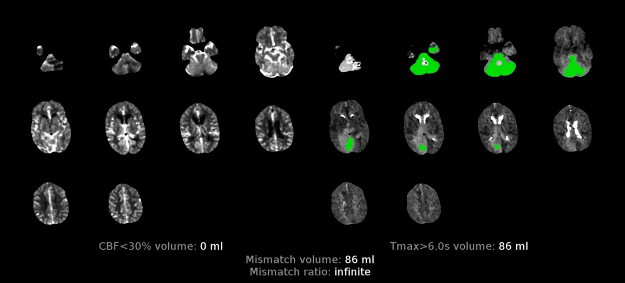
CTP Considerations in Posterior Circulation Stroke (with case example below)

  • rCBF[…] performs poorly bc the threshold was validated for anterior circulation strokes (such as DAWN/DEFUSE-3)
  • Tmax[…] performs poorly bc of skull base[…] artifact
  • Not every scanner can do whole-brain/whole-volume CTP meaning posterior circulation cuts may only have 1[…]-2[…] cross-sectional slices
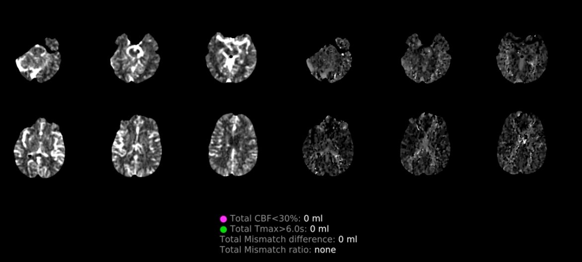
63M, IS w/abscess s/p R cranio, R MCA syndrome, NIHSS 13. CTH wnl. CTA wnl. CTP below - explain:

  • Tmax/MTT wnl, rCBV/rCBF increased (red/yellow) R MCA area > hyperemia c/f post-seizure[…]

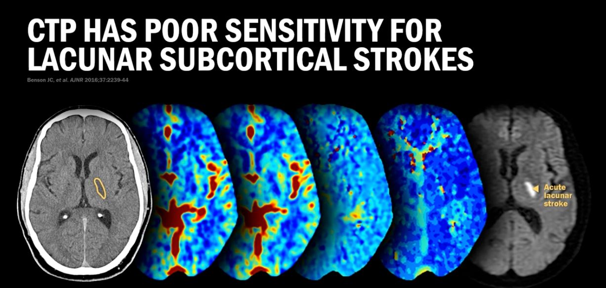
CTP has very poor sensitivity for picking up lacunar subcortical[…] strokes